Anatomy Bone Practical 1
1/100
There's no tags or description
Looks like no tags are added yet.
Name | Mastery | Learn | Test | Matching | Spaced |
|---|
No study sessions yet.
101 Terms
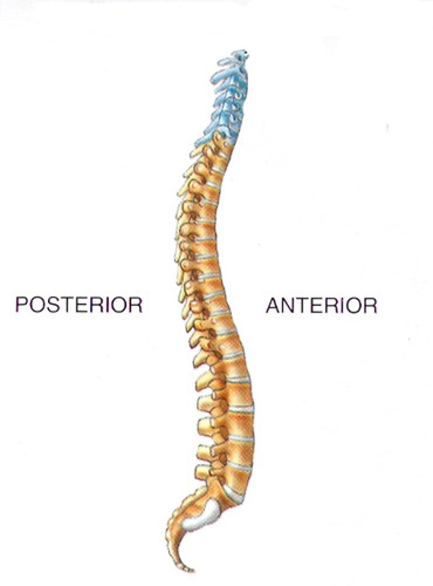
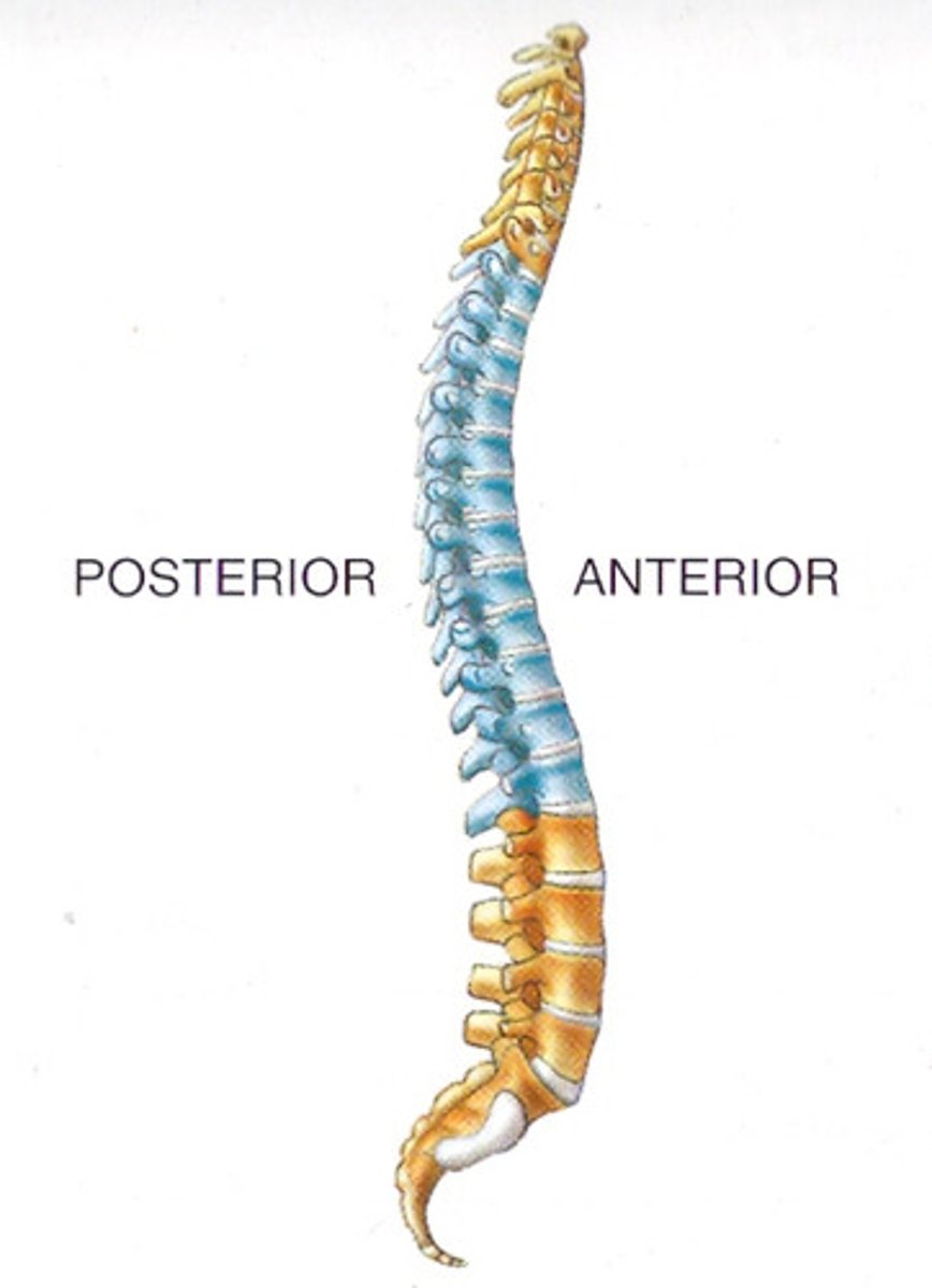
cervical ()
What type of vertebrae is shown in blue?
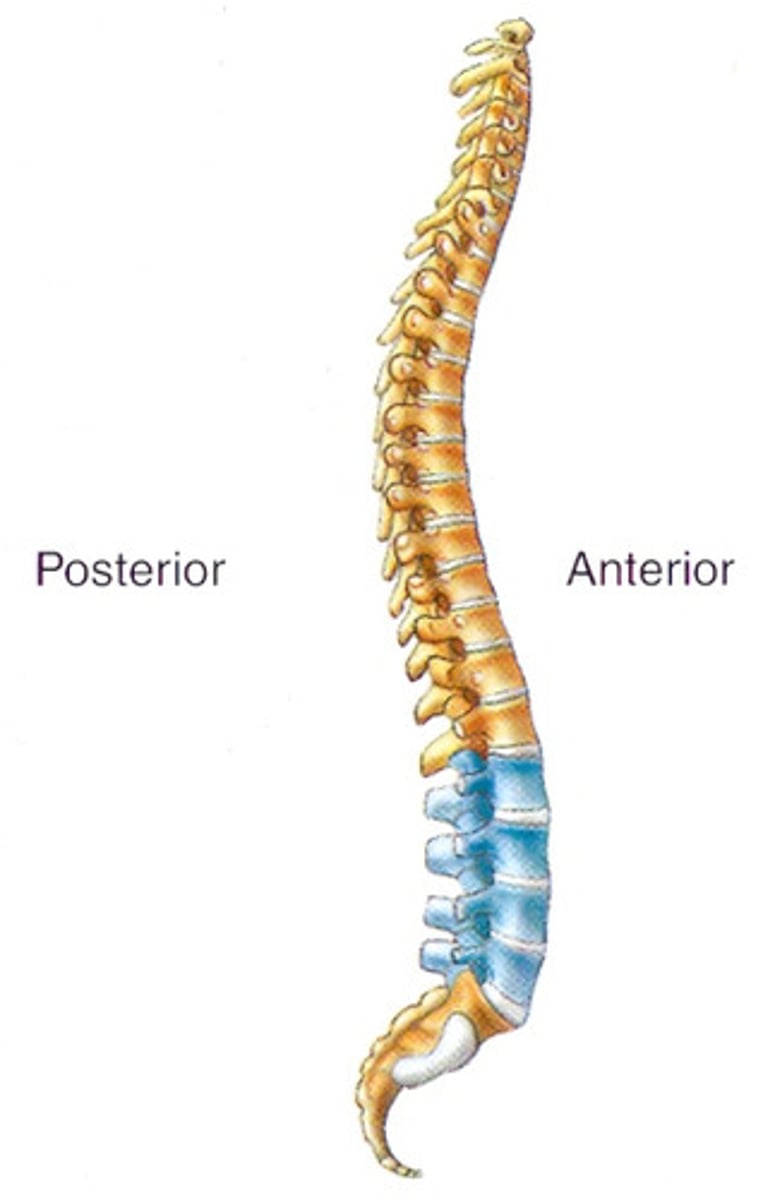
lumbar (5 largest)
What type of vertebrae is shown in blue?
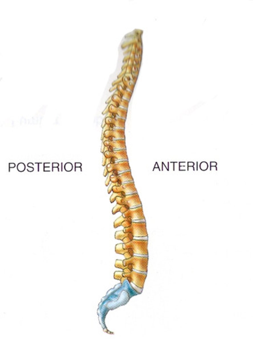
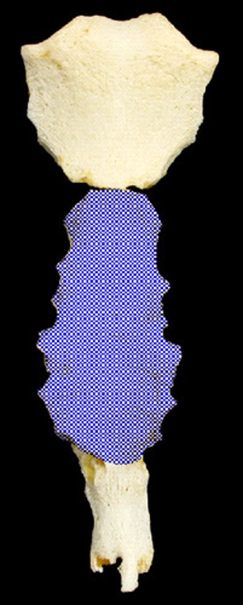
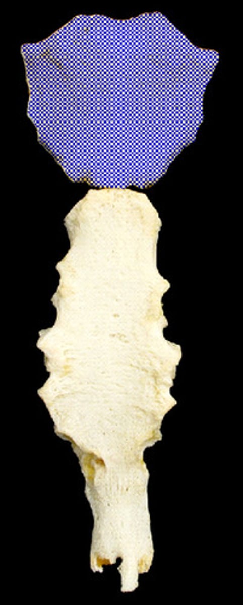
sacrum ()
What type of vertebrae is shown in blue?
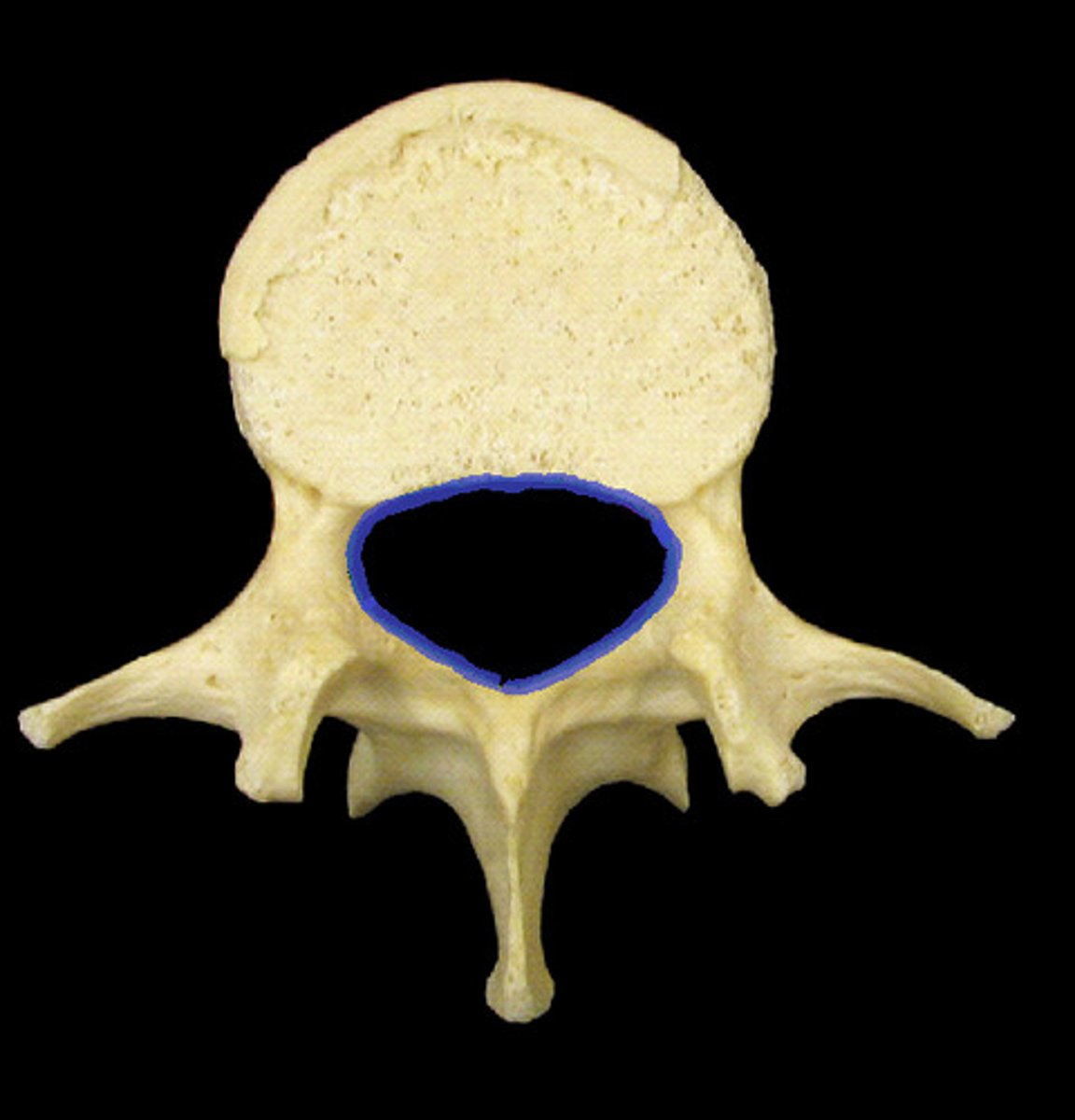
thoracic (12 vertabrae)
What type of vertebrae is shown in blue?
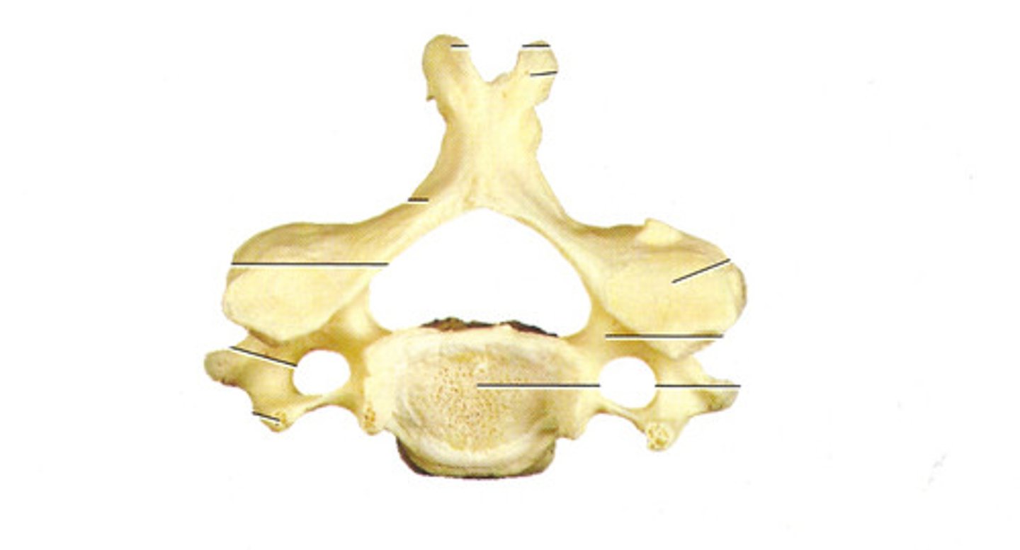
cervical
Which vertebrae is this?
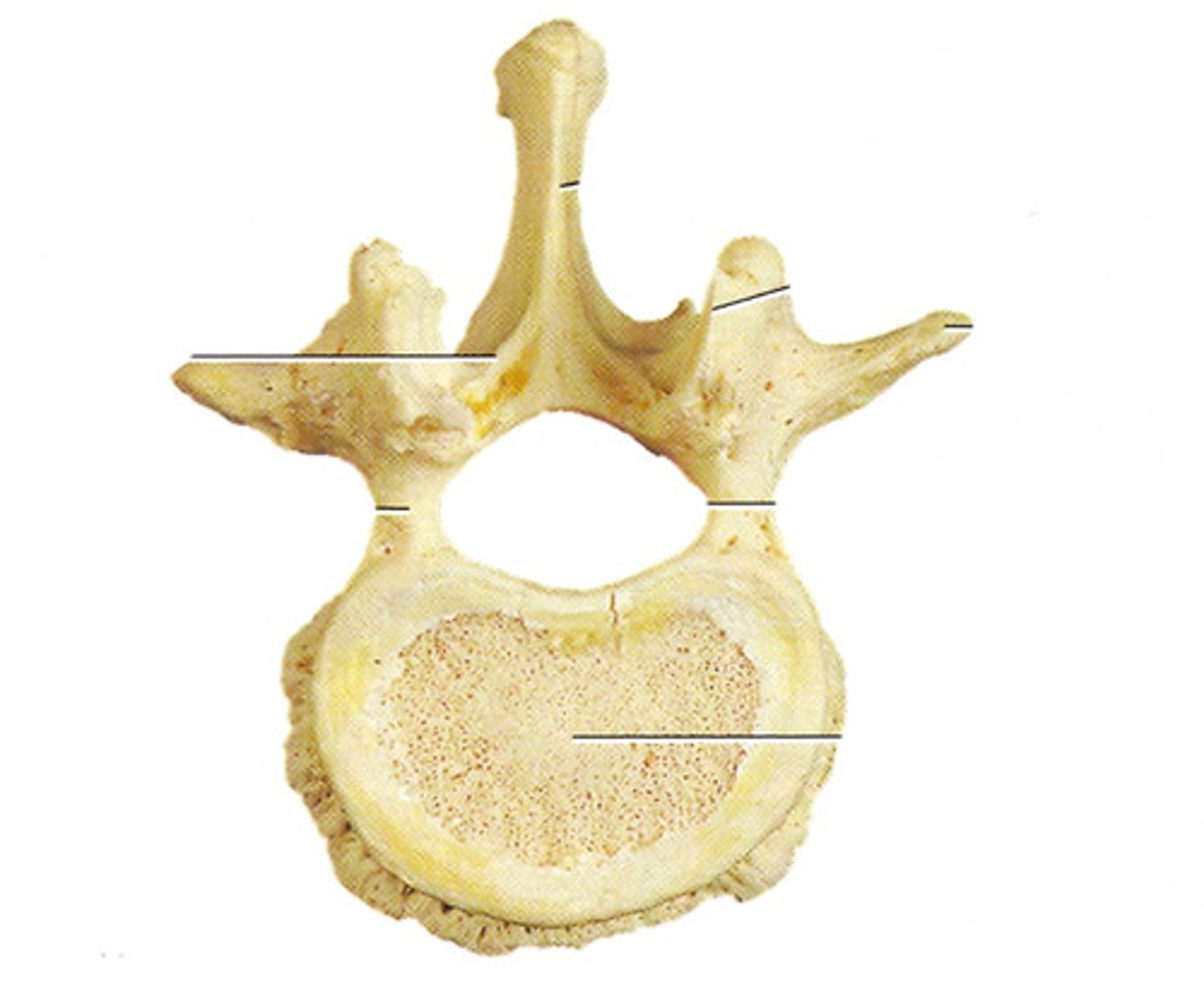
lumbar
Which vertebrae is this?
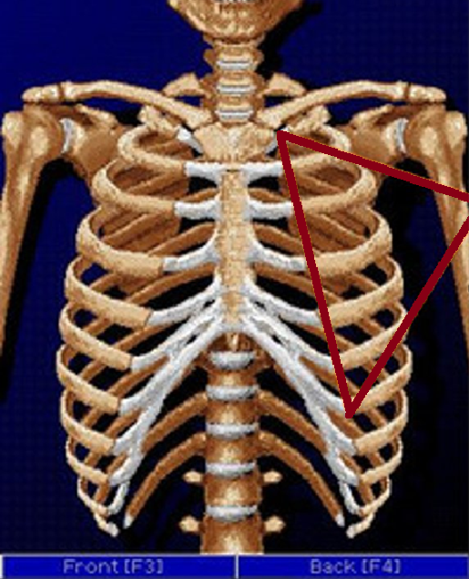
false ribs (8-12)
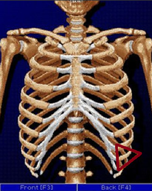
floating ribs (11-12)
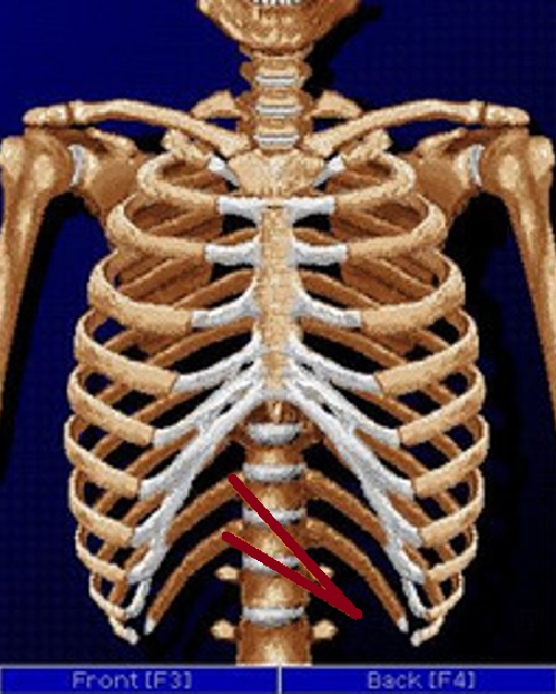
true ribs (1-7)
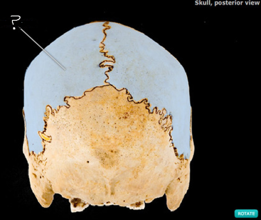
parietal
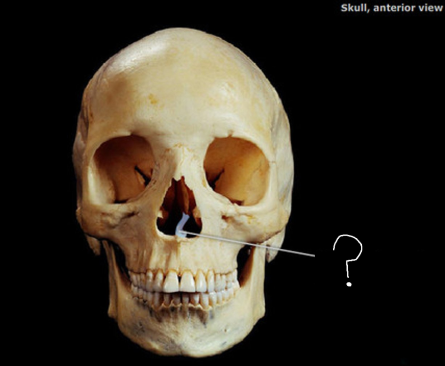
Vomer
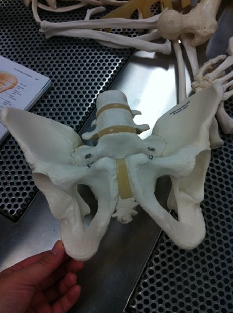
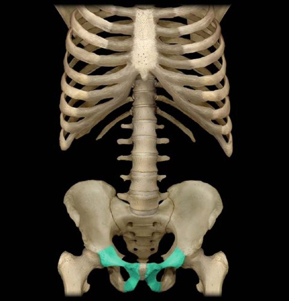
male pelvic girdle
boy or girl
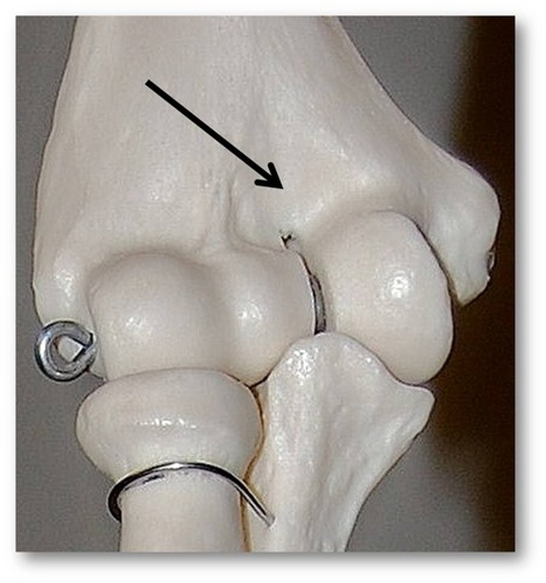
Coronoid fossa
Name this specific part of the humerus.
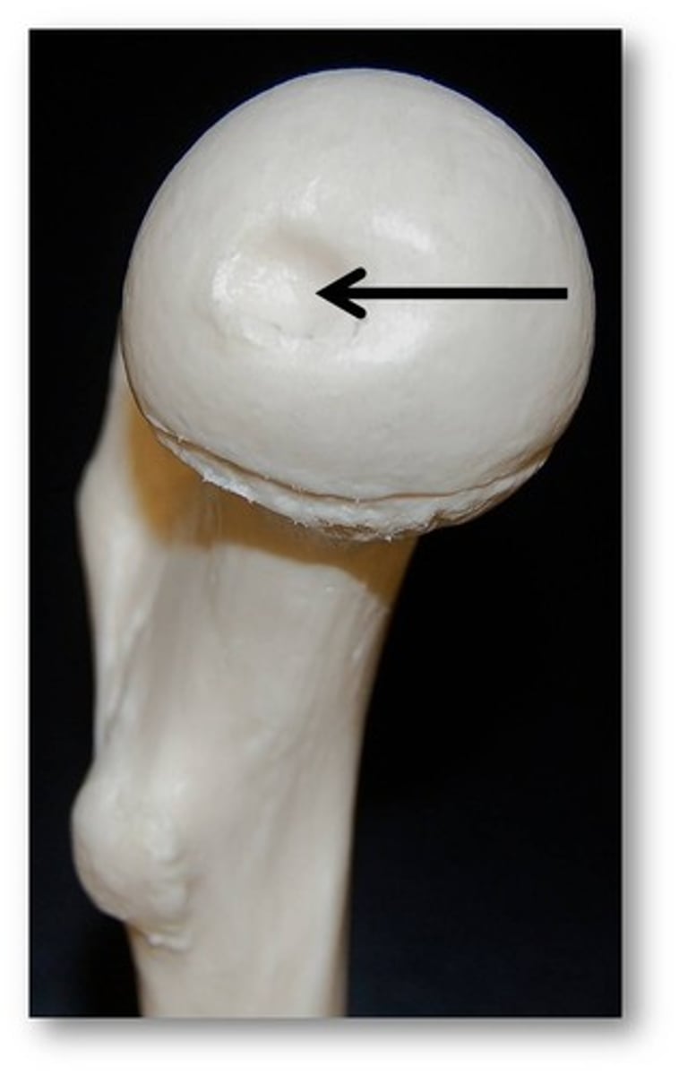
Fovea capitis
Name this specific bony landmark of the femur.
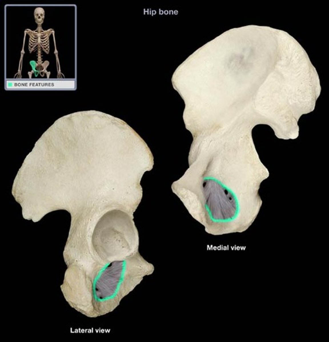
acetabulum
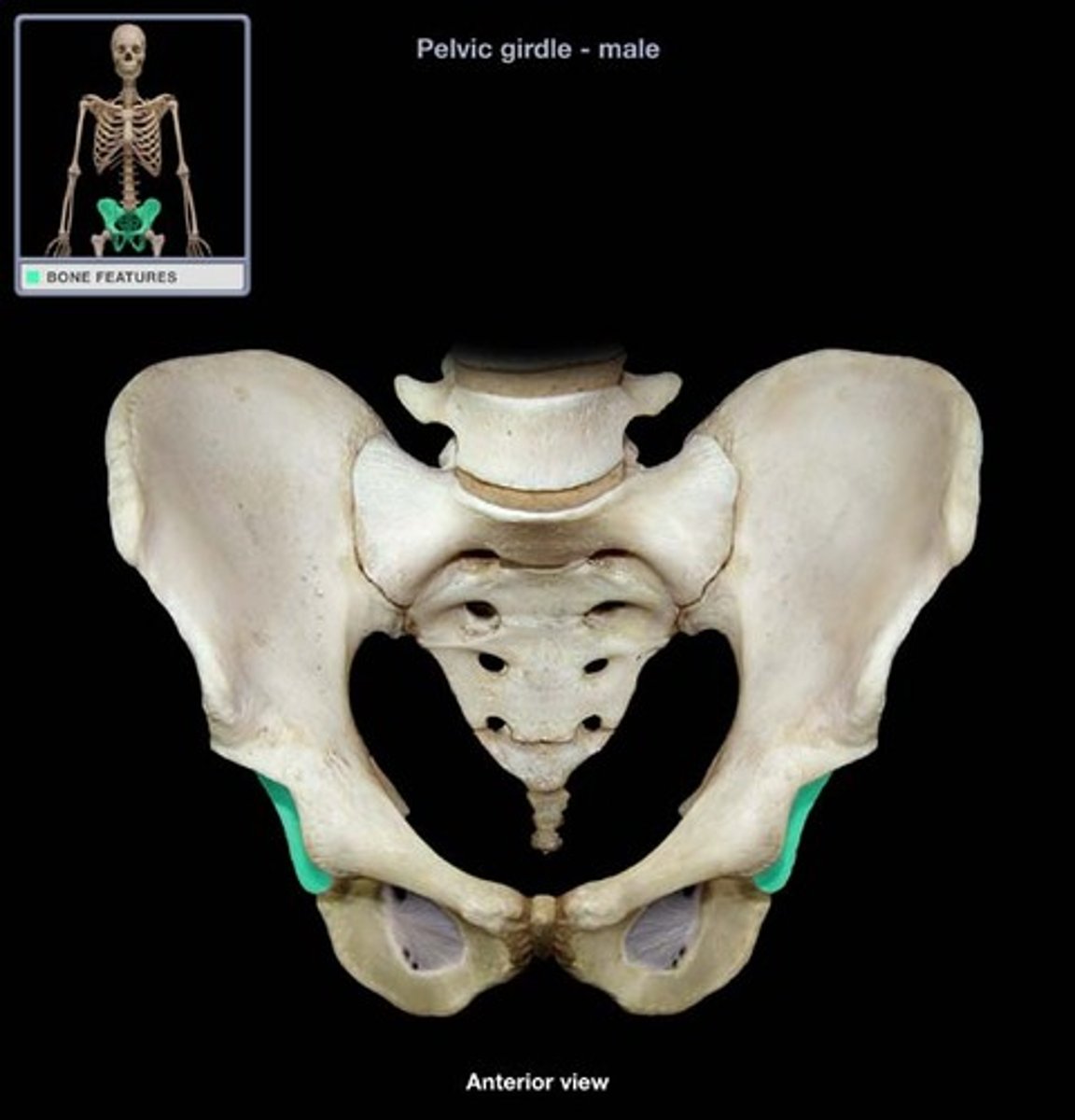
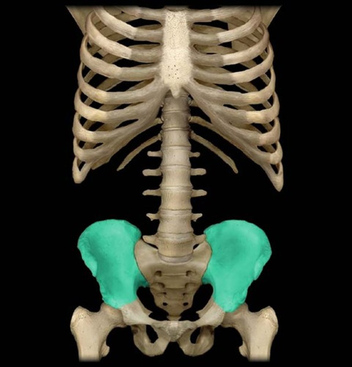
ilium
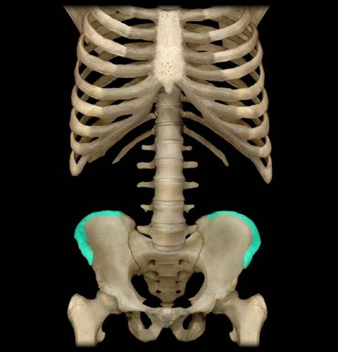
iliac crest
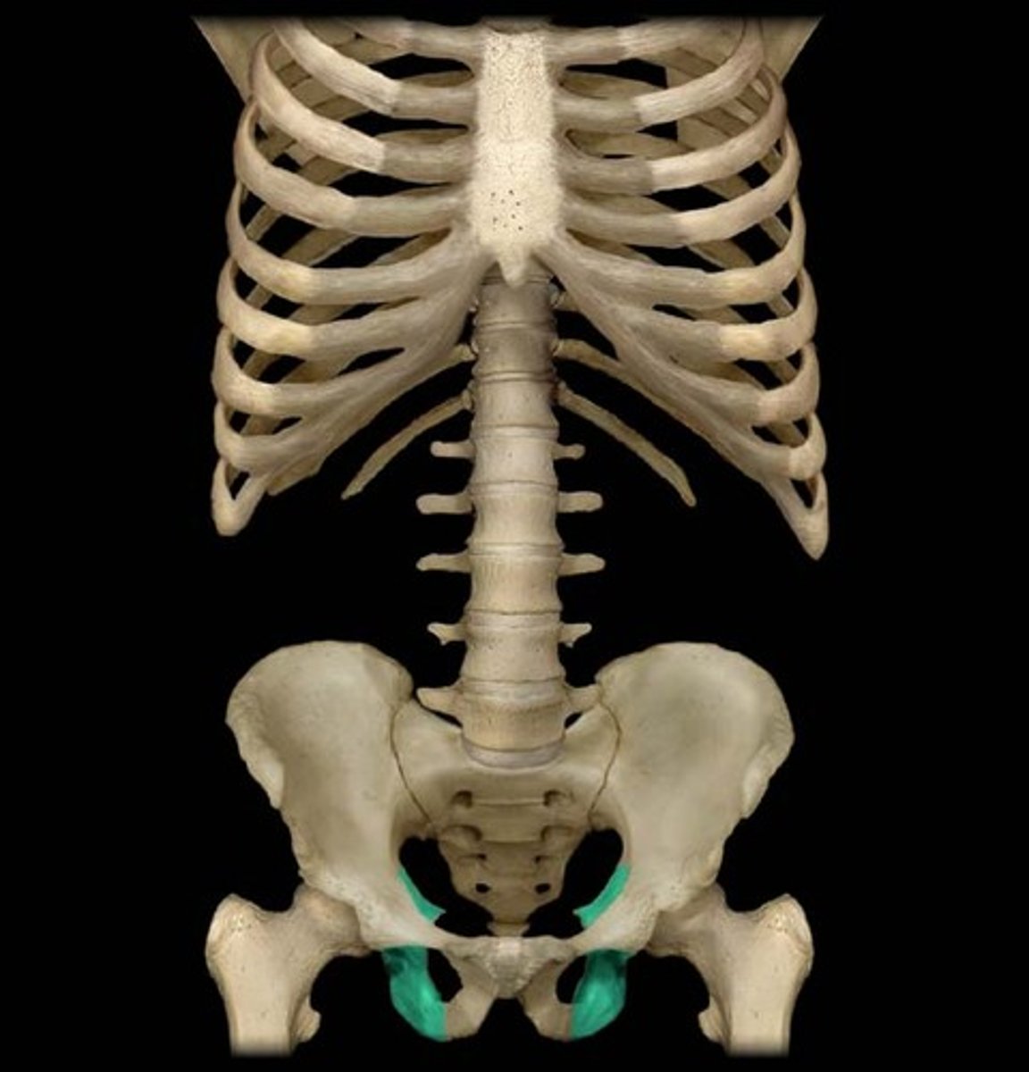
ischium
obturator foramen
pubis
ilium

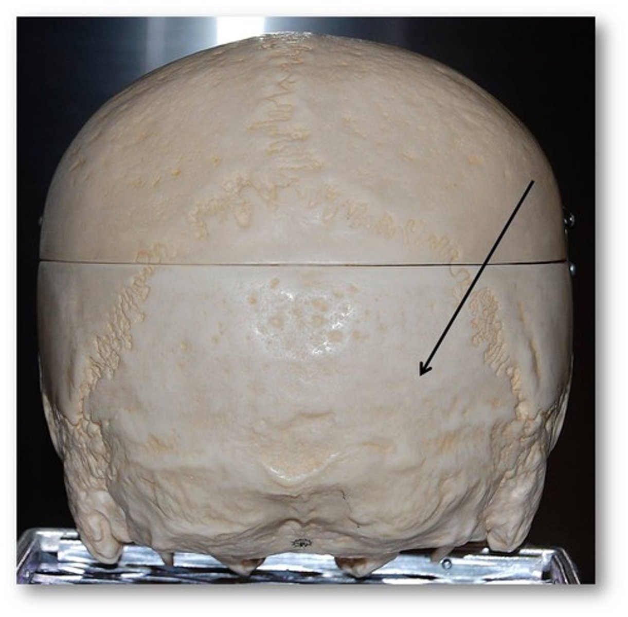
Occipital bone
Name this bone.
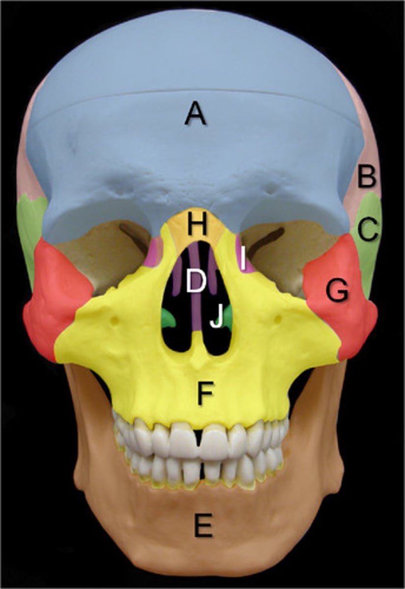
A. Frontal Bone
B. Parietal Bones
C. Temporal Bones
D. Ethmoid Bone
E. Mandible
F. Maxillary Bones
G. Zygomatic Bones
H. Nasal Bones
I. Lacrimal Bones
J. Inferior Nasal Conchae
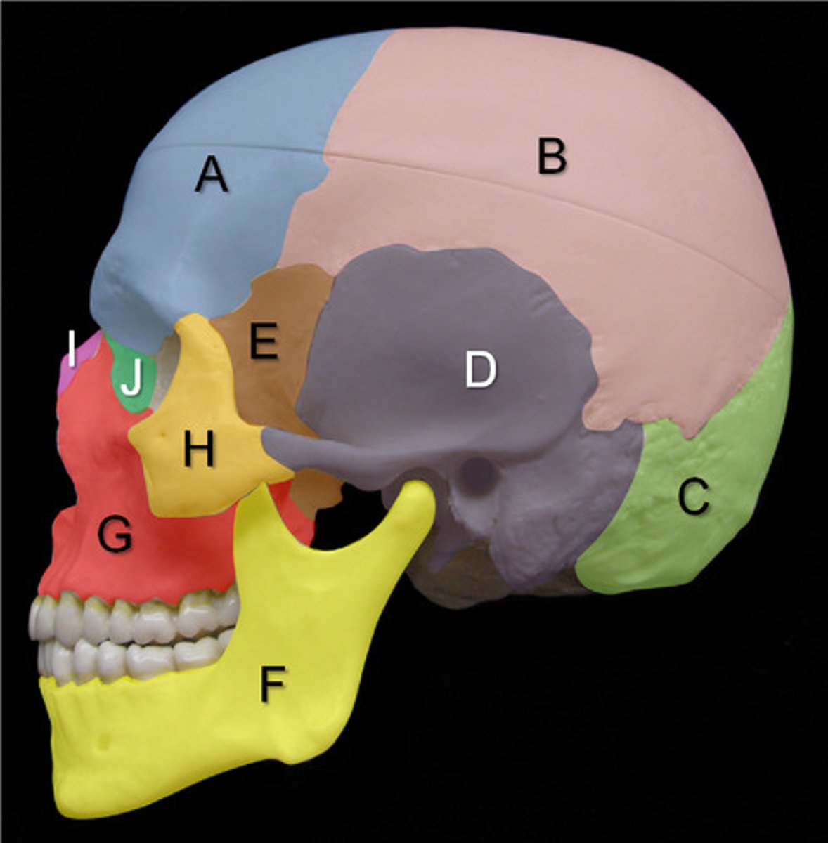
A. Frontal Bone
B. Parietal Bone
C. Occipital Bone
D. Temporal Bone
E. Sphenoid Bone
F. Mandible
G. Maxillary Bone
H. Zygomatic Bone
I. Nasal Bone
J. Lacrimal Bone
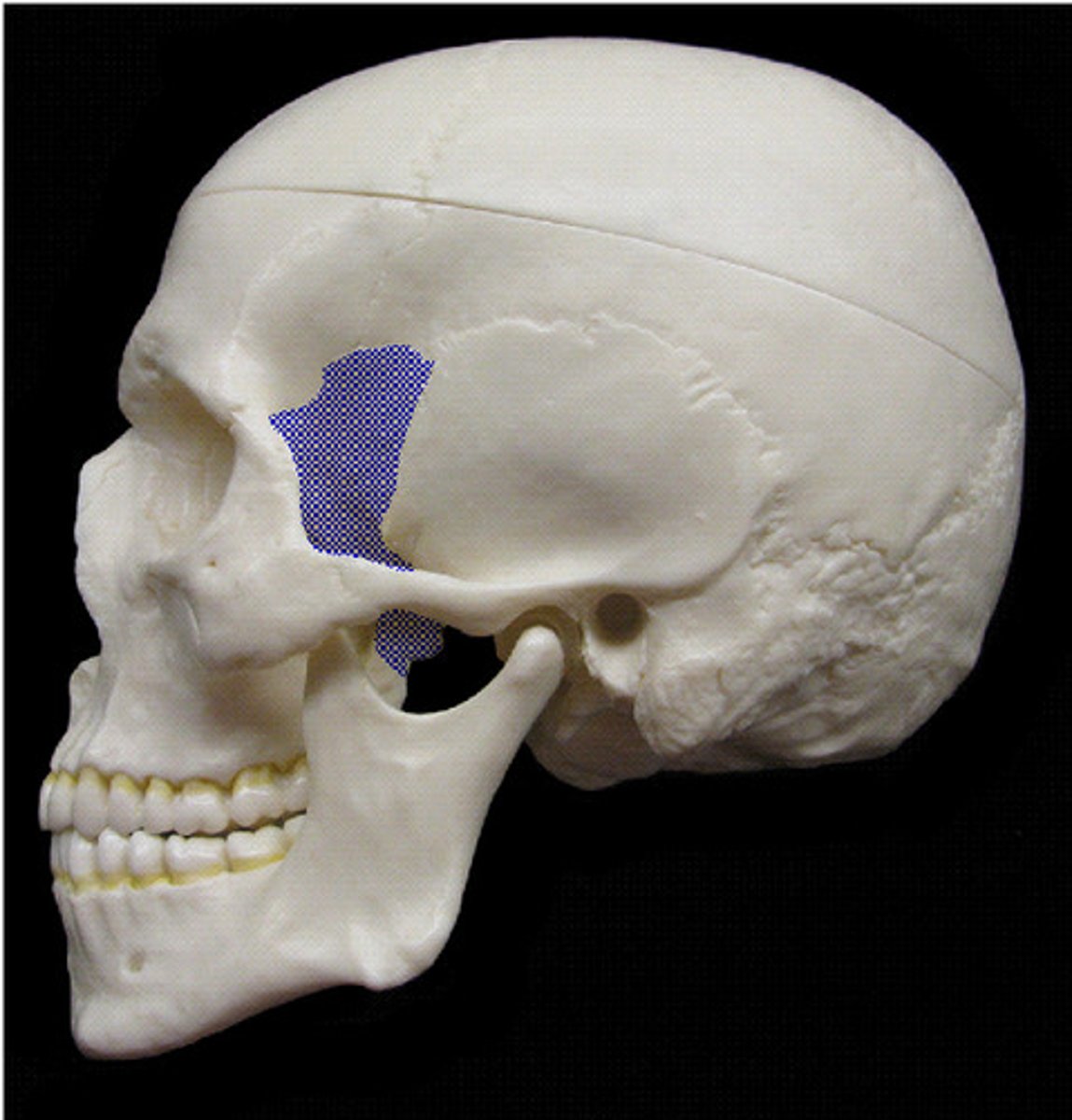
Sphenoid Bone
(lateral view)

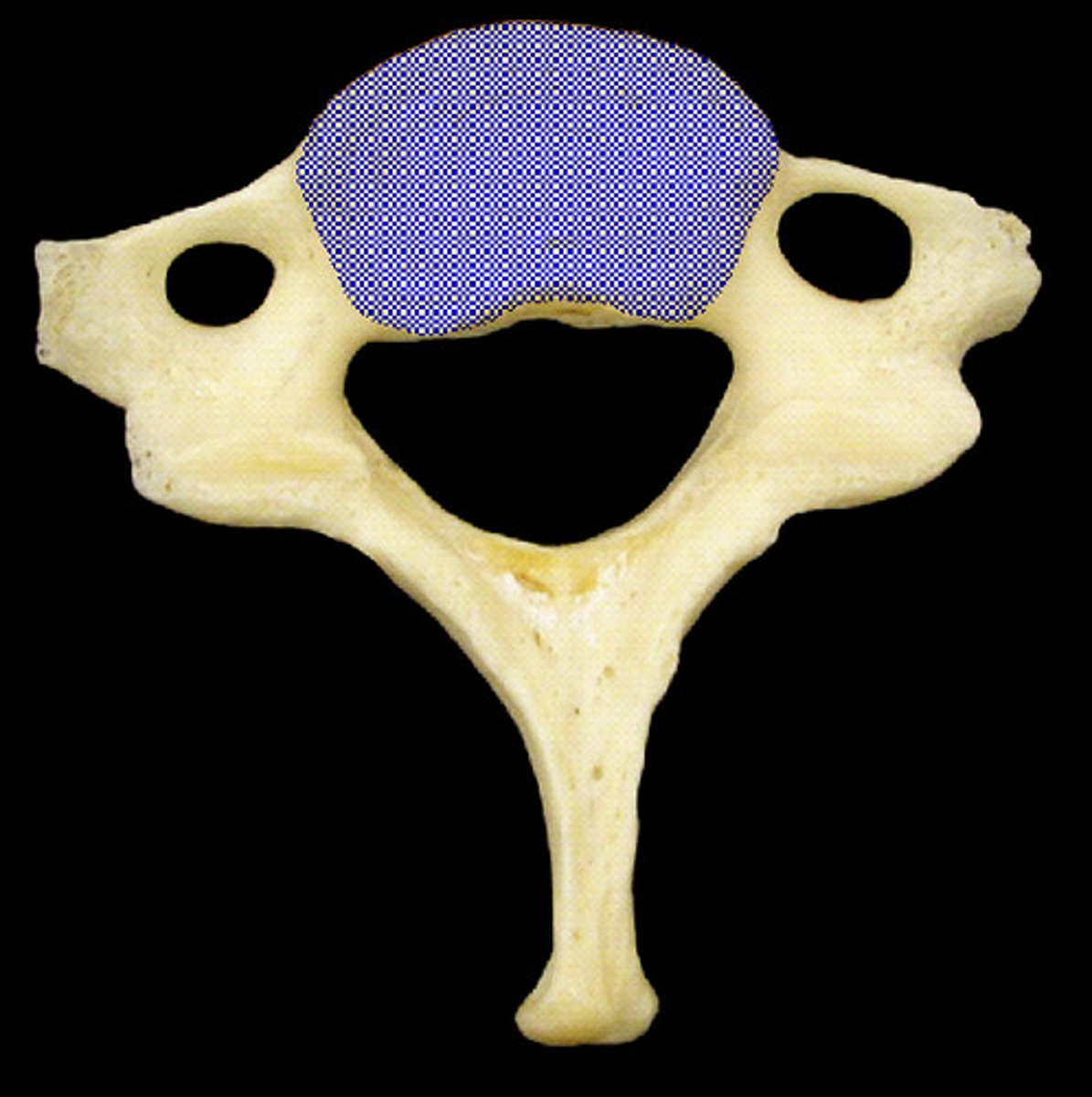
Body of Cervical Vertebrae
(superior view)
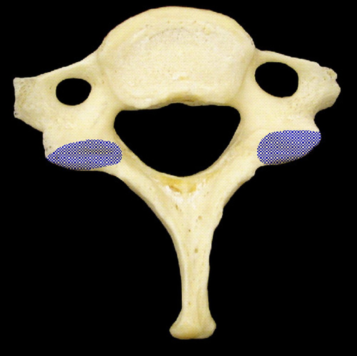
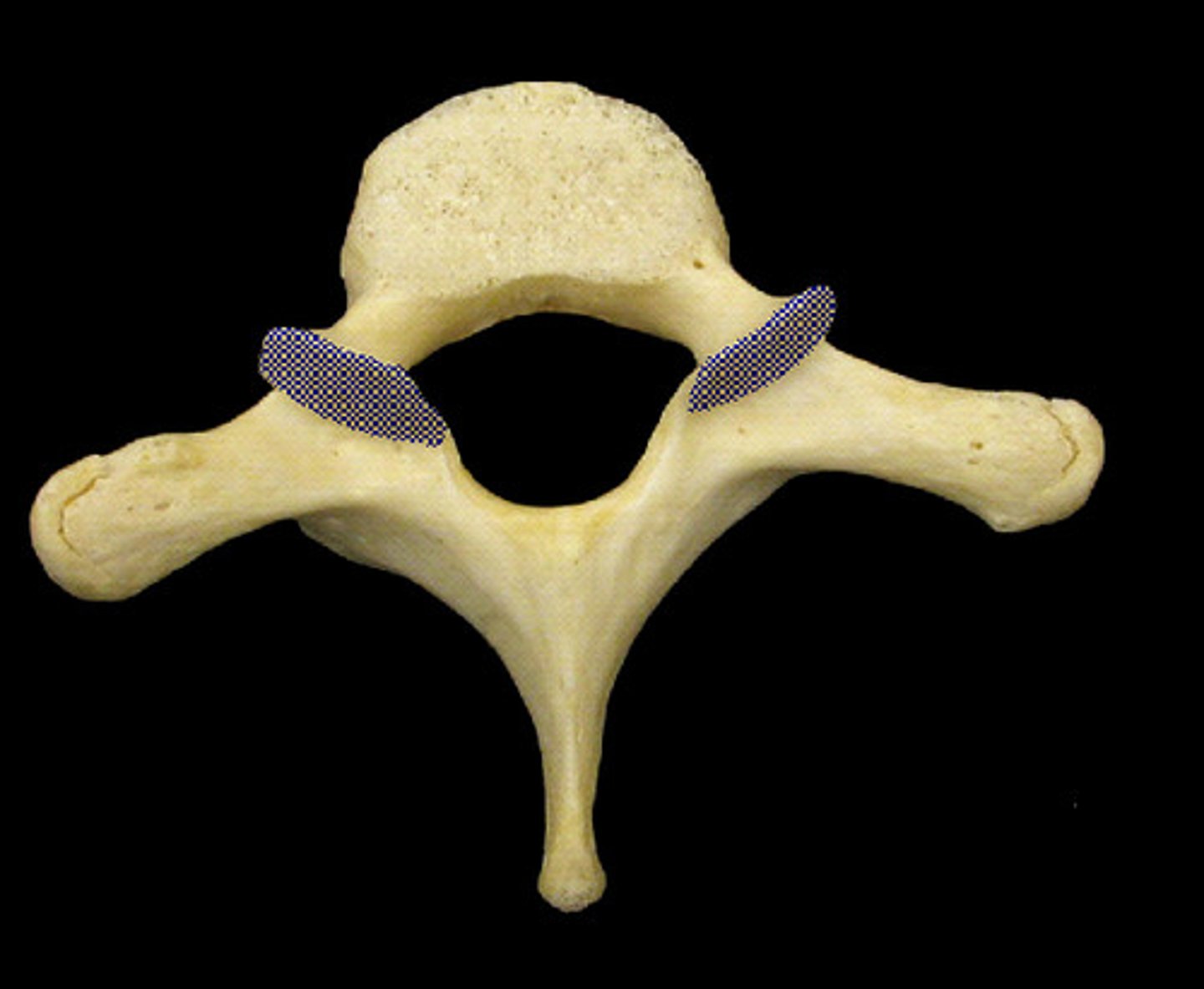
Superior Articulating Facets of Cervical Vertebrae
(superior view)
Transverse Foramina of Cervical Vertebrae
(superior view)

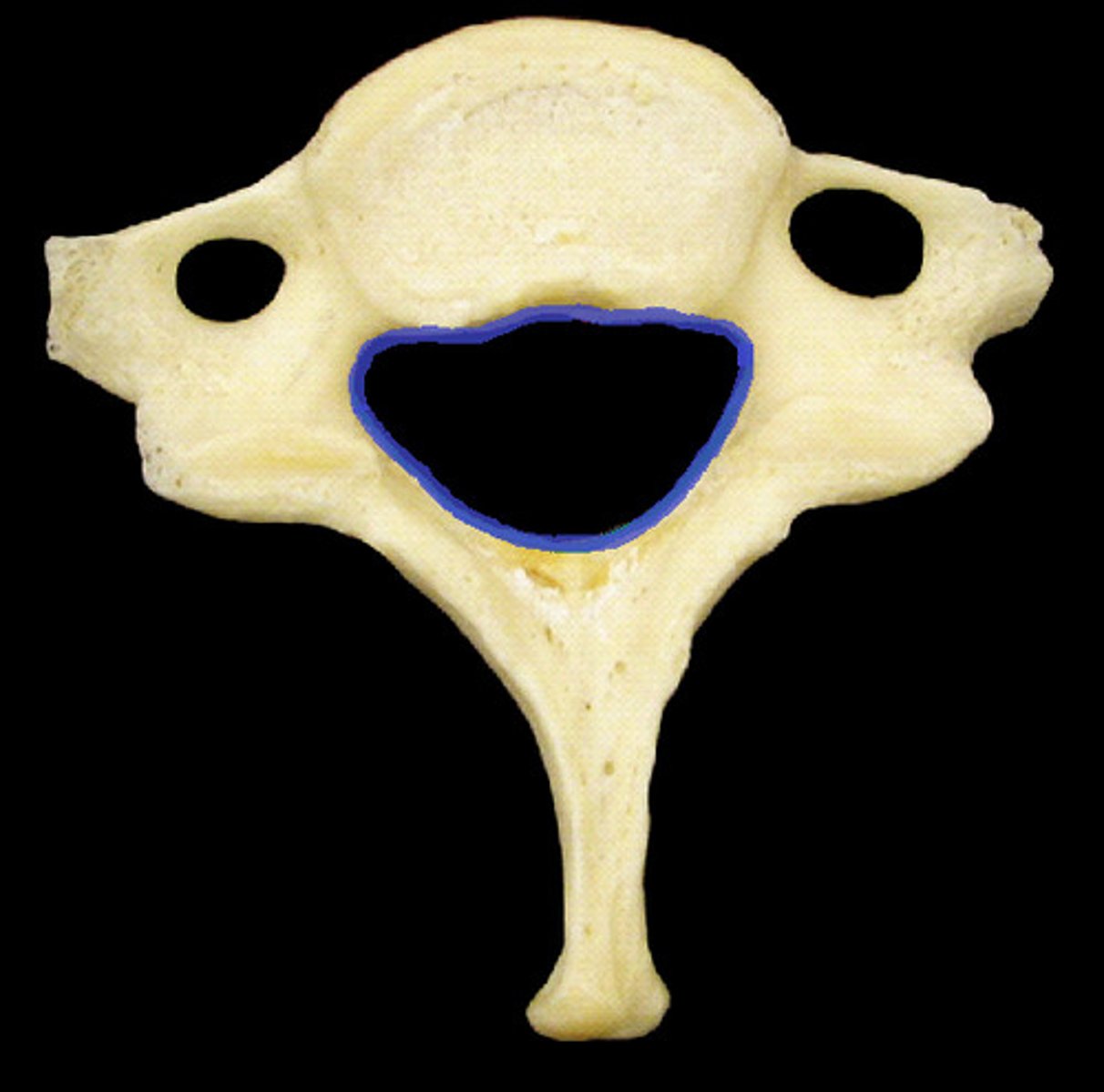
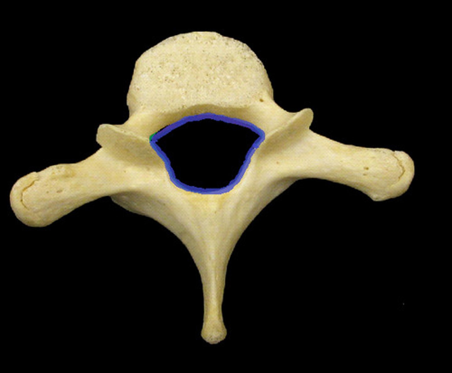
Vertebral Foramen of Cervical Vertebrae
(superior view)
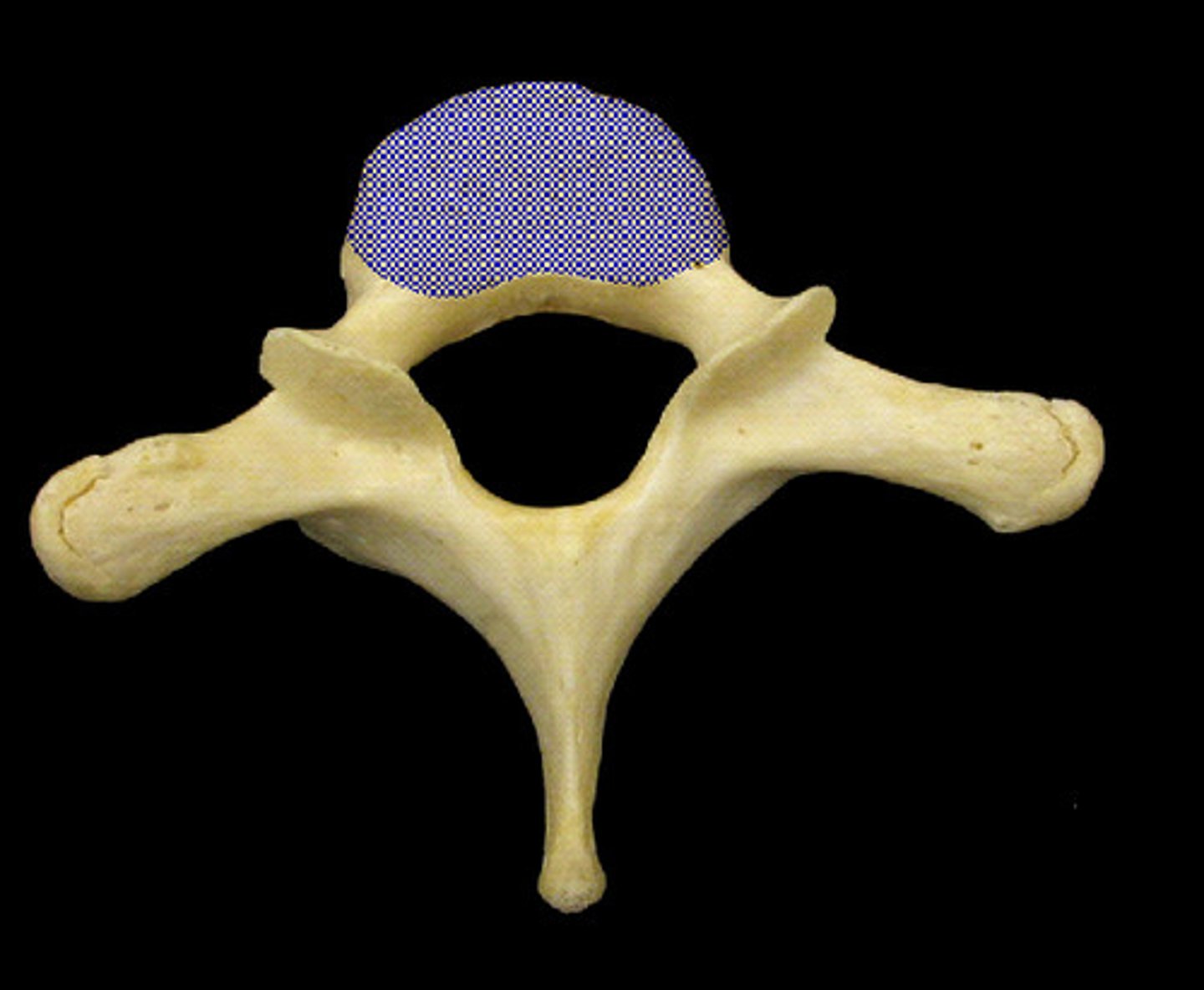
Body of Thoracic Vertebrae
(superior view)
Superior Articulating Facets of Thoracic Vertebrae
(superior view)
Vertebral Foramen of Thoracic Vertebrae
(superior view)
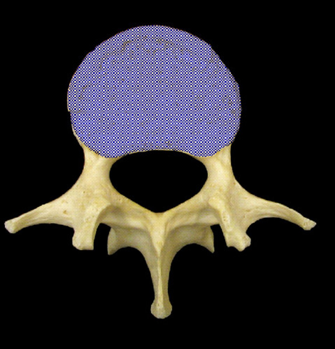
Body of Lumbar Vertebrae
(inferior view)
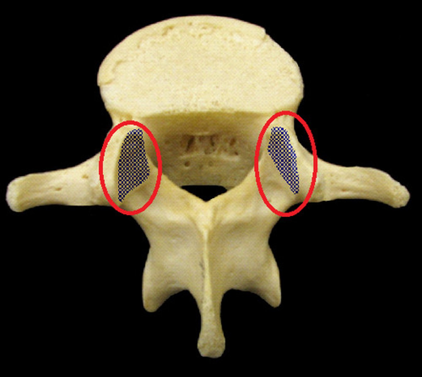
Superior Articulating Facets of Lumbar Vertebrae
(superior view)
Vertebral Foramen of Lumbar Vertebrae
(inferior view)
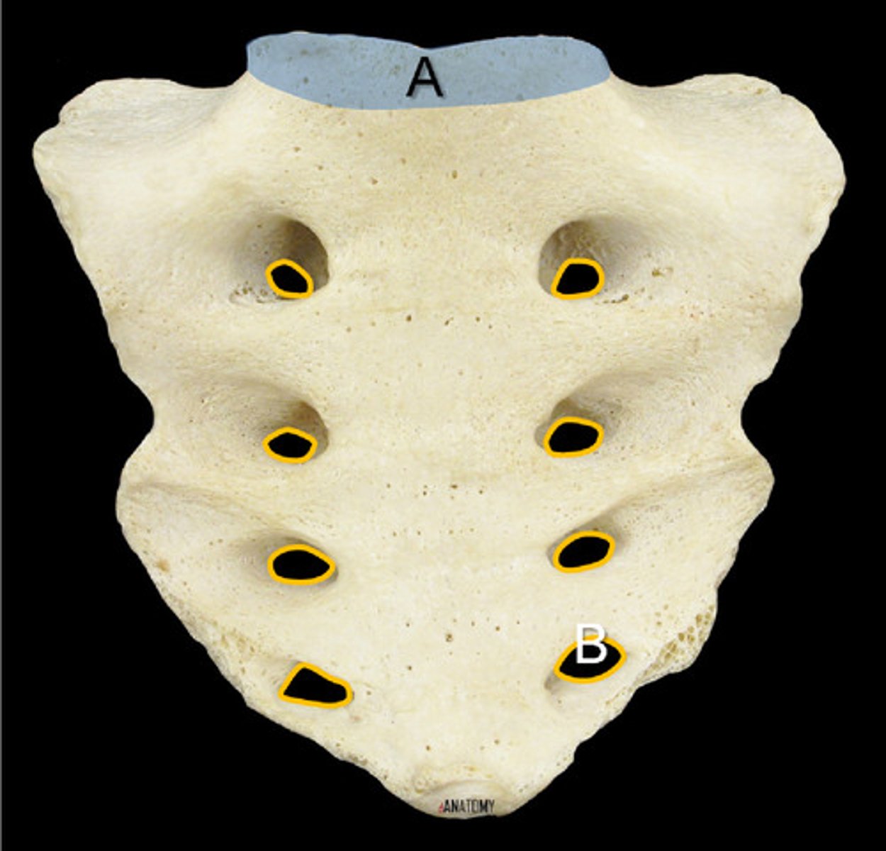
Sacrum (anterior view)
A. Sacral Promontory of the Sacrum
B. Sacral Foramina of the Sacrum
Body of the Sternum
Manubrium of the Sternum
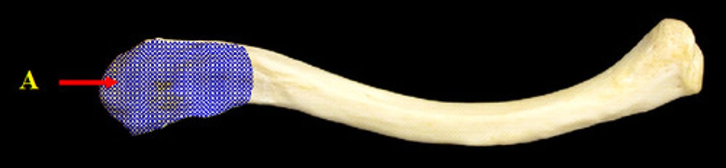
Acromial End of the Clavicle
(inferior view)
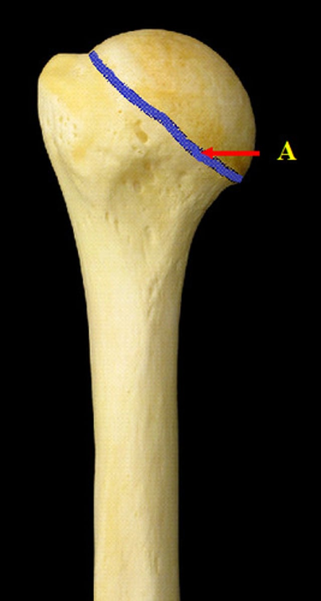
Anatomical Neck of the Humerus
(proximal end, posterior view)
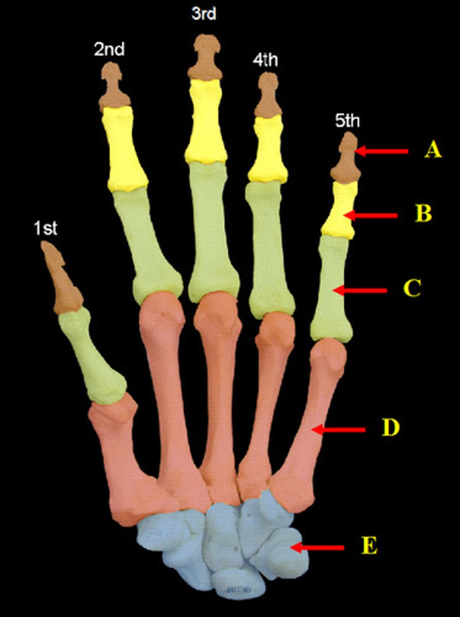
Bones of the Hand
(superior view)
A. Distal Phalanges
B. Middle Phalanges (not present in the thumb so there is no 1st middle phalanx)
C. Proximal Phalanges
D. Metacarpals
E. Carpals
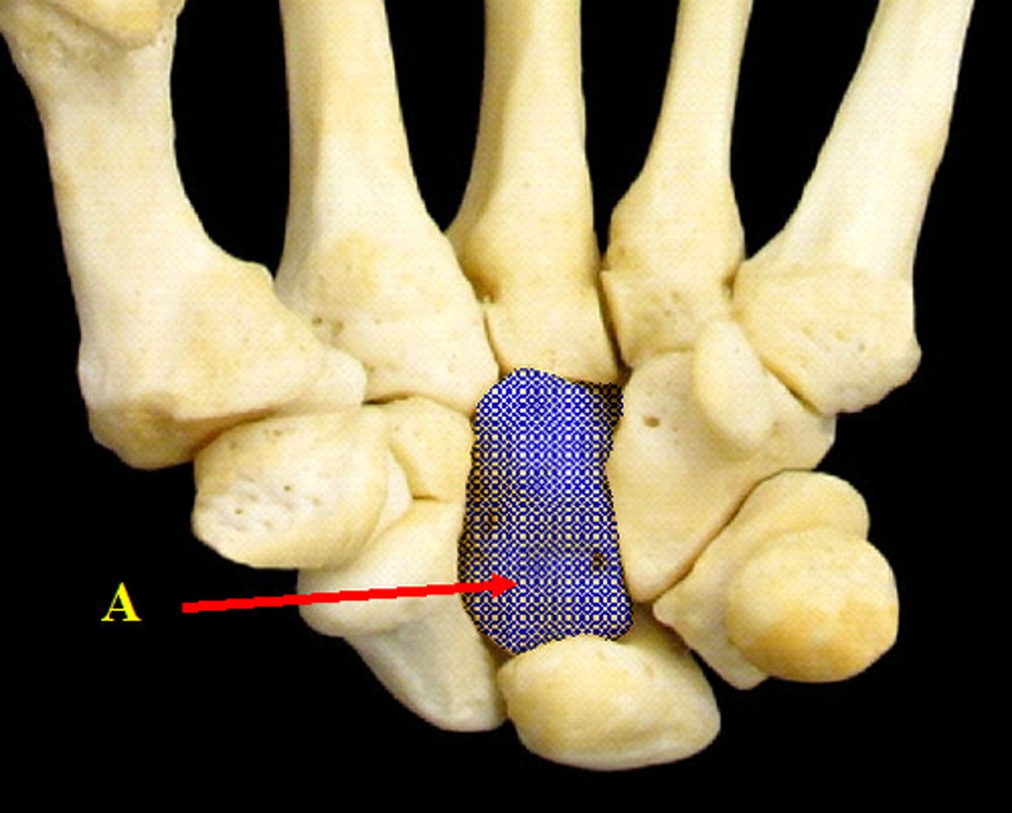
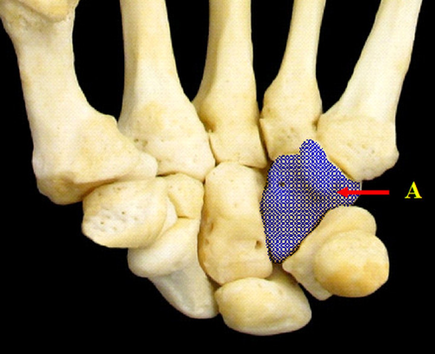
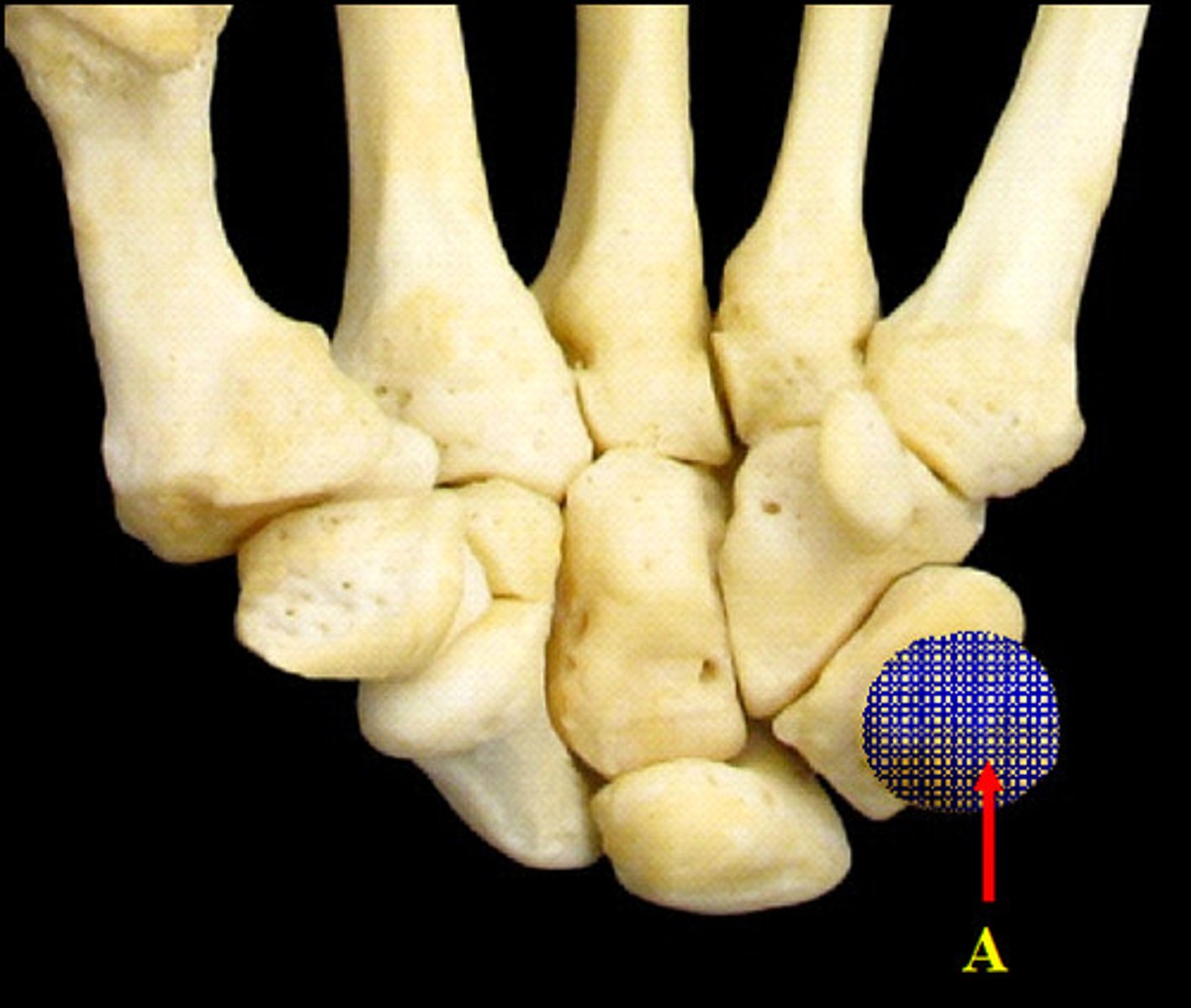
Capitate Carpal
("Can't)
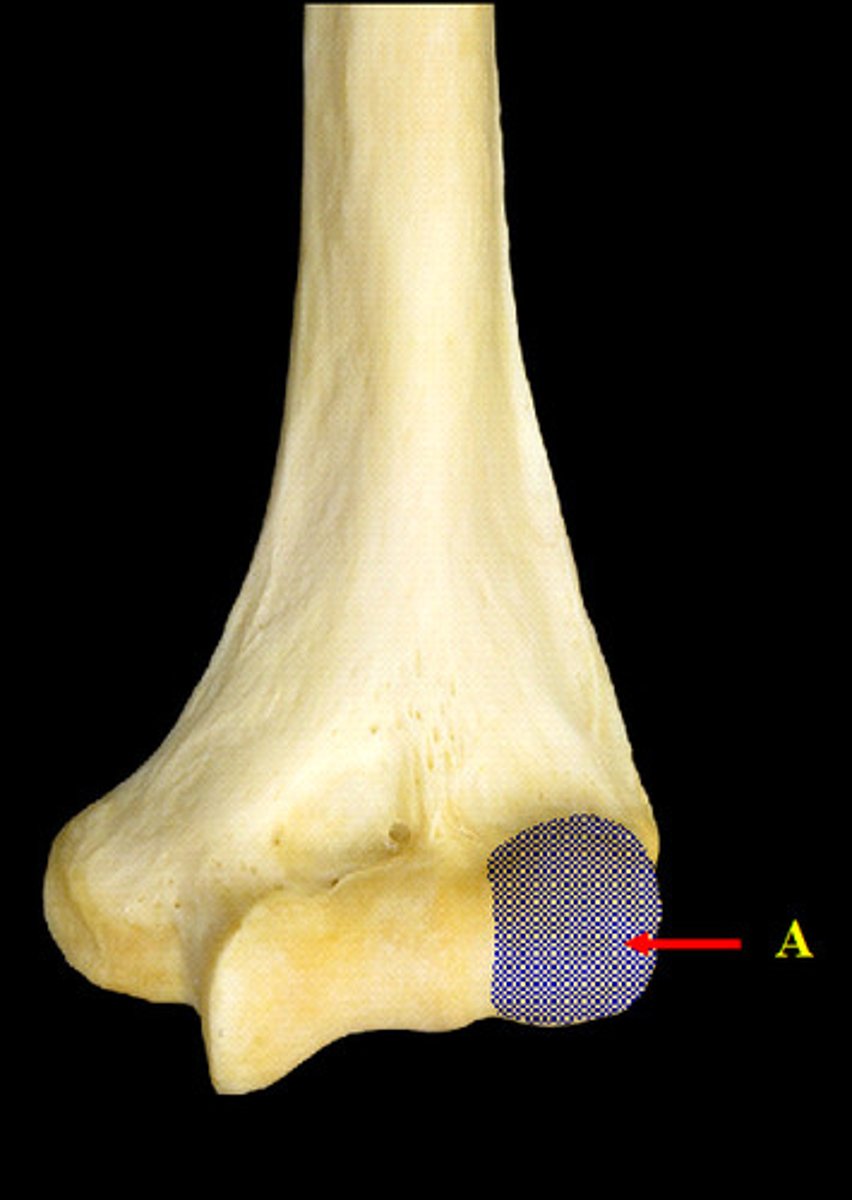
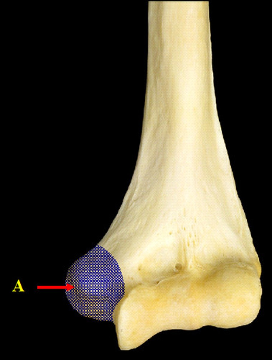
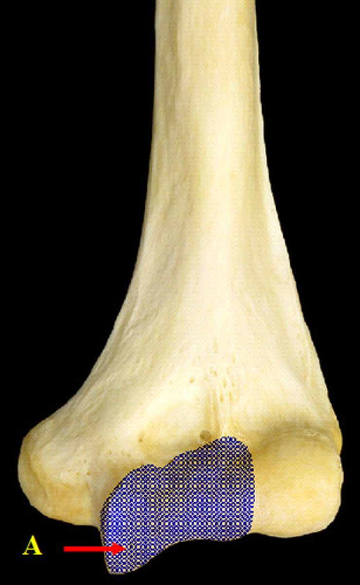
Capitulum of the Humerus
(distal end, anterior view)
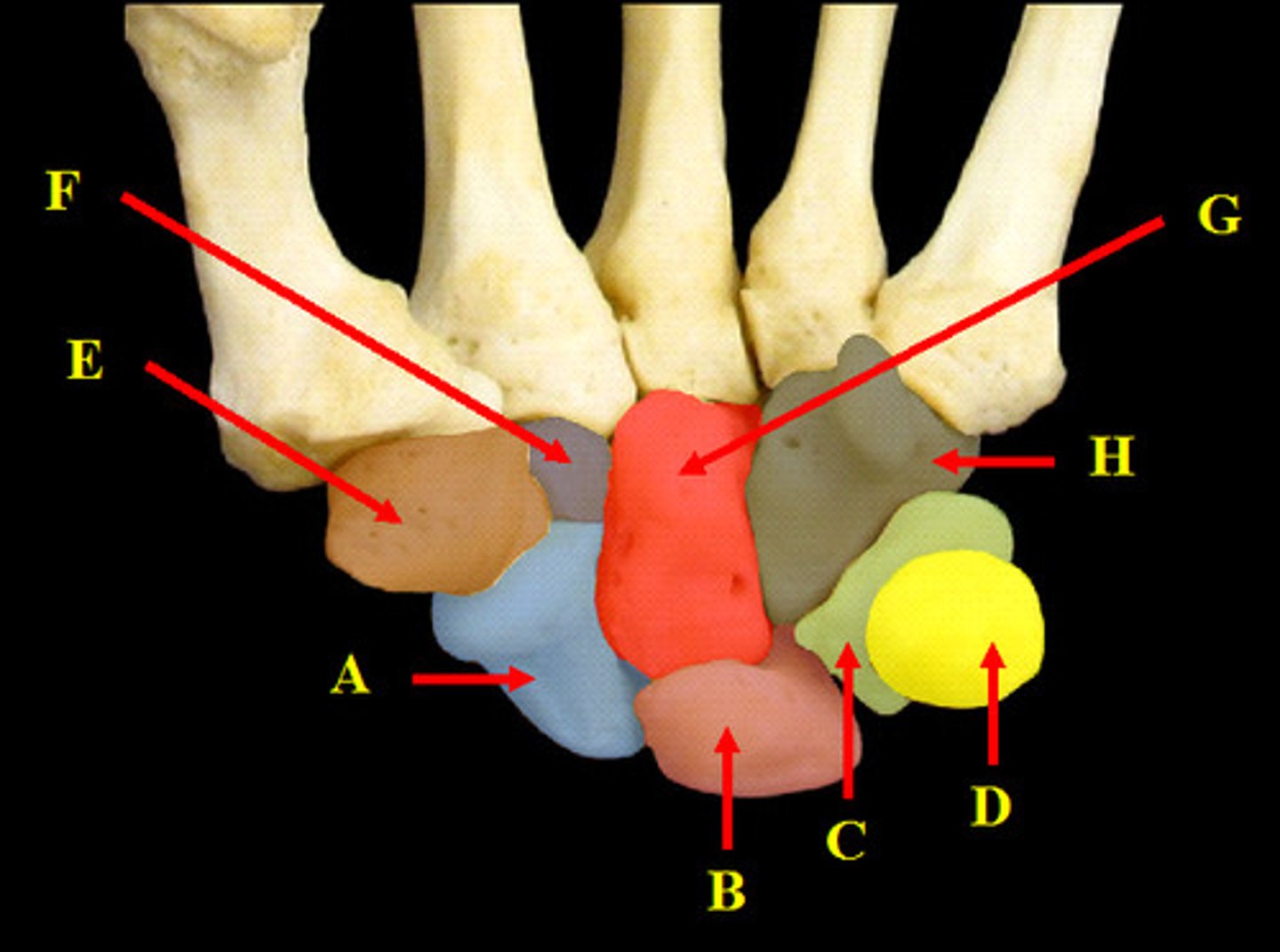
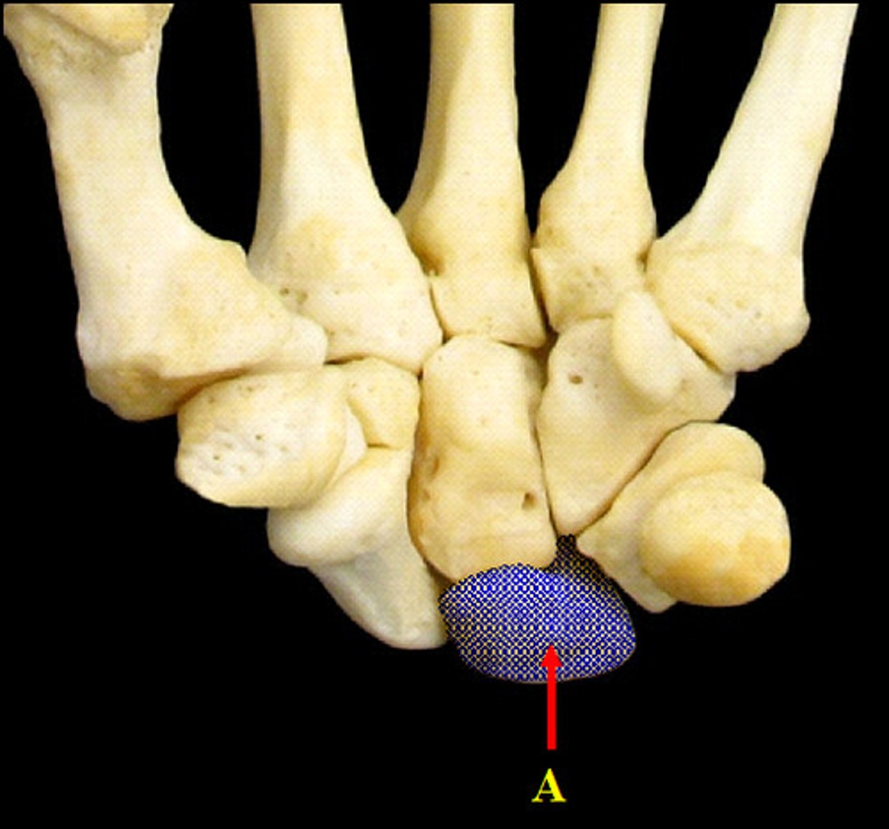
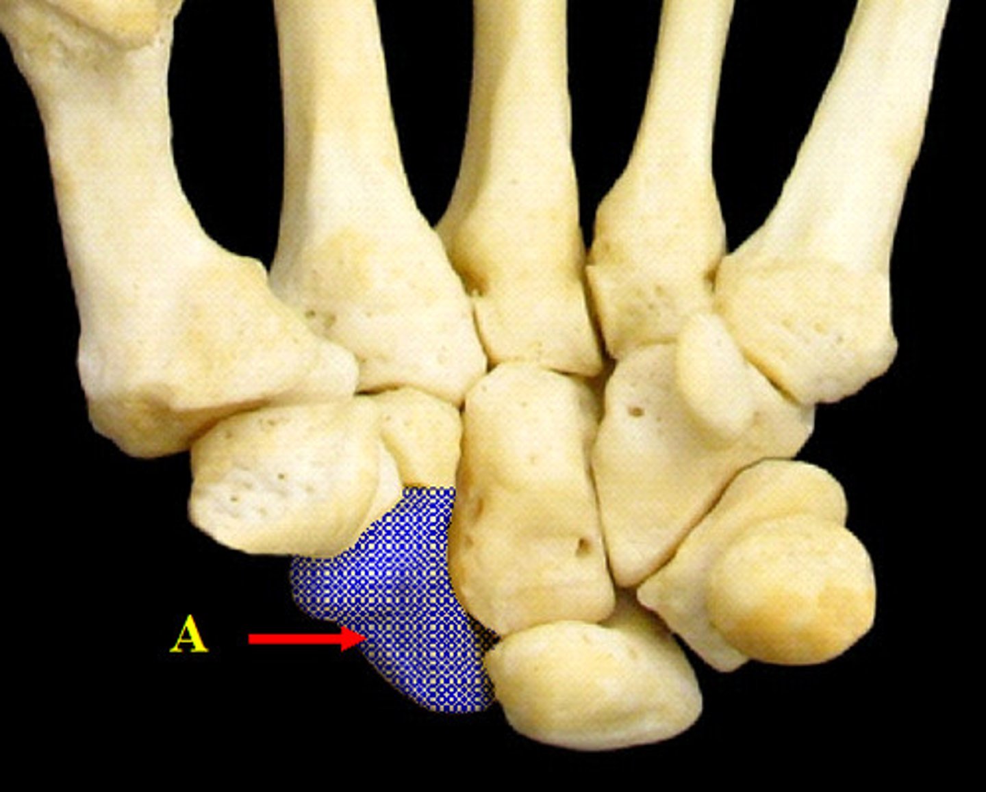
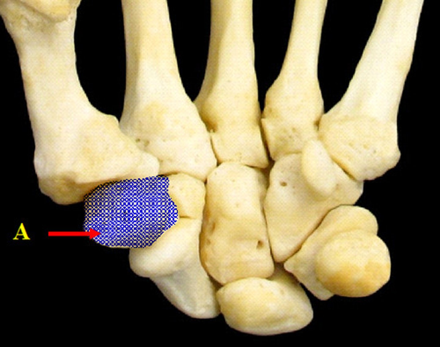
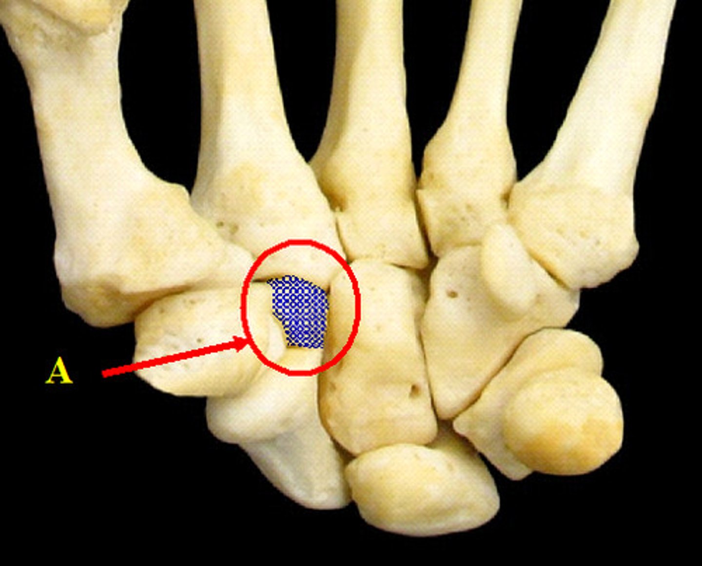
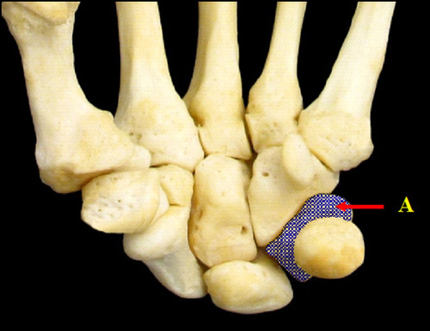
Carpal Bones of the Hand
(anterior view)
A.Scaphoid
B. Lunate
C. Triquetrum
D. Pisiform
E. Trapezium
F. Trapezoid
G. Capitate
H. Hamate
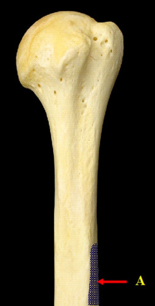
Deltoid Tuberosity
(proximal end, anterior view)
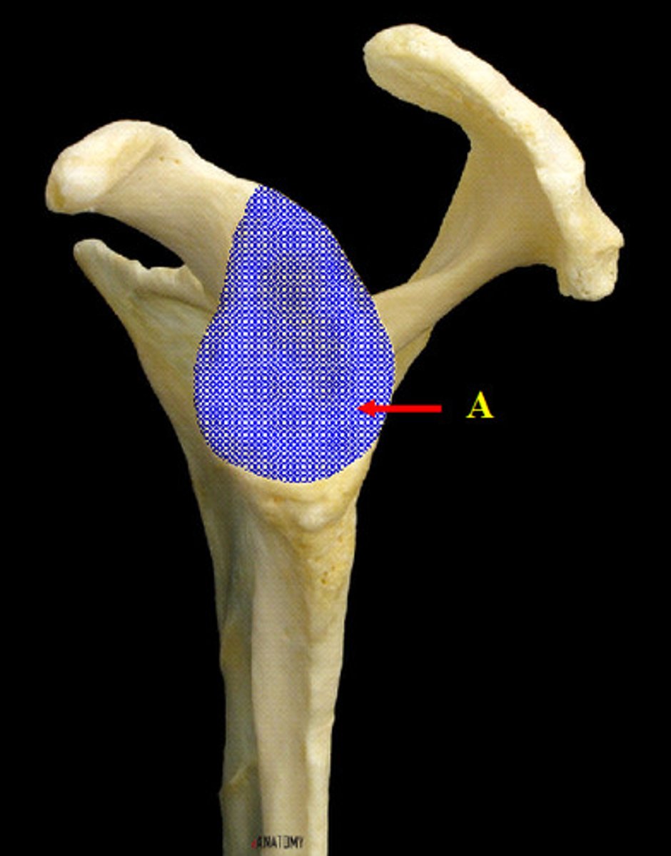
Glenoid Fossa/cavity of the Scapula
(anterior view)
Glenoid Fossa of the Scapula
(lateral view)
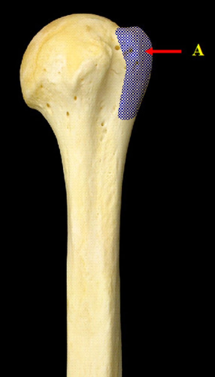
Greater Tubercle of the Humerus
(proximal end, anterior view)
Hamate Carpal
("Handle")
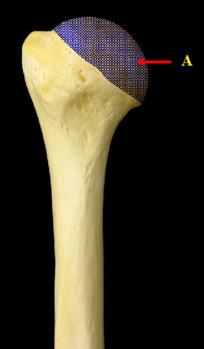
Head of the Humerus
(proximal end, posterior view)
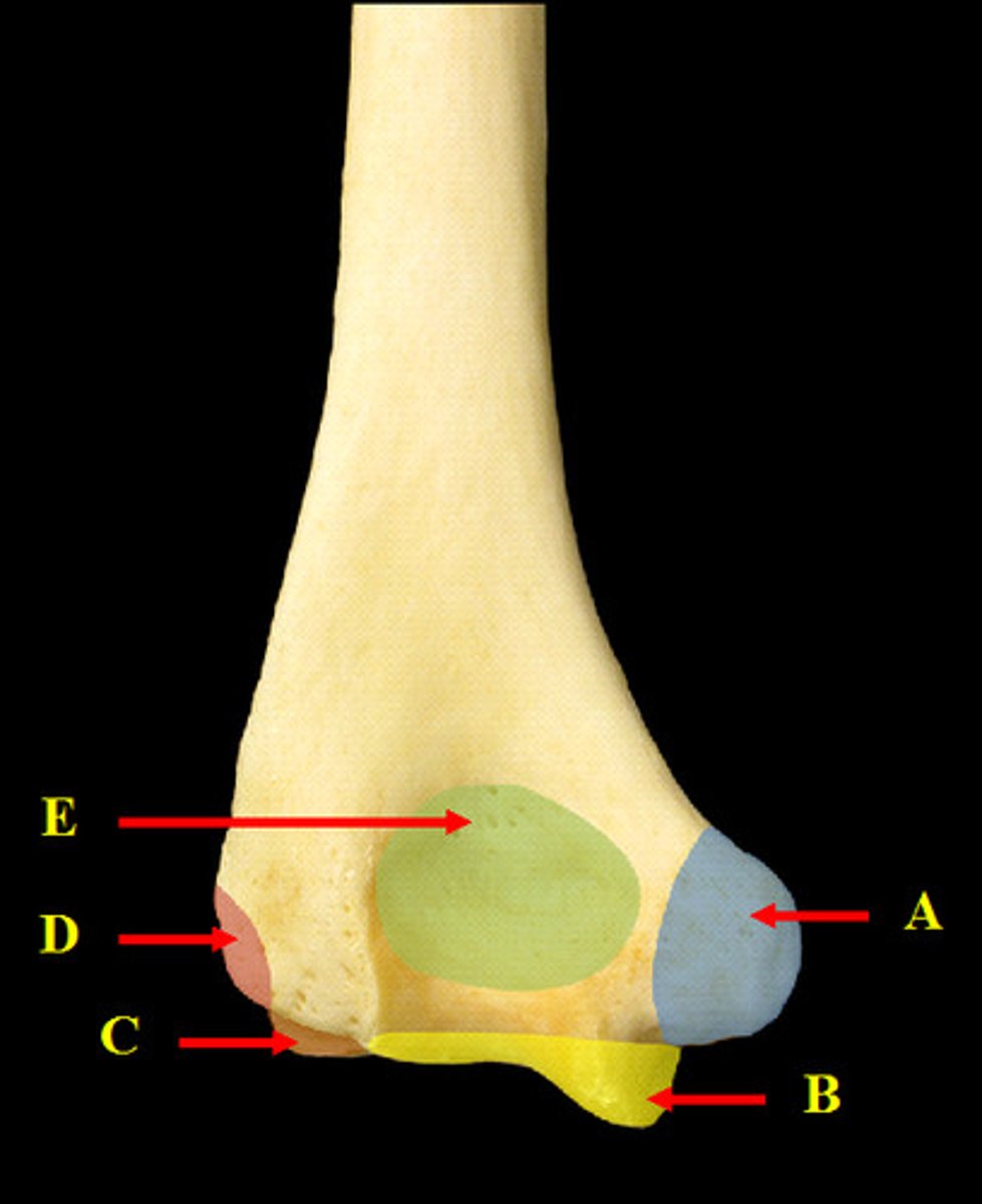
Humerus
(distal end, anterior view)
A. Lateral Epicondyle of the Humerus
B. Capitulum of the Humerus
C. Trochlea of the Humerus
D. Medial Epicondyle of the Humerus
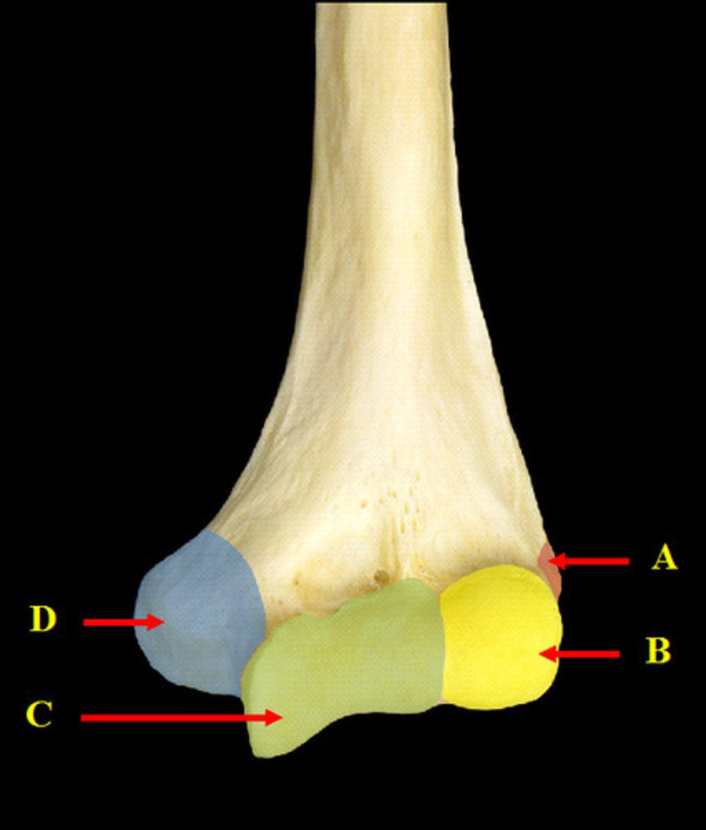
Humerus
(distal end, posterior view)
A. Medial Epicondyle of the Humerus
B. Trochlea of the Humerus
C. Capitulum of the Humerus
D. Lateral Epicondyle of the Humerus
E. Olecranon Fossa of the Humerus
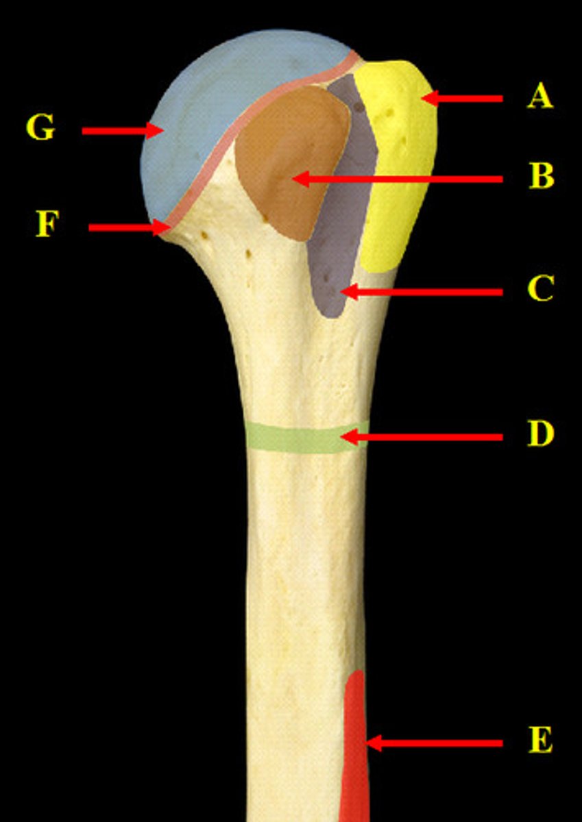
Humerus
(proximal end, anterior view)
A. Greater Tubercle of the Humerus
B. Lesser Tubercle of the Humerus
C. Intertubercular Groove of the Humerus
D. Surgical Neck of the Humerus
E. Deltoid Tuberosity of the Humerus
F. Anatomical Neck of the Humerus
G. Head of the Humerus
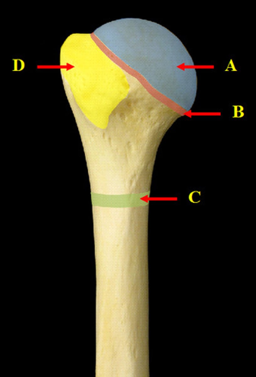
Humerus
(proximal end, posterior view)
A. Head of the Humerus
B. Anatomical Neck of the Humerus
C. Surgical Neck of the Humerus
D. Greater Tubercle of the Humerus
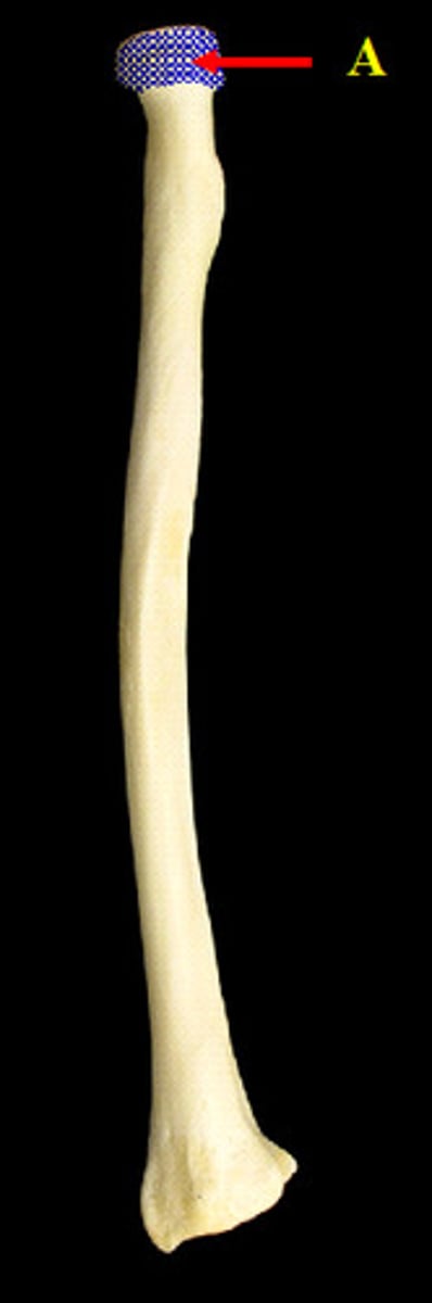
Head of the Radius
(proximal end, posterior view)
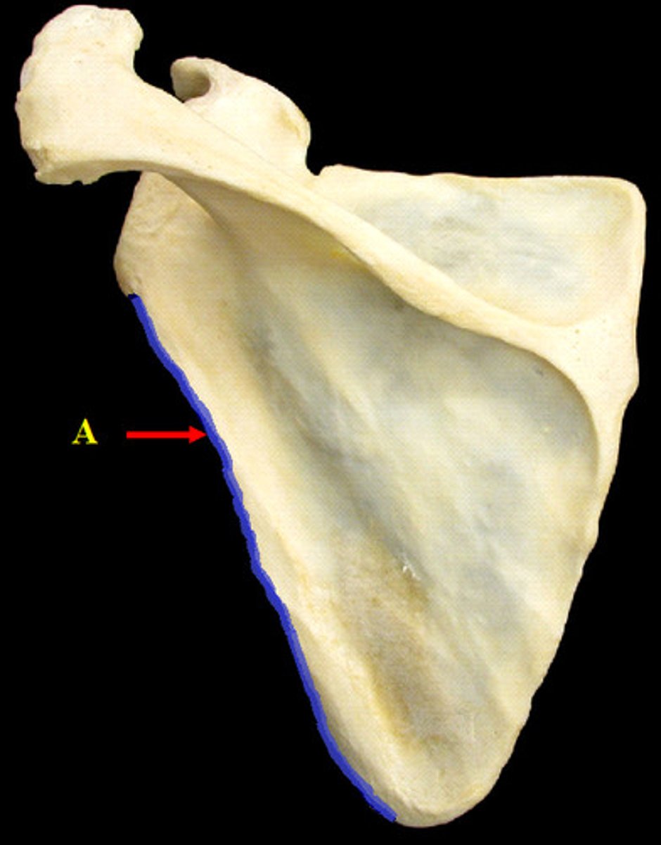
Lateral Border of the Scapula
(posterior view)
Lunate Carpal
("Lovers")
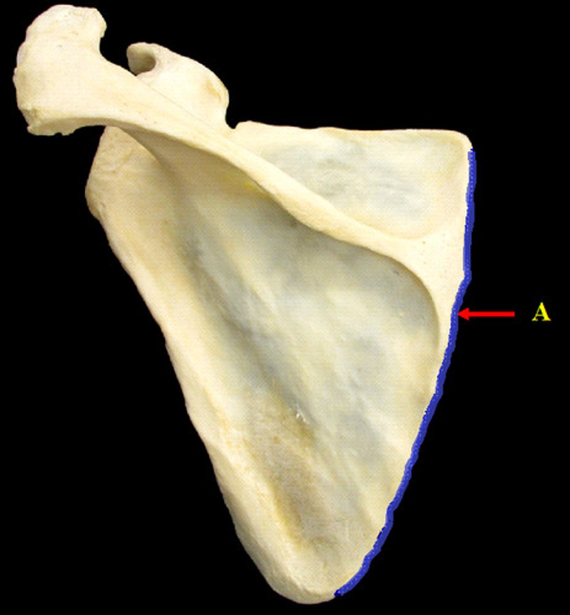
Medial Border of the Scapula
(posterior view)
Medial Epicondyle of the Humerus
(distal end, anterior view)
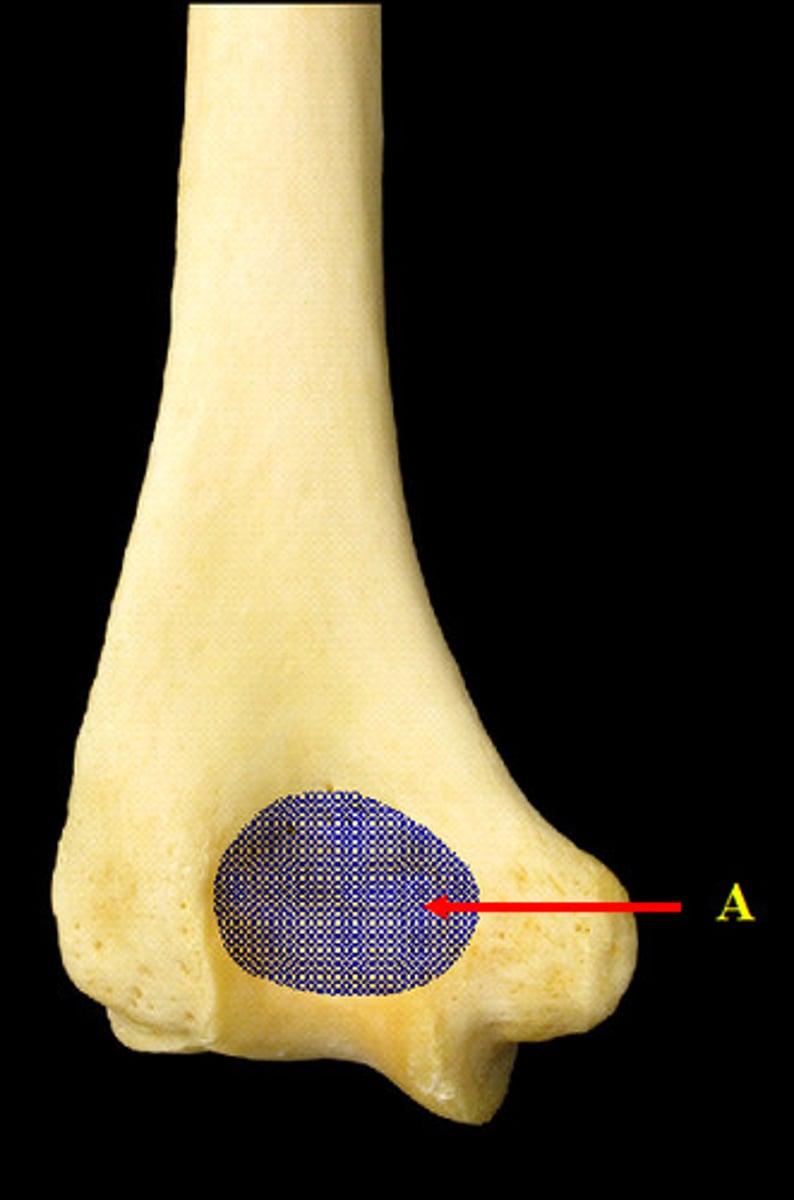
Olecranon Fossa of the Humerus
(distal end, posterior view)
Pisiform Carpal
("Positions")
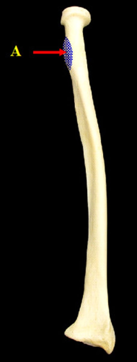
Radial Tuberosity of the Radius
(proximal end, anterior view)
Scaphoid Carpal
("Some")
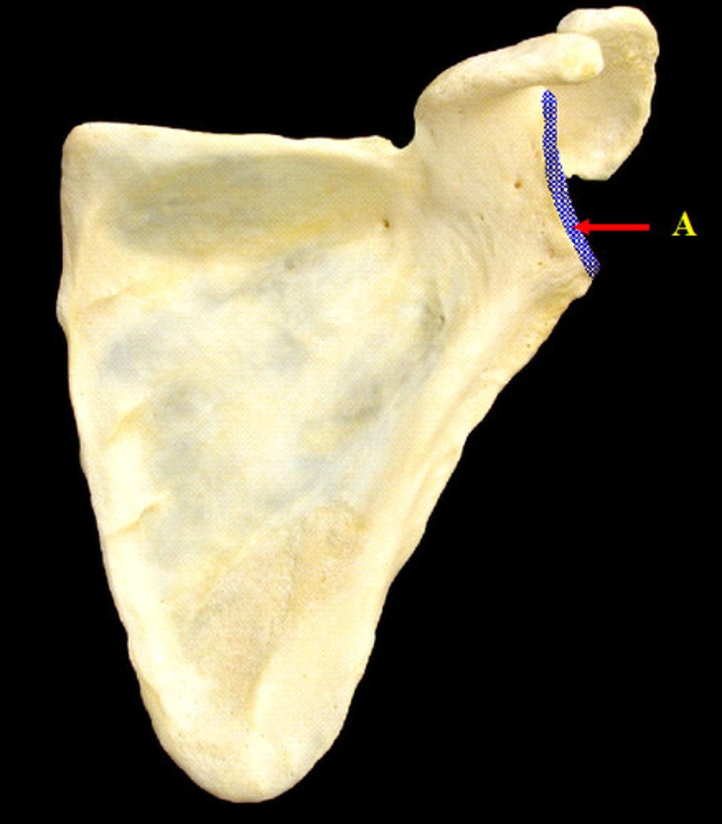
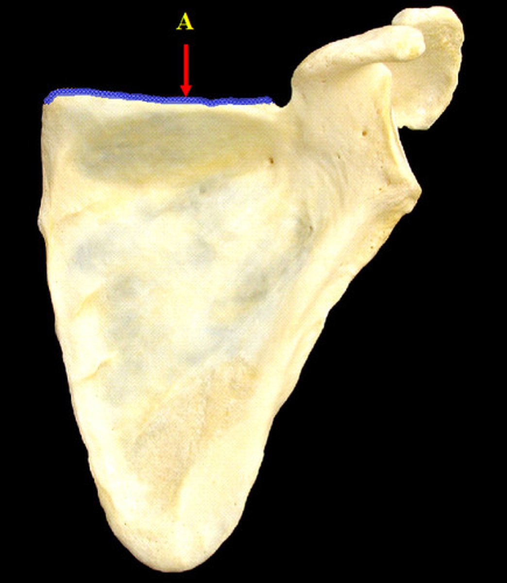
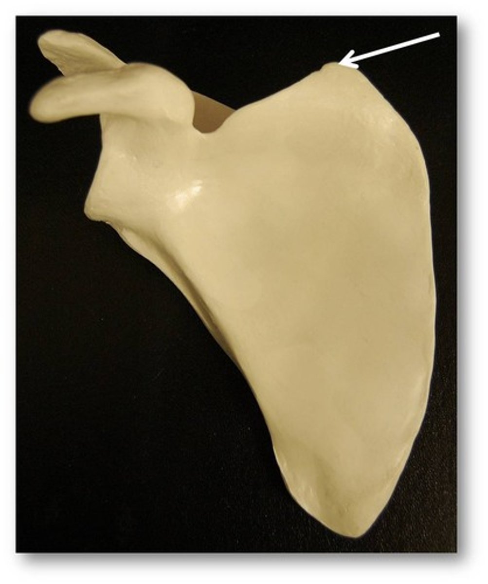
Superior Border of the Scapula
(anterior view)
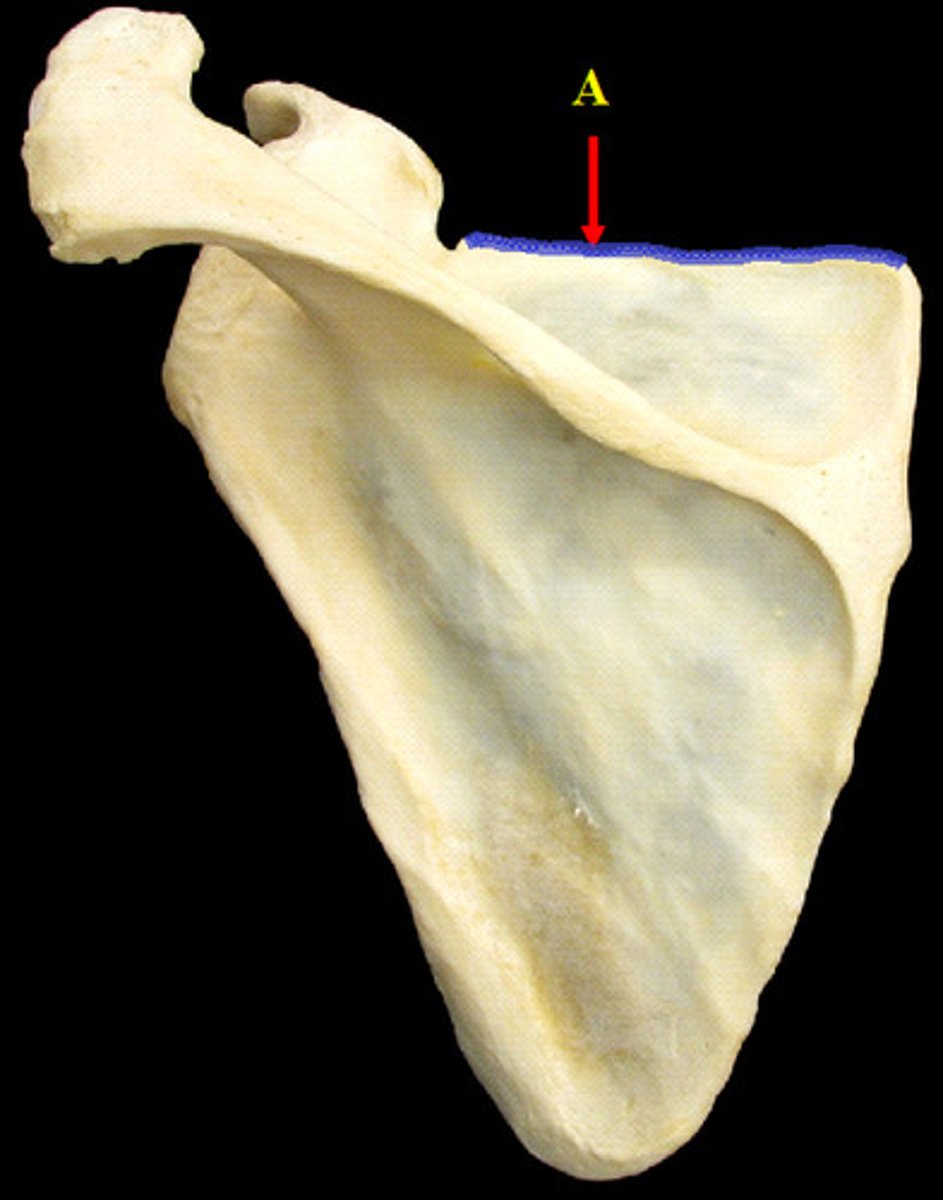
Superior Border of the Scapula
(posterior view)
Trapezium Carpal
("That")
Trapezoid Carpal
("They")
Triquetrum Carpal
("Try")
Trochlea of the Humerus
(distal end, anterior view)
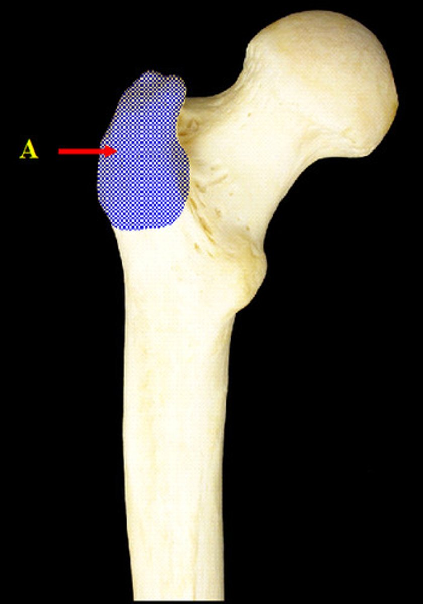
Greater Trochanter of the Femur
(posterior view)
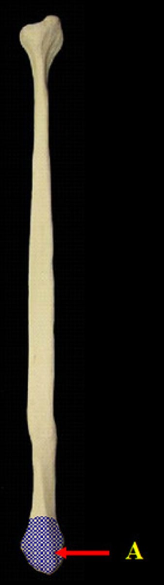
Head of the Fibula
(anterior view)
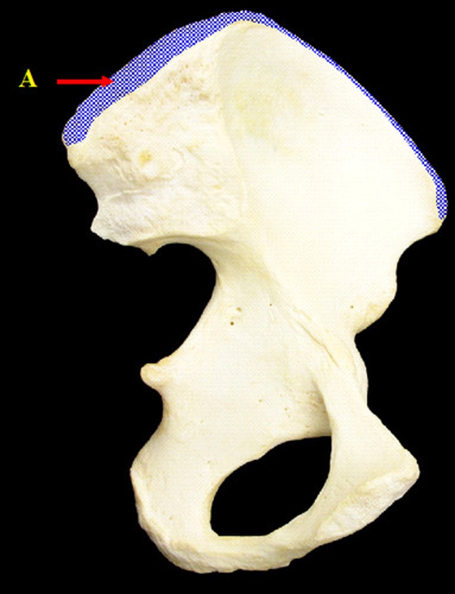
Iliac Crest of the Ilium
(medial view)
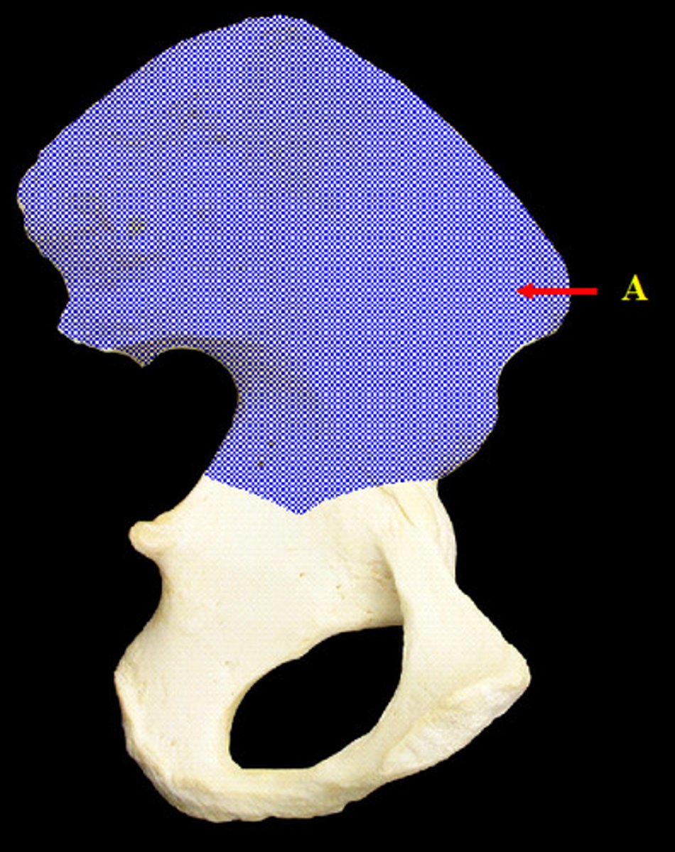
Ilium
(medial view)
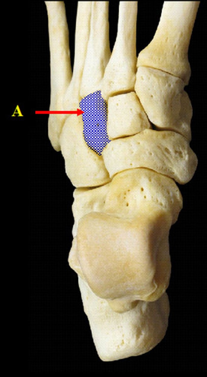
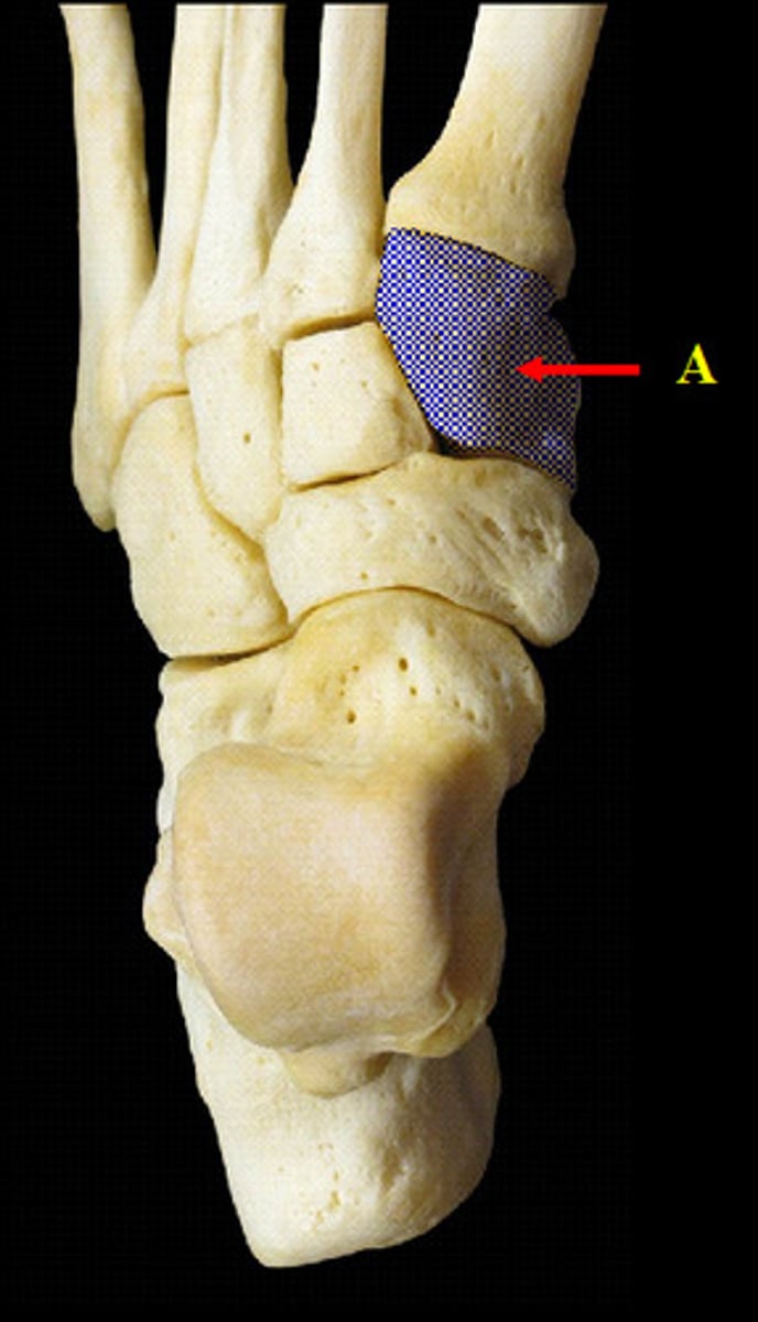
Intermediate Cuneiform Tarsal
(superior view)
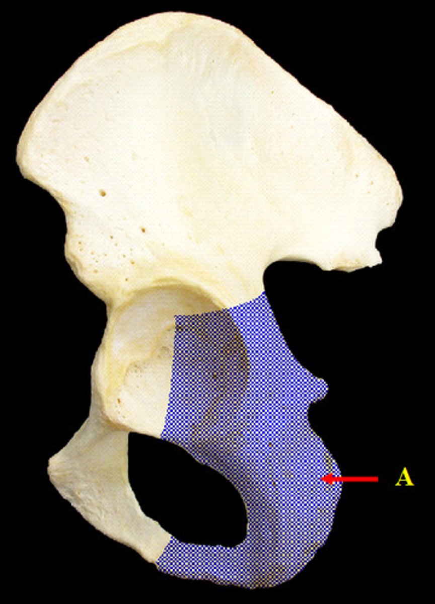
Ischium
(lateral view)
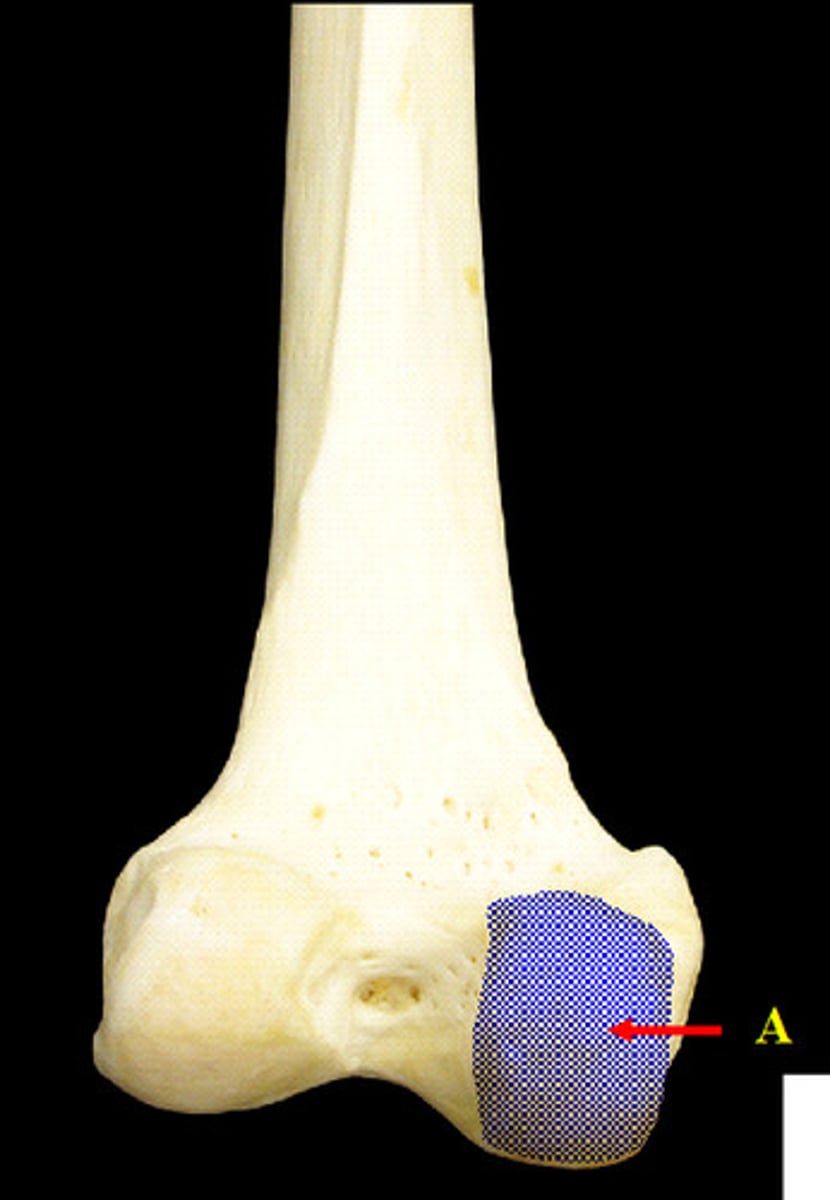
Lateral Conyles of the Femur
(distal end, posterior view)
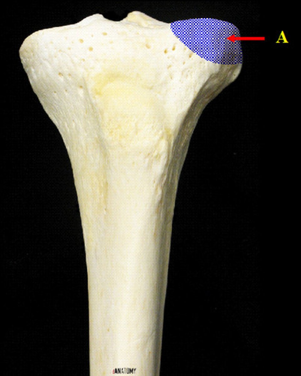
Lateral Condyle of the Tibia
(proximal end, anterior view)
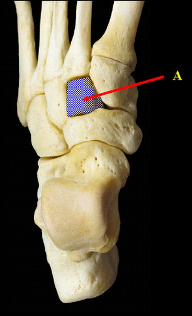
Lateral Cuneiform Tarsal
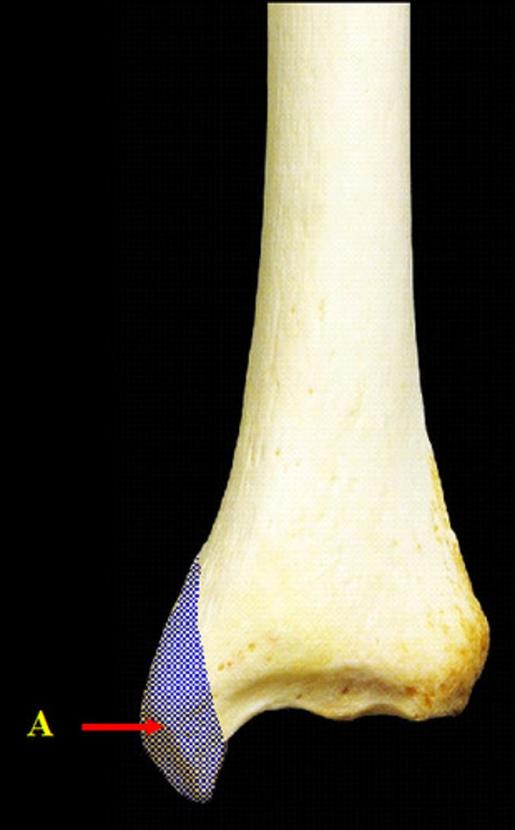
Lateral Malleolus of the Fibula
(distal end, anterior view)
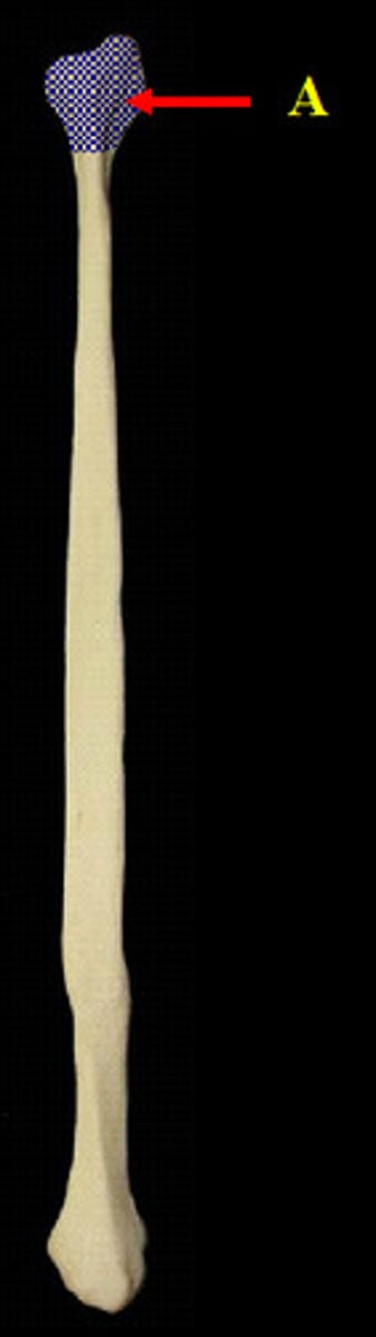
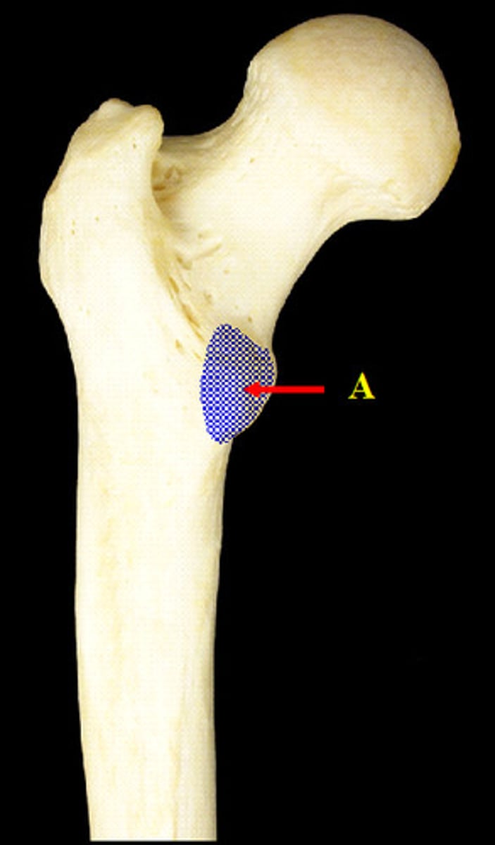
Lesser Trochanter of the Femur
(proximal end, posterior view)
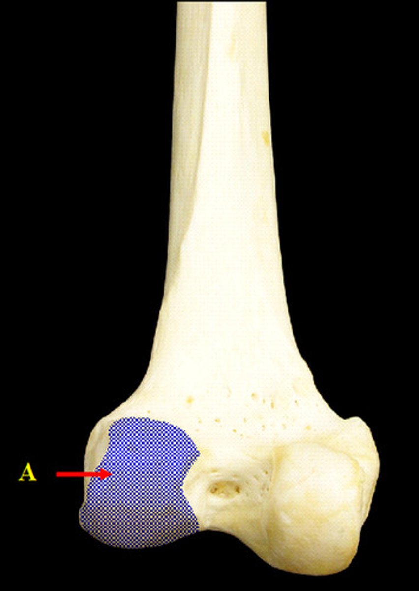
Medial Condyle of the Femur
(distal end, posterior view)
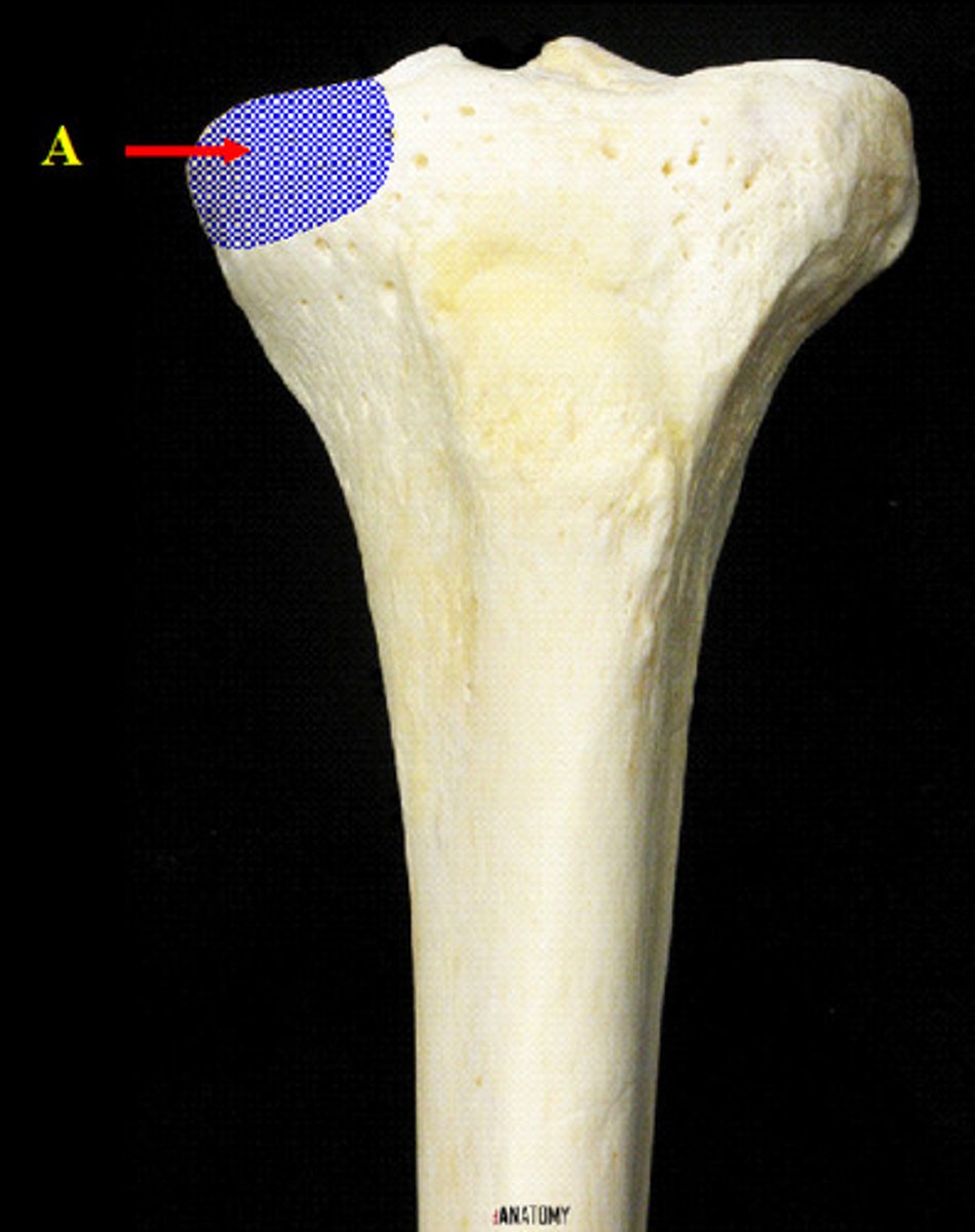
Medial Condyle of the Tibia
(proximal end, anterior view)
Medial Cuneiform Tarsal
Medial Malleollus of the Tibia
(distal end, anterior view)
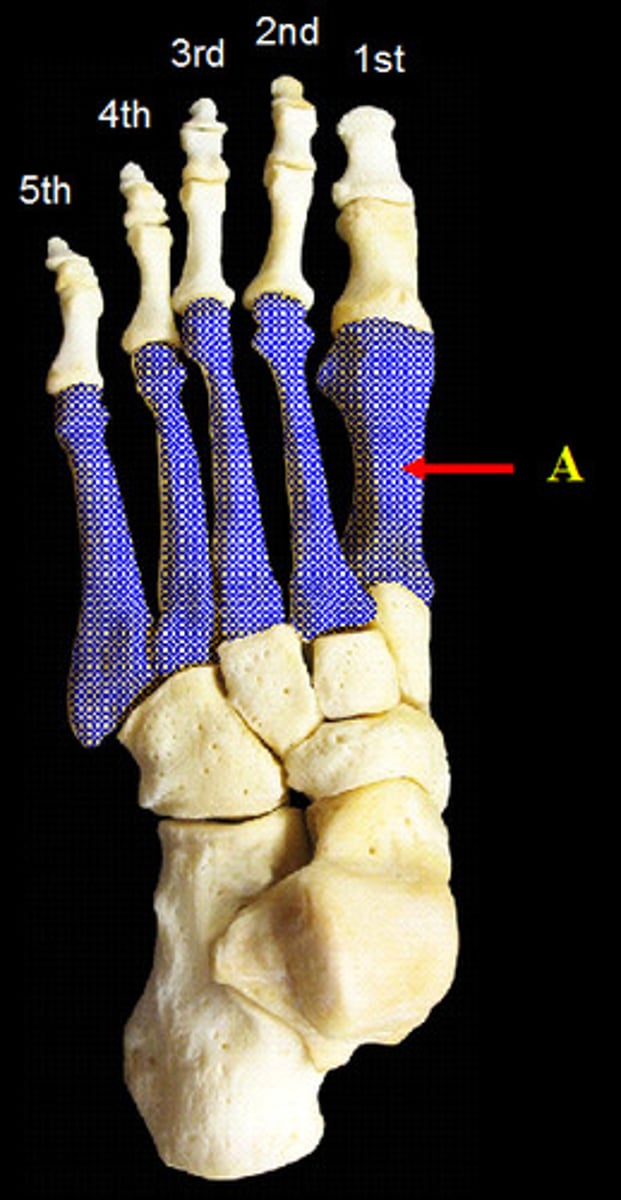
Metatarsals of the Foot
(superior view)
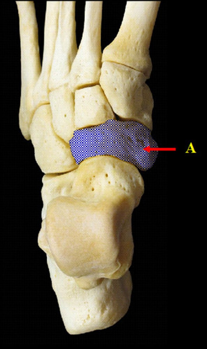
Navicular Tarsal
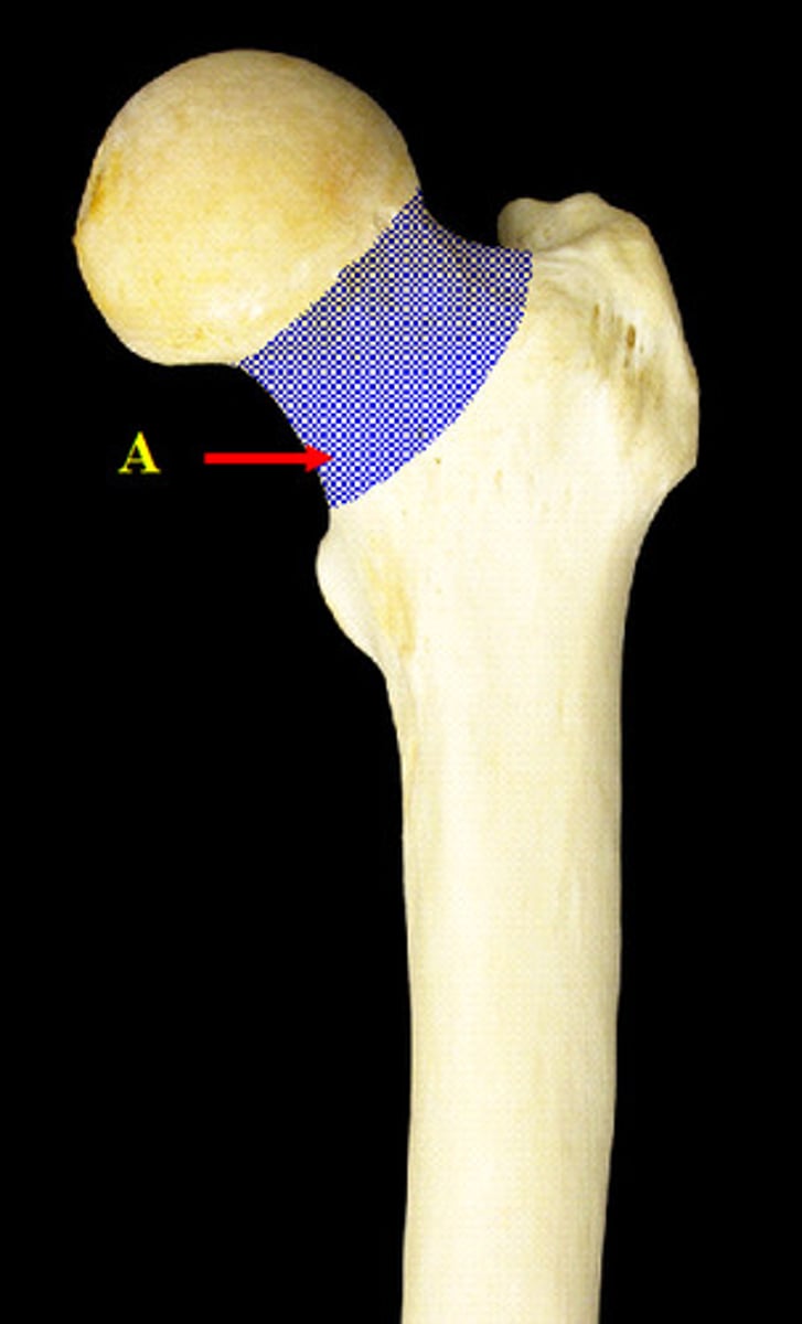
Neck of the Femur
(proximal end, anterior view)
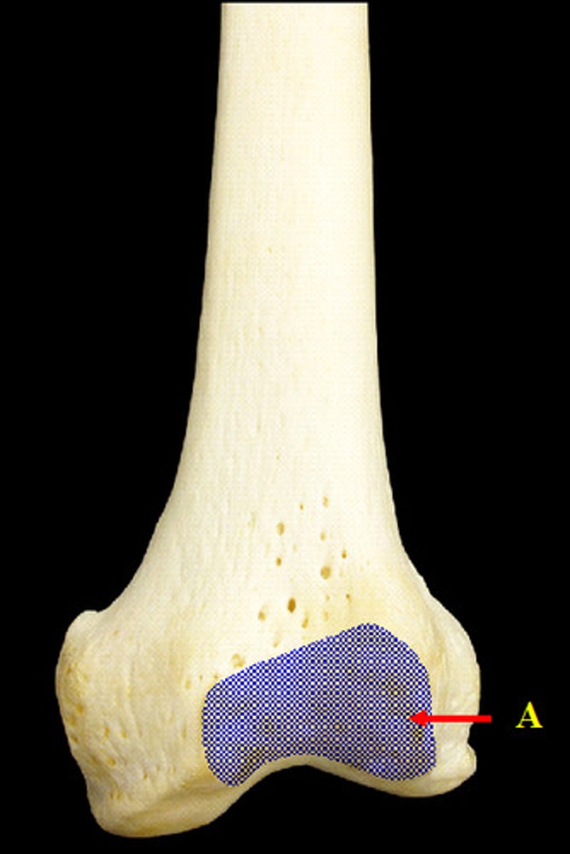
Patellar Surface of the Femur
(distal end, anterior view)
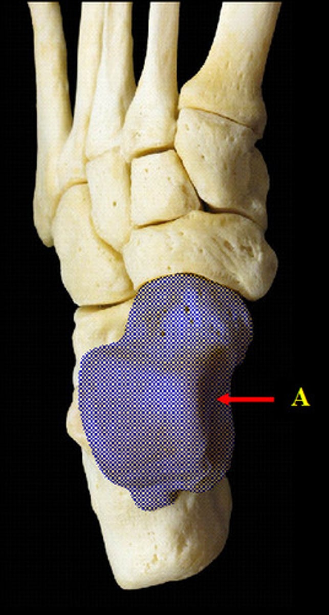
Talus Tarsal
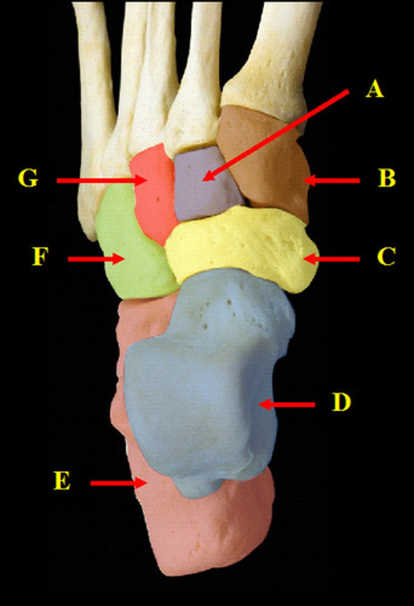
Tarsals of the Foot
(superior view)
A. Intermediate Cuneiform
B. Medial Cuneiform
C. Navicular
D. Talus
E. Calcaneus
F. Cuboid
G. Lateral Cuneiform
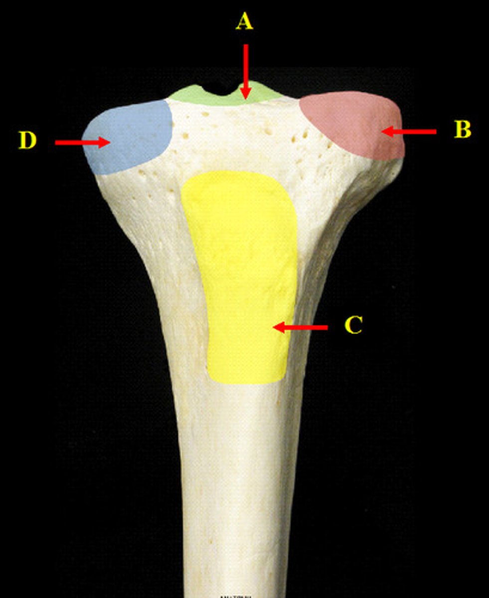
Tibia
(proximal end, anterior view)
A. Intercondylar Eminence of the Tibia
B. Lateral Condyle of the Tibia
C. Tibial Tuberosity of the Tibia
D. Medial Tuberosity of the Tibia
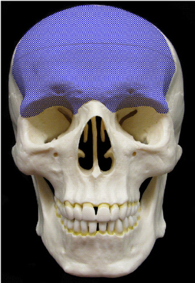
Frontal Bone
(anterior view)
Inferior Nasal Conchae
(anterior view)
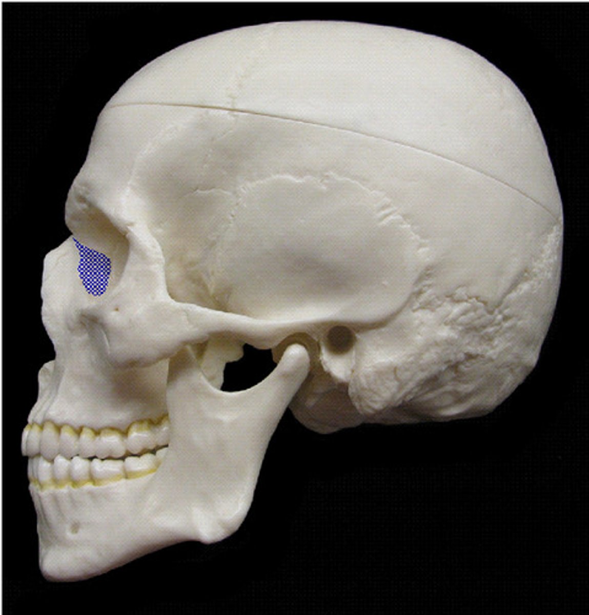
Lacrimal Bone
(lateral view)
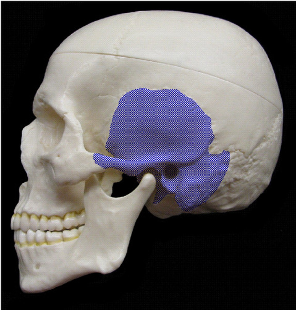
Sphenoid Bone
(lateral view)
Temporal Bone
(lateral view)
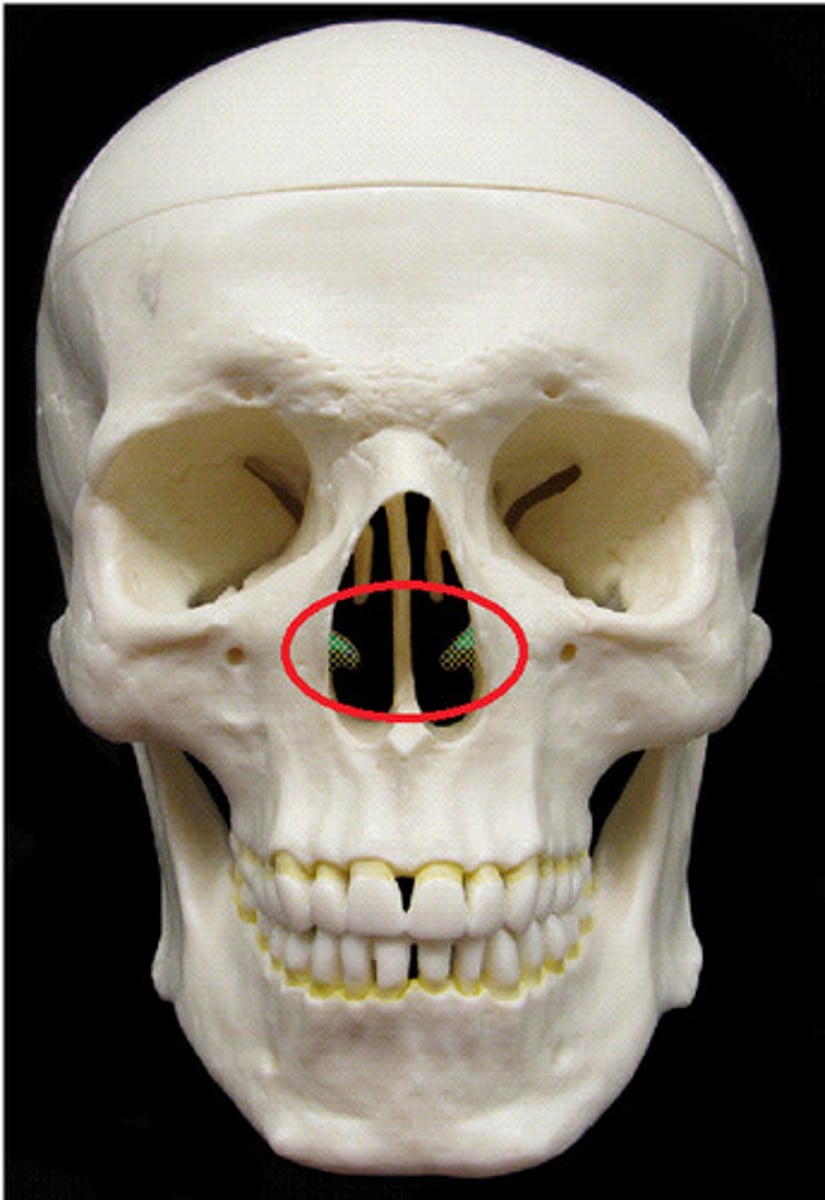
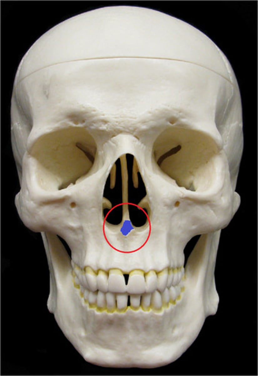
Vomer
(anterior view)
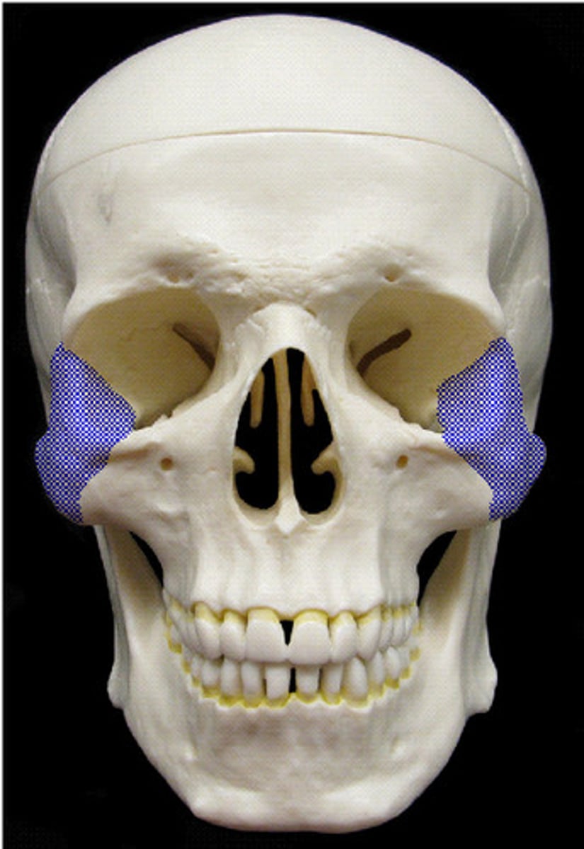
Zygomatic Bones
(anterior view)
Superior angle
Name this specific part of the scapula.
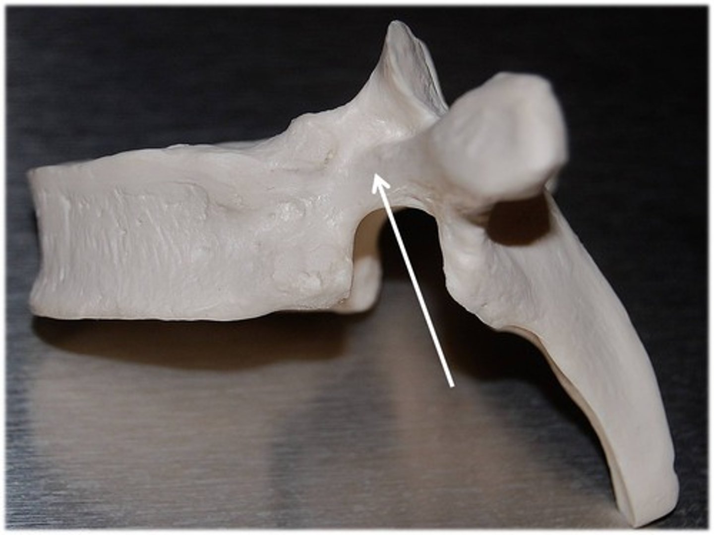
Pedicle
Name this specific part of the thoracic vertebra.