OFFICIAL PPOM2 OMM Practical 1
 Call Kai
 Learn
Learn Practice Test
 Spaced Repetition
 Match
Match Flashcards
 Knowt Play
1/170

There's no tags or description
Looks like no tags are added yet.
Name | Mastery | Learn | Test | Matching | Spaced | Call with Kai | Chat |
|---|
No analytics yet
Send a link to your students to track their progress
171 Terms
dx oa (cervical spine)
OA joint = “opposite always” (deep sulcus = side of rotation so SB will be opposite of that)
for examination: translate head from left - right and right to left with head in neutral position
dx: determine translation (gives S/R) check with slight flexion/extension
ex: if motion is greater from L to R, freedom is side-bent L and rotated right (restriction in right side-bending), if restriction of lateral translation is MORE signficant in flexion, but goes away in extension, then the segment is extended
dx aa (cervical spine)
AA joint = rotation only
you must be standing
1. ensure pt nods head forward to lock out OA joint
2. flex pt next to approx 45 degrees until locking occurs below the AA joint in the rest of the cervical spine
3. slowly rotate head from midline to left and then midline to right
if head rotates more freely to the right the diagnosis is AA Rr
dx c2-c7
follows type 2-like (rotation & side bending = same)
check for flexion/extension
cervical landmarks:
C2 - mandible
C3 - hyoid
C4 - superior aspect of thyroid
C5 - thyroid cartilage
C6 - cricoid ring
C7 - vertebral prominence
hvla of cervical spine (c2-c7)
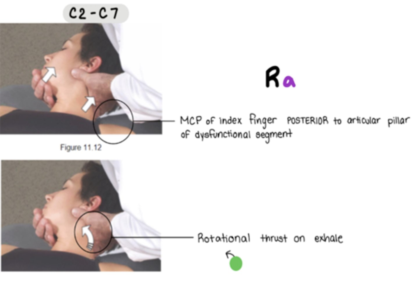
Ra
stand on SAME side of dysfunctional rotation component
MCP of index finger POSTERIOR to articular pillar of dysfunctional segment
**must lock out
can slightly flex to point and also side bend towards to initiate lockout
rotational thrust on exhale
met of cervical spine (c2-c7)
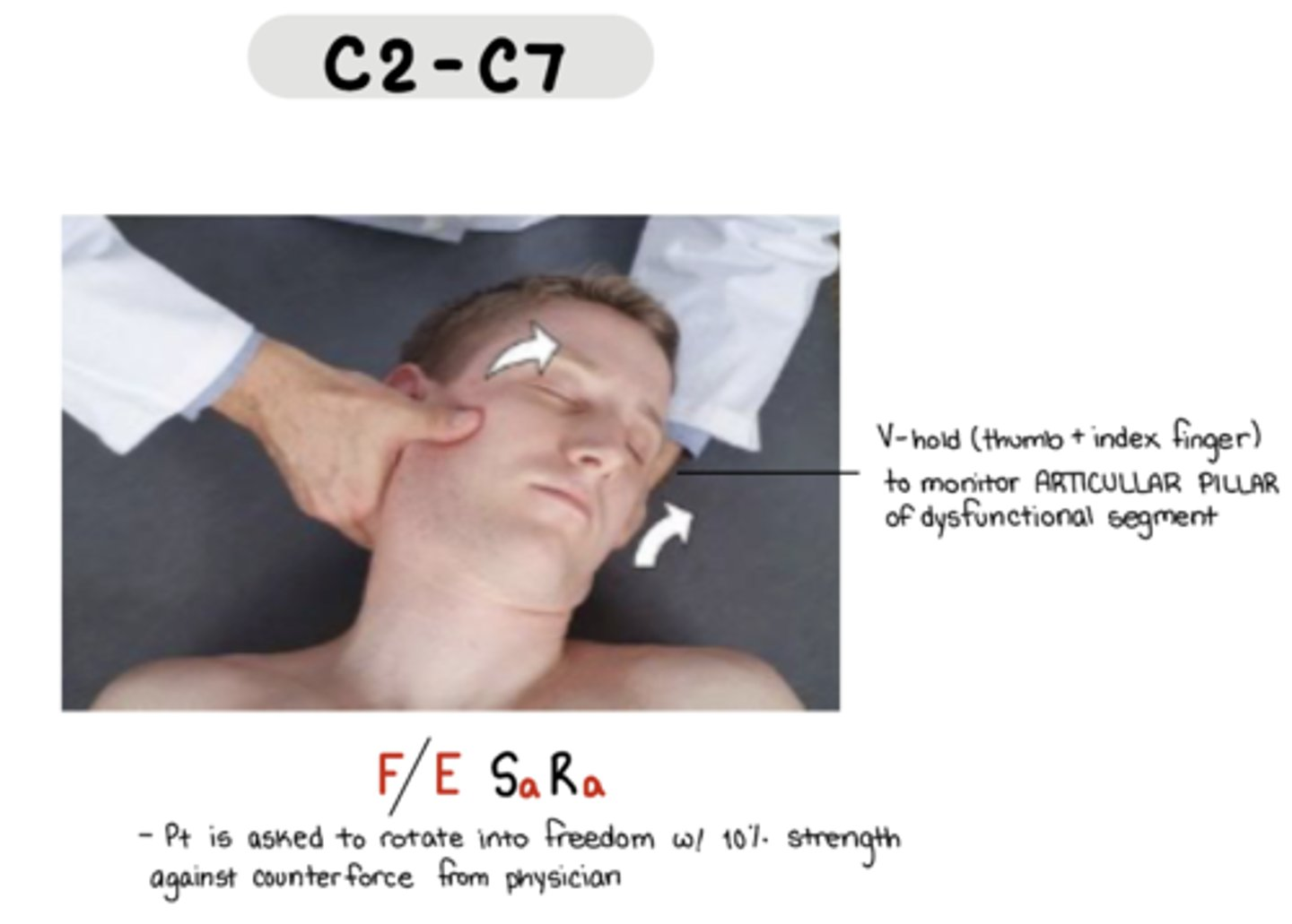
F/E Sa Ra
V-hold: thumb and index fingers to monitor articular pillae
pt is asked to rotate into freedom with 10% strength against counterforce
cs of anterior cervical spine
C1 = Ra (TP or mandible)
C2-C6 = FSaRa (posterior to SCM)
C7 = FStRa (posterior/superior surface of clavicle)
C8 = FSara (medial end of clavicle)

dx of thoracic spine (t1-4 or t5-12)
screen (TART changes)
T1-T3 = TP is same level as SP
T4-T6 = TP ½ level up
T7-T10 = TP whole step up
T11 = TP ½ level up
T12 = TP same level
thumb rolling superior = flexion, inferior = extension
hvla of thoracic spine
type 1 SD = neutral, elbow down
type 2 SD = f/e, elbow up
stand on opposite side of rotation
arms opposite over adjacent
thenar eminence on TP, inhale, thrust on exhale
met of thoracic spine
osteopathic hug or pt arm on neck
monitor TP
t1-4 = move head to engage in barrier
t5-12 = move body to engage in barrier
cs of anterior thoracic
Patient is supine for T1-6
T1 = F (episternal notch)
T2 = F (angle of louis)
T3-6 = F to dysfunctional level
Patient is seated for T7-9
FStRa (slouch to F, move leg out to SB pt, pt wraps arms around leg to R)
use the OPPOSITE leg on table
T7 = ¼ from xiphoid process
T8 = halfway xiphoid and umbilicus
T9 = ¼ from umbilicus
Patient is supine for T10-12
FStRa (stand on same side, flex legs, knees and ankles to me)
T10 = ¼ from umbilicus to pubic symphysis
T11 = halfway umbilicus and pubic symphysis
T12 = anterior superior aspect of iliac crest

dx of rib cage
TART changes (i.e. red reflex, skin drag)
spring test while pt is prone
focus on side with + spring test
assess breathing
inhalation dysfunction = likes to stay inhaled (will have trouble moving caudad in exhalation)
exhalation dysfunction = likes to stay exhaled (will have trouble moving cephalad in inhalation)
BITE = inhale (bottom rib = key rib); exhale (top rib = key rib)
side w/ less motion = dysfunctional side, name for freedom
rib 1-5 = pump handle
rib 6-10 = bucket handle
hvla ribs 1, 2 exhalation sd
RaSt
pt seated
physcian leg opposite of SD to stabilize
one hand on pt head, forearm against head to stabilize
mcp of index finger on posterior aspect of dysfunctional rib
thrust directed down and slightly diagonally (as if you were aiming at ur opposite knee)
hvla ribs 3-10
pt supine, arms opposite over adjacent
stand on OPPOSITE side of dysfunctional rib
thenar eminence on ___ angle of rib angle to push rib back
inferior aspect of rib angle for exhalation sd → ribs stuck down
superior aspect of rib angle for inhalation sd → ribs stuck up
(basically thoracic HVLA other than hand placement)

hvla ribs 11, 12 exhalation sd
exhalation sd
Sa = legs away from dysfunctional rib (towards me)
stand OPPOSITE of dysfunctional rib
active hand: hand pulls UP on ASIS to push rib back down
other hand: hypothenar eminence on inferior aspect of rib ABOVE dysfunctional rib to stabilize rib
ribs stuck UP

hvla ribs 11, 12 inhalation sd
inhalation sd
St = legs away from me
standing OPPOSITE side of dysfunctional rib
hypothenar eminence on inferior aspect of dysfunctional rib - to push rib back UP
(other hand stabilizes on ASIS)
ribs stuck DOWN

met of rib inhalation sd
Inhalation
push down on exhale. repeat 5-7 times
rib 1 = FStRa
ribs 2-6 = FSt
ribs 7-10 = St
ribs 11-12 = on the dysfunctional rib, stabilize hip, push up on rib on exhale, pt legs position away from you

met of rib exhalation sd
Exhalation
stand on same side
make sure to hook index and middle finger on rib, pulling inferior and lateral
woe is me (1-2) = scalenes
pt head is rotated away for this one
have pt push head up
wave hi (3-5) = pec minor
pt elbow should be above their shoulder
apply pressure to the elbow area
have pt push elbow up
shut the gate (6-8) = serratus anterior
have pt push elbow up
outstretched arm (9-10) = latissimus dorsi
have pt push arm down (against your hip/leg)
11-12 = on the inferior aspect of rib above, pull ASIS down, have pt hike hip up towards ear, pt legs position towards you

cs anterior rib
anterior = exhalation sd
AR 1,2 = FStRt
AR 3-10 = FStRt
ASS! - anterior same side -> pt mermaid legs same side as TP
Arm back = Rt
Physician moves leg out = St
physician leg on opposite side

cs posterior rib
posterior = inhalation sd
PR 1 = ESaRt
PIE is OP
posterior
I = 1
E = ESaRt
OP = use opposite leg for stabilizing
PR 2-10 = FSaRa
pt mermaid legs opposite of TP
use same side leg
Arm back = Ra
Physician moves leg out = Sa

dx of lumbar spine
dx can be done seated
sphinx = testing for extension
child’s pose = testing for flexion
landmarks = iliac crest btwn L4/L5
Type 1 = opposite N S R
Type 2 = same side F/E R S
hvla of lumbar spine
side of rotation faces DOWN (on table); pt faces physician
localize SP of dysfunction AND level below (monitor throughout setup)
flex pt until dysfunctional segment is in neutral
if type I = keep neutral
if type 2 = adjust for F/E by moving legs
arm closest to table is pulled TOWARD physician to engage rotation
pt grabs their own elbows; physician’s cephalad forearm loops through and braces ribcage (switch monitoring hand prn)
caudad arm placed on greater trochanter
if type I → keep neutral (no need to adjust)
if type II extension SD → flex legs further until felt at level of dysfunction
if type II flexion SD → tell pt to straighten bottom leg
drop top leg down
THRUST
type 1
arms come APART horizontally (caudad arm moves down; cephalad arm moves up to resist torso moving down)
arms come apart perpendicularly (caudad arm moves pelvis towards you; cephalad arm moves slighly forward/kind of away from you)
type 2
arms come TOGETHER horizontally
arms come apart perpendicularly
like ur punching yourself

met of lumbar spine
osteopathic hug or pt arm on neck
monitor TP
(same as thoracic sitting MET)

cs anterior lumbar
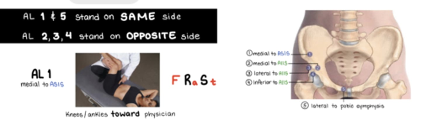
1, 5 same side
2, 3, 4 across the floor (opposite side)
AL 1 = FRaSt (medial to ASIS) → knees and ankles to me
AL 2-4 = FSaRt (my little igloo) → knees and ankles to me
AL 2 = medial to AIIS
AL 3 = lateral to AIIS
AL 4 = inferior to AIIS
AL 5 = FSaRa (lateral to pubic symphysis) → knees to me, ankles AWAY
cs QL
QL = EXTEND, ABDUCT, EXTERNALLY ROTATE HIP
stand on same side of TP
attachments = inferior aspect of 12th rib, lateral aspect of lumbar TPs, superior aspect of iliac crest

Sacral diagnosis steps
- verbally indicate landmarks as you diagnose (e.g., “left sacral base posterior”, “left ILA posterior”)
Seated flexion test
Place thumbs on inferior aspect of PSIS
Positive for side that moves first/forward
Spring or sphinx test
+ spring = extension
- spring = flexion
Identify deep sulcus
Locate PSIS and roll your thumbs so they’re more medial and superior
Identify posterior ILA
Palpate down sacral bone until you feel the ledge and find the points on both ends
met forward sacral torsion
Pt’s side of axis faces DOWN
Monitor sacral sulcus (slightly medial to PSIS, hook thumbs medial to help locate it)
Flex hips and knees of pt towards you until motion is felt
Pt turns away and hugs table
Drop pt knees off table
Engage in barrier by pushing pt legs down while continuing to monitor + passive stretch

met backward sacral torsion
Pt side of axis faces DOWN
Monitor sulcus
Flex hips and knees until motion is felt
Straight bottom leg and hook top leg over
Grab pt shoulder to rotate them onto their back
Drop top leg off table, but keep it parallel to the floor.
Push top leg DOWN and pt resists by move leg UP + passive stretch

met unilateral sacral flexion
Pt prone
Stand on opposite side
Walk fingers/palpate down to ILA
ABduct leg and int rot side of dysfunction
Hypothenar on SD ILA, cover sacrum, place cephalad hand on top
Push down on inhale for ILA, as they exhale maintain pressure on SD ILA (push on inhale, resist exhale)
5-7 breaths
Test both sacral bases for reassessment

met unilateral sacral extension
Pt prone, physician stands on opposite side
Hand monitor PSIS and sacral bases of side of dysfunction
Abduct and int rot leg of SD
Place hypothenar on sacral base of side of dysfunction (points caudad), other hand on top (points cephalad)
Ask pt to Sphinx pose
Resist on inhale and follow on exhale for 5-7 times
Reassess sulci

met bilateral sacral flexion
Pt prone
Hypothenar and thenar on BOTH ILAs
ABduct and int rot BOTH legs
Push on inhale and resist exhale 5-7 times
Reassess sacral bases and ILA and spring test

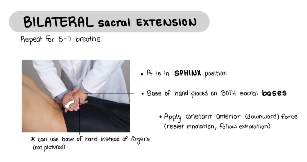
met bilateral sacral extension
Pt sphinx pose
Locate sacral bases with two fingers of one hand. Other hand covers the two fingers.
Optional: loose pack the legs
Resist on inhale and follow through on exhale 7-10 times.
Reassess.
dx pelvis/innominate
standing flexion test (+ is side where thumb moves first)
pt reseats pelvis (observe anterior and posterior)
asis compression test (+ on side with more resistance)
locate landmarks (asis, pubic tubercles, medial malleoli. psis)
naming is based off of side of + standing flexion test
use the weird longhorn gang sign to help in naming dx (thumb is psis, index is asis, pinky is pubic tubercle)
hvla of anteriorly rotated innominate
Patient lies dysfunctional side up
Monitor medial to PSIS and flex both hips and knees up until motion is felt
Extends bottom leg
Grasp the patient’s arm that is in contact with the table and rotate their torso away from table until motion is felt
Drop upper leg off the table and held between the physician’s knees
With the patient’s arms crossed, pass the cephalad hand through the patient’s arms and stabilize caudad (thrusting) arm behind greater trochanter
Check lockout
Instruct patient to inhale deeply and exhale fully. At the end of exhalation, exert a quick, rotational, slightly downwards thrust through the iliac crest through the patient’s top leg. Greater thrust with arm on trochanter
Reassess

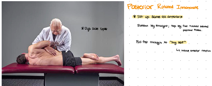
hvla of posteriorly rotated innominate
Patient lies on nondysfunctional side with the posteriorly rotated innominate facing up
Monitor medial to PSIS and flex both hips and knees up until motion is felt at monitoring hand
Patient straightens bottom leg and hooks foot of top leg over the popliteal fossa
Grab pt arm closest to table and bring that arm towards you till motion is felt at the monitoring hand over the SI joint
Pt cross arms, weave your hand through to monitor SI joint. Thrusting arm on posterior iliac crest.
Maintain sufficient rotation localized to level of the somatic dysfunction
CHECK FOR LOCK OUT.
Instruct patient to inhale deeply and exhale fully. At the end of exhalation, exert a quick, rotational thrust through the dysfunctional ilium in an anterior superior direction (LIKE YOU’RE PUNCHING/HUGGING YOURSELF)
Reassess
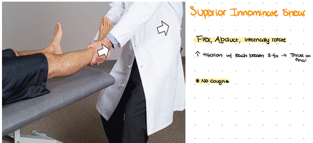
hvla of superior innominate shear
Abd IR
Grasp the tibia and fibula above the ankle on the side of the innominate dysfunction
Abduct and internally rotate the hip and leg until movement is felt at hip
Leaning back gently to provide an axial traction and instruct the patient to inhale and exhale. Provide more traction with each exhale
Repeat this 5-7 times
On the last exhalation tug on the leg
Reassess
hvla of inferior innominate shear
Pt lies with dys side up
Standing in front of the patient, monitoring the lumbosacral junction
Flex the legs and knees until motion at the lumbosacral junction
Induce rotation towards you by pulling pt’s arm
Extend the bottom leg and place the foot of top leg over the popliteal space of other leg
Tell pt to box their arms. Stabilize the trunk while monitoring the sacroiliac joint on the side of the dysfunction
Firmly contact the inferior aspect of the ischial tuberosity with the caudad forearm
Provide a cephalic thrust through the ischial tuberosity parallel to the table toward pts head (basically straight up to the pt’s head)
Reassess

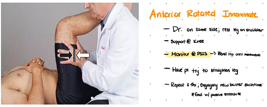
met anteriorly rotated innominate
Pt is supine.
Sit on edge of table.
Pt’s ankle (side of dysfunction) is placed against your shoulder.
Hold onto thigh above knee.
Monitor PSIS on dysfunctional side (not shown in image)
Flex leg up until motion is felt at PSIS (this is the barrier)
Pt resists by pushing leg down against your shoulder
Flex leg further
New barrier is when movement is felt at PSIS again
Repeat 3-5 times + passive stretch
reassess (reseat pelvis right before), can look at ASIS and possible compression test
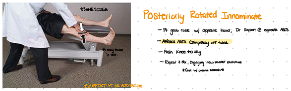
met posteriorly rotated innominate
Pt is supine.
Raise table as needed.
Drop leg (ischial tuberosity of dysfunctional side) off table. Instruct pt to hold onto edge of table with opposite hand. PT FOOT SHOULD NOT TOUCH FLOOR
Hand on opposite ASIS to stabilize.
Push leg DOWN (hand above knee)
Pt resists by pushing leg back up
Repeat 3-5 times + passive stretch
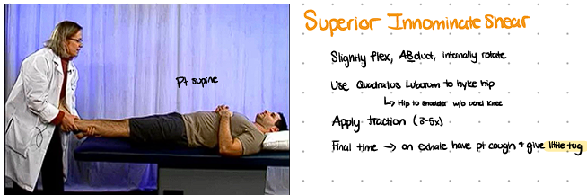
met superior innominate shear
Abd IR
Pt is supine.
One hand holds heel and the other cups the bottom of the foot of side of dysfunction.
ABduct leg until hip is loose packed (feels loose) + internally rotate leg. Add traction by pulling leg toward yourself.
“Bring your foot towards your shoulder without bending knee as you inhale”
Traction is maintained during inhalation. (You lean back to keep the traction)
Traction is ADDED at end of each exhale to reach new barrier
Articulatory component: thrust at the end of the last exhalation.
Give a nice tug as pt coughs “on your final exhale cough”
Respiratory assist method so NO passive stretch
met inferior innominate shear
Pt is prone.
Pt at edge of table, leg (on side of dysfunction) drops off table with knee bent (rests on your thigh)
Monitor SI joint until you get pt into position THEN move hand to monitor IT.
Monitor SI until movement is felt at joint, then switch hand to IT
Hand pushes ischial tuberosity UP
Ischial tuberosity is pushed further up at the end of each exhale to reach new barrier.
As they inhale, I’m resisting, as they exhale, I’m applying force to pt head for 3-5 seconds
“Inhale deeply and exhale forcefully”
Internal rotation of hip is added by moving knee slightly under table.

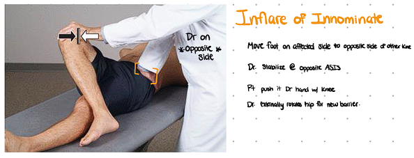
met innominate inflare
Pt is supine.
Leg (on same side as inflare) is crossed over the other knee.
Can stand on either side of patient. One hand on bent knee and the other on opposite ASIS
Hand on opposite ASIS to stabilize or monitor ASIS on side of dysfunction.
Engage in barrier by pushing knee OUT (toward table)
Pt resists by pushing knee in against your hand. OR BRING THEIR KNEE INWARDS
Leg is pushed out further each time to engage new barrier.
Repeat 3-5 times + passive stretch
met innominate outflare
Pt is supine.
Leg (on same side as outflare) is crossed over.
Can stand on OPPOSITE side (can stand on same but set up would be harder). Reach out and hold knee.
Hand on opposite ASIS to stabilize (not pictured) or monitor PSIS on side of dysfunction.
Engage in barrier by pushing knee IN
Pt resists by pushing knee OUT against your hand
Leg is pushed in further each time to engage new barrier.
Repeat 3-5 times + passive stretch

met superior pubic shear
Same set up as posterior innominate rotation MET EXCEPT:
Ischial tuberosity stays on table (localizes to pubic tubercle)
Pt is supine.
Hand on opposite ASIS to stabilize.
Ischial tuberosity (on side of dysfunction) ON table
Push leg DOWN (hand above knee)
Pt resists by pushing leg back up and slightly medially (as if they’re trying to get back onto the table)
Repeat 3-5 times + passive stretch
Stand with your hip against pt’s hip to stabilize.

met inferior pubic shear
Pt is supine. You stand on opposite side.
Monitor PSIS on dysfunctional side.
Flex pt’s leg up until motion is felt at PSIS (this is the barrier)
Pt resists by pushing up to ceiling
Flex leg further
New barrier is when movement is felt at PSIS again
Repeat 3-5 times + passive stretch

met pubic compression
Pt is supine.
Hips are flexed at 45 and knees at 90
Make sure pts ankles are touching!
Push knees apart until you feel resistance.
Pt pushes knees back together (toward freedom)
Start with fist between knees, then forearm; gradually increasing distance between knees
Can do with palm, then fist, then additional hand for length IF YOU FIT FOREARM BETWEEN LEGS.

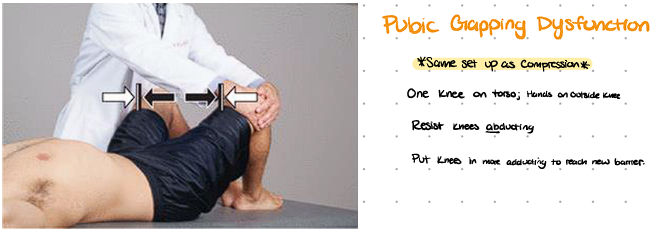
met pubic gapping
Pt is supine.
Other knee against your abdomen
Make sure pts ankles are touching
Push knees together (into barrier)
Pt pushes knees apart (into freedom)
Distance between knees is decreased each time
cs of anterior pelvic points

cs iliacus
F ER (hips)
Pt supine
Stand on same side of Tender pt which is located ⅓ from ASIS to midline (straight line across)
Cross ankles opposite over adjacent
Flex pt hip/knees bilaterally and rest legs on your thigh (FROGGY LEGS)
Finetune with further flexion and external rotation.

cs psoas
F St er (hips)
Pt supine
Stand on same side of tender pt
Tender pt is ⅔ distance from ASIS to midline (straight line across)
Cross pt ankles (opposite over adjacent)
Flex pt hips/knees bilaterally and rest legs on your thigh
Bring knees towards me
add slight external rotation and pull knees towards tender pt slightly side bend lumbar spine
Fine tune with more or less hip flexion, side bend and rotation

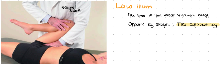
cs low ilium
F
Use iliac crest to find ASIS, then move to AIIS.
Follow from AIIS down inguinal ligament until you feel a bulb OR feel pubic tubercle and go ½ in lateral and ½ in superior
Physician stands on the same side of the tender point
Markedly flex patient’s hip
Fine tune with more/less hip flexion
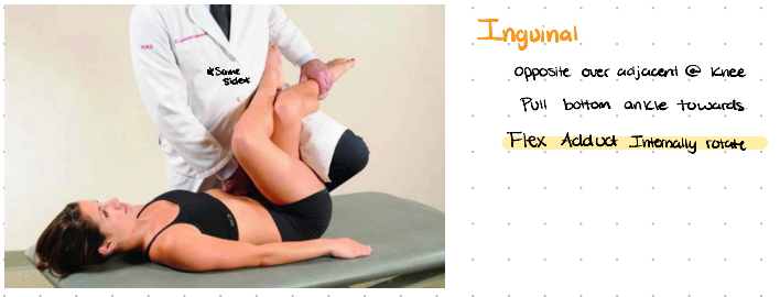
cs inguinal
F ADduct Internally Rotate
Tender point is located on the lateral aspect of the pubic tubercle; associated with the attachment of the inguinal ligament and/or pectineus muscle
Standing on the same side of the tender point
Cross the patient’s thighs opposite over adjacent. flex hips/knees bilaterally and place legs on physician’s thigh
Pull patient’s ipsilateral leg towards physician to induce adduction and internal rotation of the hip
Fine tune with more/less hip flexion, internal rotation and adduction
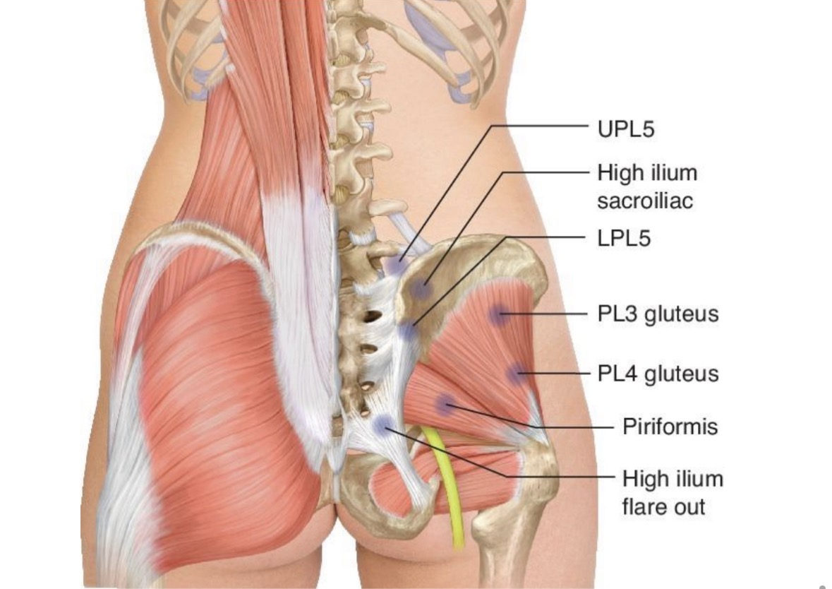
cs of posterior pelvic points
we just need to know UPL5 and Piriformis
cs piriformis (posterior pelvic)
Point is between sacral ILA and greater trochanter
Stand on same side
Ask pt to move to edge of table
Move leg down and hold between your knees
Flex and ABduct leg on affected side
Hold just below ankle and fine tune with internal or external rotation

cs UPL5 (posterior pelvic)
Point is superior and medial to PSIS
Pt is prone
Stand on opposite side
Hold pt leg of the tender point above knee
Extend and ADduct

dx sc joint
proximal clavicle
spring test (superior to inferior, anterior to posterior)
the side that has less springing is the side you’ll diagnose
check motions by placing two fingers at proximal clavicle
have patient move (abduct, adduct, flex, extend)
name for direction of ease
shoulder elevation = inferior/abduction sd
shoulder depression = superior/adduction sd
shoulder retraction = anterior/extension sd
shoulder protraction = posterior/flexion sd
dx ac joint
spring test superior to inferior in distal clavicle
you can ask pt which side they’re having issues with
dx side with most restriction
check range of motion by passively moving pt arm
compare both sides (check one arm at a time)
movement pairings
adduct, external rotation, inferior glide
abduct, internal rotation, superior glide
dx gh joint
assess active ROM (ballerina pose - dysfunctional side is the one with the lower arm) or ask pt which side bothers them
Place digits 3 and 4 on ball anteriorly and thumb on humerus posteriorly
do passive ROM and check for restriction
name for direction of ease
shoulder extension (swings back) = anterior sd
shoulder flexion (swings forward) = posterior sd
shoulder elevation = superior sd
shoulder depression = inferior sd
dx ulnohumeral joint
Motions
aBduction SD (medial glide) = arm is more angulated = increased carrying angle.
aDduction SD (lateral glide) = arm appears straighter = decreasing the carrying angle (producing a “gunstock deformity”).
tell pt to do active forearm pronation and supination
ballerina pose (arms go overhead)
is there’s a side that’s bothering you?
Feel for both medial and lateral olecranon process.
Two-hand hold possible OR place thenar/hypothenar on one end then cup using fingers while other hand grabs wrist rather than hand instead.
Naturally elbow should be 5-45°
Test for ROM (Abduct, adduct, flex, extend)
greater angle with carrying position = prefers aBduction

dx radial head / radioulnar joint
Easy posterior glide on pronation = posterior SD
Test active ROM bilateral
Locate medial epicondyle then go to lateral epicondyle then more distal
Divot when arm is extended and go to the BOTTOM of the divot. Feel the bone and PINCH
Pt arm flexed 90
Test for right area using pronation and supination
Dx focused on more proximal area
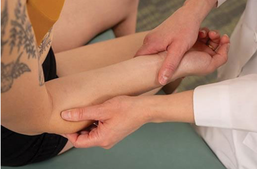
dx of wrist (radiocarpal)
test active ROM bilateral
Put thumbs on anterior/palmar surface of proximal carpals. On backside, fingers are monitoring posteriorly.
Compare side to side with wrist flexion and extension
Now do radial and ulnar deviation.
On practical just need to name one of the SD if you find two.

hvla anterior radial head
Pt seated, physician stands facing pt
Thumb on anterior head
Other hand is shaking hands
Rotate forearm into pronation until barrier
Flex into barrier
Thrust is a flex from hand holding wrist
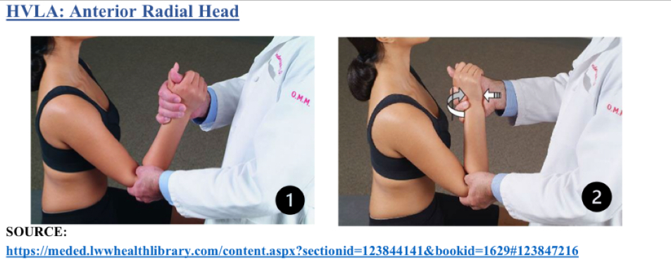
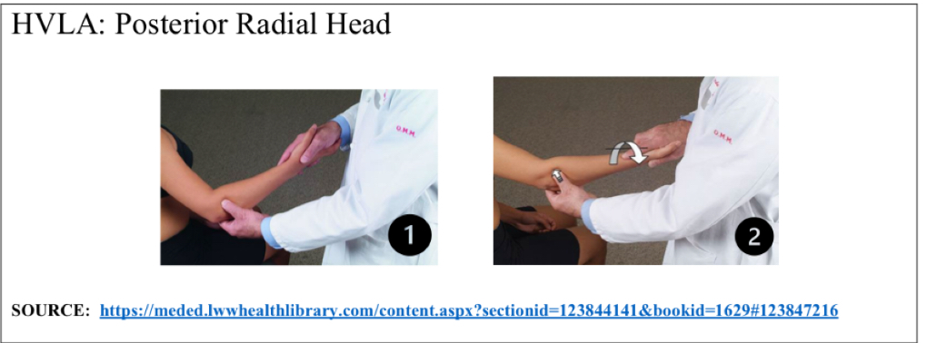
hvla posterior radial head
Pt seated, physician stands facing pt
Thumb on posterior aspect of radial head
Other hand holds pt hand like hand shake
Supinate forearm into barrier
Extend forearm into barrier
Thrust into extension
hvla displaced carpal (standard)
Dorsal displaced = flexed sd
Ventral displaced = extended sd
Pt seated, physician faces pt
Hold wrist like dx
Extend/flex into barrier while maintaining pressure
Thrust is baby whip further into barrier

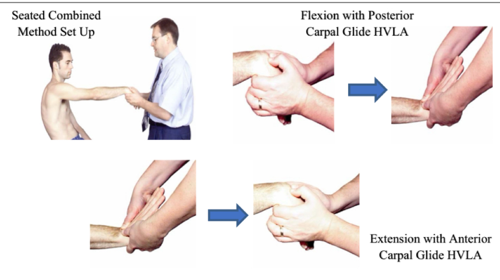
hvla seated combined method for wrist
Pt seated, physician faces pt
Hold wrist like dx
Extend/flex into barrier while maintaining pressure
pt will lean back for slightly back for traction
Thrust is baby whip into barrier
Flex SD w posterior glide
Extension SD w anterior glide
met sc joint - adduction sd (superior glide)
patient is supine
stand on same side of dysfunction
place one hand on proximal clavicle
other hand grabs wrist to extend and internally rotate arm
patient raises arm to ceiling
engage in barrier by moving arm down (no need to further internally rotate)
repeat 3-5 times + passive stretch

met sc joint - extension sd (anterior glide)
patient is supine
stand on same side of dysfunction
with one hand monitor SC joint
other hand cups scapula
ask patient to hold onto your shoulder with their SD arm
pull scapula up which moves distal clavical anterior (kind of balances the dysfunction out since SC joint is anterior)
patient pulls shoulder back
engage in barrier by pulling scapula further up
repeat 3-5 times + passive stretch

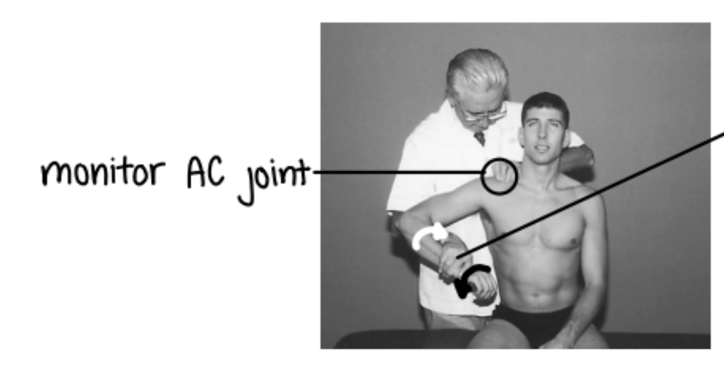
met ac joint - internal rotation sd
patient is seated + you stand behind them
stabilize lateral end of clavicle
monitor AC joint
flex 30, abduct 90
hold distal forearm
EXTERNALLY rotate
pt will internally rotate

met ac joint - external rotation sd
patient is seated + you stand behind them
stabilize lateral end of clavicle
monitor AC joint
flex 30, abduct 90
weave through to hold distal forearm
INTERNALLY rotate
pt will externally rotate
met ac joint - adduction sd
patient is seated + you stand behind them
compress distal clavical towards AC joint
flex 30 + abduct into barrier
patient adducts/moves to freedom

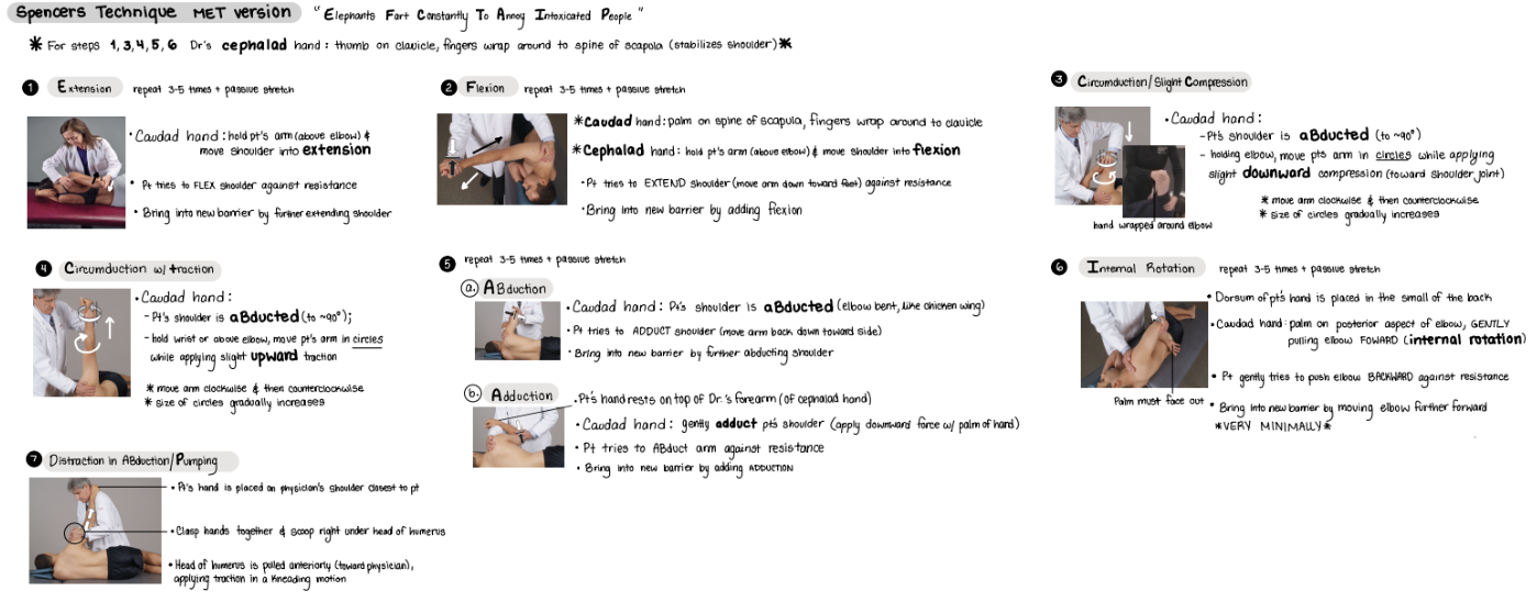
full spencer (7 steps) articulatory technique without met
elephants fart constantly to annoy intoxicated people
note that the techniques are where you position the patient
extension
flexion
compression (circumduction)
traction (circumduction)
abduction/adduct
internal rotation
pumping
spencer technique - extension
patient on lateral recumbent (on their side)
cephalad hand grabs patient’s shoulder to lock AC and SC joint
caudad hand holds patient’s arm above elbow and moves shoulder into extension
patient resists by flexing shoulder
repeat 3-5 times + passive stretch
if articulatory (not MET), then just pulse

spencer technique - flexion
patient on lateral recumbent (on their side)
caudad hand’s palm on spine of scapula + fingers wrap around clavicle
cephalad hand holds patient’s forearm and moves shoulder into flexion
patient resists by extending (moving arm toward the feet)
repeat 3-5 times + passive stretch
if articulatory (not MET), then just pulse
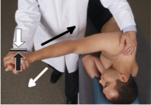
spencer technique - compression w/ circumduction
patient on lateral recumbent (on their side)
abduct patient’s shoulder to 90
cephalad hand holds shoulder
caudad hand on elbow, applying downward pressure, circumducting

spencer technique - traction w/ circumduction
patient on lateral recumbent (on their side)
abduct patient’s shoulder to 90
cephalad hand holds shoulder
caudad hand on wrist, applying traction (upward tension), circumducting

spencer technique - abduct
patient on lateral recumbent (on their side)
patient shoulders abduct (chicken wing) to restrictive barrier
cephalad hand is placed on shoulder
patient grabs physician’s arm (same one that’s holding shoulder)
patient resists by adducting
repeat 3-5 times + passive stretch
if articulatory (not MET), then just pulse
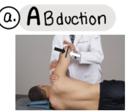
spencer technique - adduct
patient on lateral recumbent (on their side)
patient shoulders adduct (chicken wing) to restrictive barrier
cephalad hand is placed on shoulder
patient grabs physician’s arm (same one that’s holding shoulder)
patient resists by abducting
repeat 3-5 times + passive stretch
if articulatory (not MET), then just pulse

spencer technique - internal rotation
patient on lateral recumbent (on their side)
internally rotate patient’s arm + palms face out + hand placed on back
cephalad hand holds patient’s shoulder
caudal hand is behind patient’s elbow to induce internal rotation
patient resists by externally rotation (pushing elbow backwards)
new barrier is engaged by moving elbow forward
repeat 3-5 times + passive stretch
if articulatory (not MET), then just pulse

spencer technique - pumping
patient on lateral recumbent (on their side)
patient’s hand on physician’s shoulder
both of your hands clasp and “scoop” under head of humerus
pull head of humerus anteriorly (towards you)

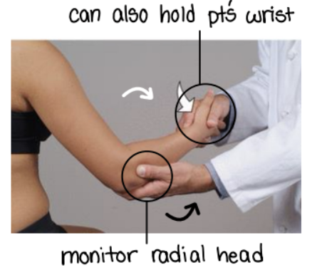
met elbow (radial head)
pronation sd = posterior radial head
monitor radial head with one hand
other hand holds pt’s wrist
supinate pt forearm to engage in barrier
pt tries to pronate forearm against resistance
bring into new barrier by adding supination
repeat 3-5 times + passive stretch
supination sd = anterior radial head
same set up just opposite movement
pronate pt’s forearm
pt tries to supinate
cs shoulder - supraspinatus
F Abd ER
Location is in belly of supraspinatus muscle
Pt supine
Physician sits by pt at level of shoulder girdle
Palpate tender point and use other hand to move arm
Flex arm 45 degrees, abduct 45 degrees, externally rotate (like a parade wave or Statue of Liberty)

cs shoulder - biceps brachii (long head)
F Abd IR
Location is over the tendon of the biceps muscle in the bicipital groove
Pt supine
Physician sits or stands same side of point
Point is found at the bicipital groove (flex to feel for tendon)
Flex, abduct, and internally rotate (like you’re scratching head)
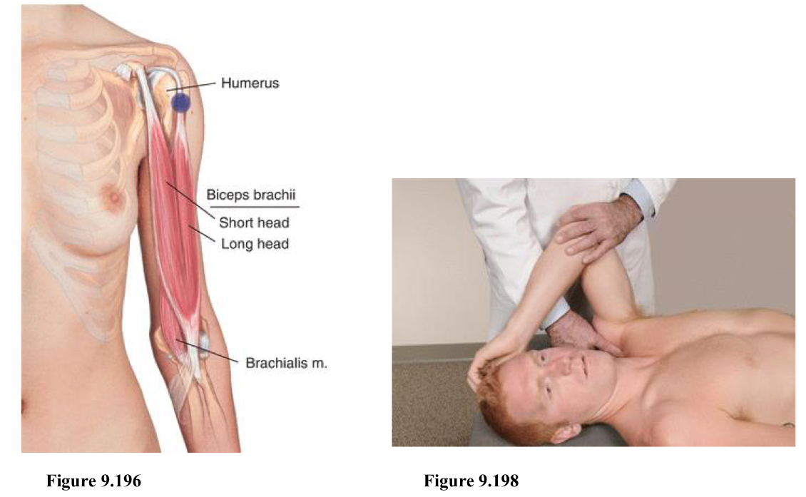
cs shoulder - biceps brachii (short head) / coracobrachialis
F Add IR
Location is at the inferolateral aspect of the coracoid process
Pt supine
Physician can stand from either side
Start at AC joint and follow down until you feel a bony prominence (will need to move through muscle). Then go inferior and lateral of coracoid process to locate point.
Tip - feel for humeral head and point will be slightly medial to that. Or just cup and thumb should feel it.
Shoulder stays on table!!!!
elbow and shoulder are flexed, shoulder minimally adducted and internally rotate (same side arm crosses to grab onto opposite shoulder)

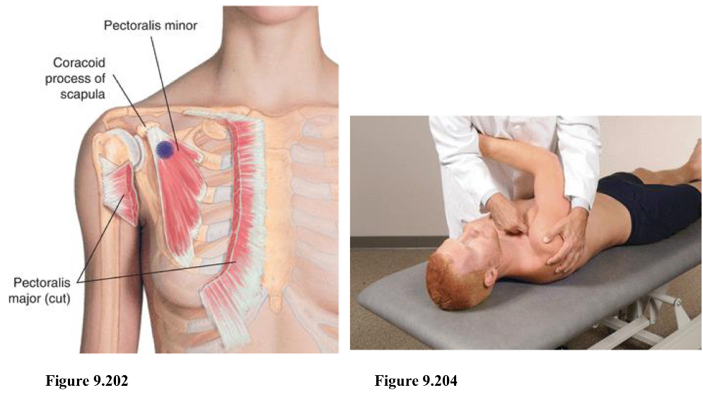
cs shoulder - pec minor
F Add
Location is inferior and medial to the coracoid process
Pt supine
Physician stands opposite of tender point
Point is inferior and medial to coracoid process
Arm is adducted across chest, shoulder/scapula is pulled anterior, inferior, and medial
Shoulder should be OFF table
Can stabilize by holding arm or forearm
cs shoulder - subscapularis
E IR
Location is at the anterolateral border of the scapula on the subscapularis muscle pressing from an anterior lateral to posteromedial direction
Pt supine
Physician sits or stands on same side
Locate Scapula, follow the lateral border, press deeper as your go superior (must go through muscle layers)
Tender point is on anterior surface of scapula, palpating in a posterior and medial direction. (Deep enough as you should not be able to see your finger tip)
Shoulder is extended and internally rotated

cs shoulder - levator scapulae
IR Abd traction
Location is on the superior medial border of the scapula at the attachment of the levator scapula
Pt prone with head looking away from point and arms at the sides
Physician sits on same side
Find spine of scapula, follow border up, point is superior and medial of the border
Physician caudad hand grabs pt wrist while other hand holds point.
Internally rotate pt shoulder and add mild to moderate traction and minimal abduction

cs shoulder - rhomboid minor/major
E Add
Location is along the medial border of the scapula at the attachment of the rhomboid muscles
Pt prone
Physician stands on either side (Cornick stood on opposite side)
Point is along medial border of scapula. Press medial to lateral. Shoulder be between spine of scapula and angle of scapula.
One hand on point, other hand holds elbow to set into position.
Shoulder is extended and adducted by pulling arm/elbow posterior and medial
Try to have pt hand stay on side and not on their back.

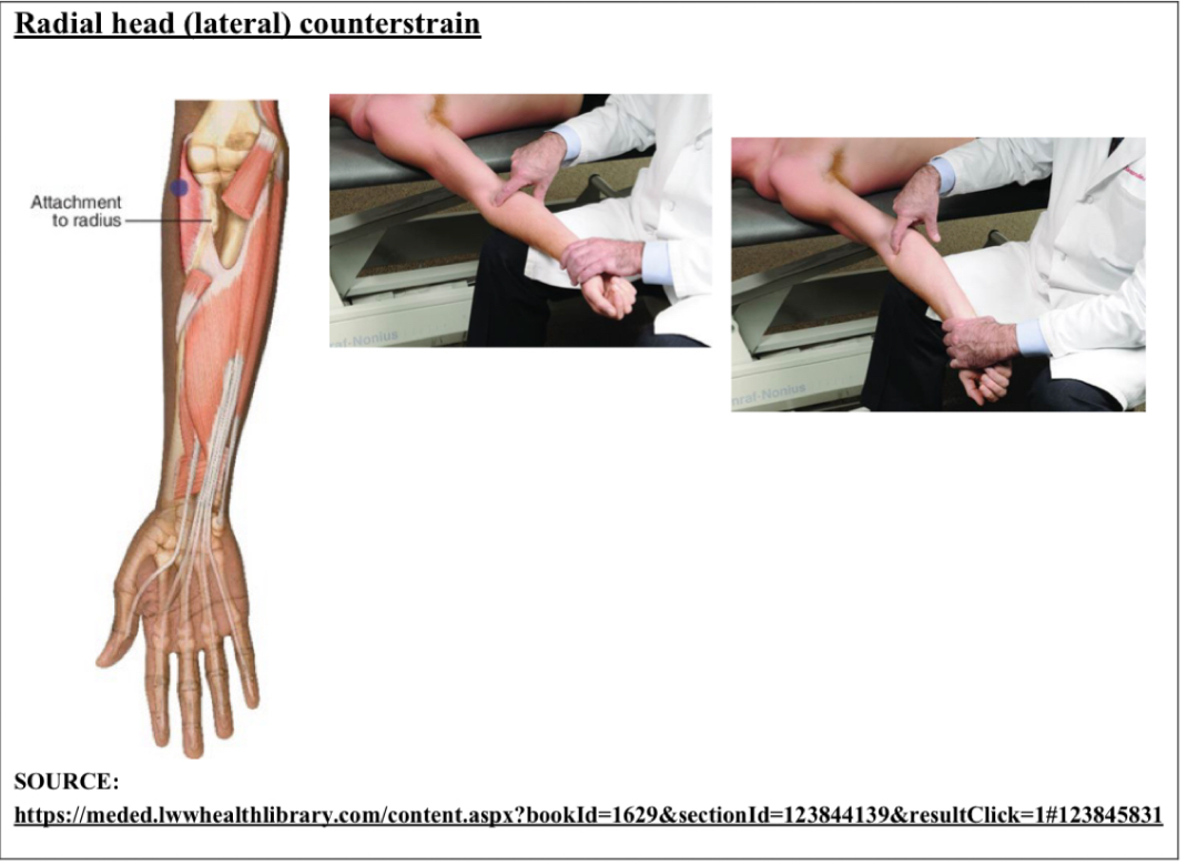
cs radial head
E Sup Val
Point is anterolateral aspect of radial head at attachment of supinator
pt supine, physician same side as point
pt elbow in full extension, forearm supinated
fine tune w/ supination and valgus force
cs lateral epicondyle
E Sup Abd
Point is lateral epicondyle of humerus
pt supine, physician on same side of point
pt elbow fully extended. Use table as fulcrum to put arm into position
arm supinated and abducted with varying amounts of force

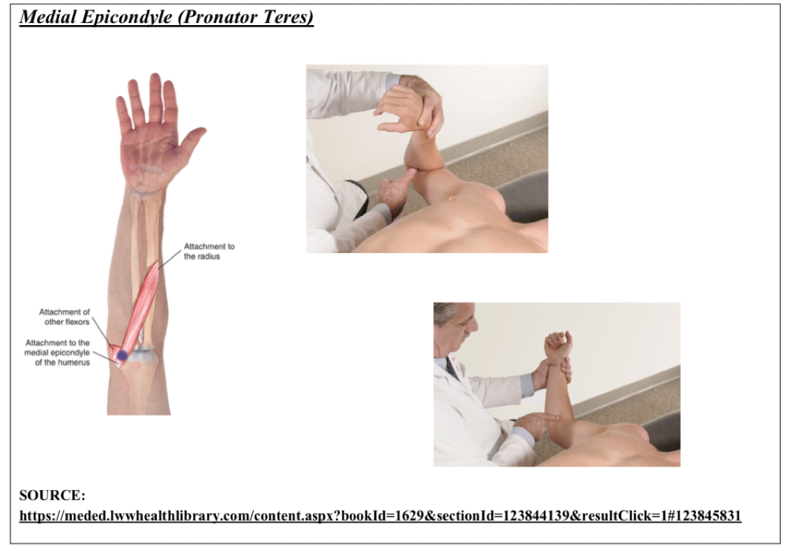
cs medial epicondyle
F Pro ADd with slight flexion of wrist
Point is medial epicondyle of humerus at common flexor tendon and attachment of pronator teres
pt supine, physician on same side of point
pt elbow flexed, wrist pronated, forearm slightly adducted, wrist slightly flexed
fine tune with elbow flex, wrist pronation, forearm adduction
cs extensor carpi radialis (dorsal)
Wrist extension, slight abduction (radial side)
Distal end and Dorsal surface of 2nd metacarpal in extensor carpi radialis
Like if you palpate 2nd MC joint then go down to tendon
pt either seated or supine, physician faces pt
pt wrist extended and abducted (radial deviation)

cs extensor carpi ulnaris distal attachment to 5th metacarpal
Wrist extension, slight Add
dorsal surface of 5th MC joint in extensor carpi ulnaris muscle
pt either seated or supine, physician faces pt
pt wrist extended and adducted (ulnar deviation)

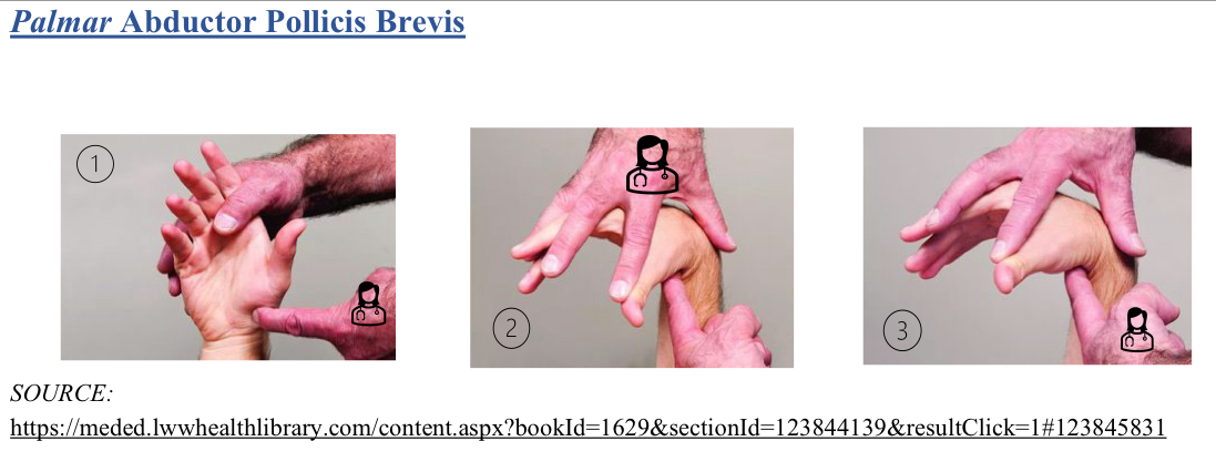
cs abductor pollicis brevis
F wrist, ABd thumb
Radial aspect of palmar base at 1 MC joint (abductor pollicis brevis)
pt seated or supine
locate point w/ index finger
pt wrist flexed, thumb abducted
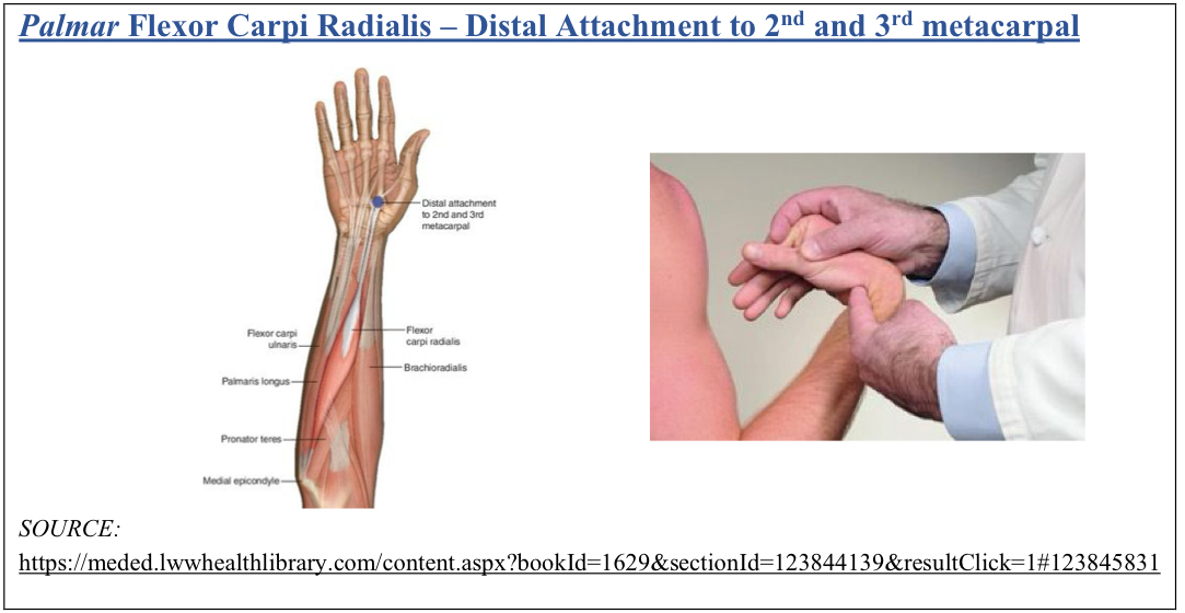
cs flexor carpi radialis
F ABd(radial)
distal attachment to 2nd and 3rd MC joint
pt seated or supine, physician faces pt
pt wrist flexed and abducted (radial deviation)
cs flexor carpi ulnaris
F aDd (ulnar)
Located t palmar base of 5th MC in flexor carpi ulnaris
pt seated or supine, physician faces pt
pt wrist flexed and adducted (ulnar deviation)

if patient moves easily with shoulder elevation, what is SC dx?
inferior/abduction somatic dysfunction
if patient moves easily with shoulder depression, what is SC dx?
superior/adduction somatic dysfunction
if patient moves easily with shoulder retraction, what is SC dx?
anterior/extension somatic dysfunction
if patient moves easily with shoulder protraction, what is SC dx?
posterior/flexion somatic dysfunction
name the motion pairings for AC joint
adduct, external rotation, inferior glide
abduct, internal rotation, superior glide
step up indicates which glide
inferior
clavical sits inferior to acromion, so you step up