Clinical Imaging - Exam 1: My Lung Pattern is Plaid, What's Yours? (Organs)
 Call Kai
 Learn
Learn Practice Test
 Spaced Repetition
 Match
Match Flashcards
 Knowt Play
1/82

There's no tags or description
Looks like no tags are added yet.
Name | Mastery | Learn | Test | Matching | Spaced | Call with Kai | Chat |
|---|
No analytics yet
Send a link to your students to track their progress
83 Terms

ID the lesion
Skin fold

ID the lesion
Pleural effusion

ID the lesion
Neoplasia
What are the four radiographic criteria of an alveolar lung pattern?
Air bronchogram
Uniform soft tissue
Lobar sign
Border effacement
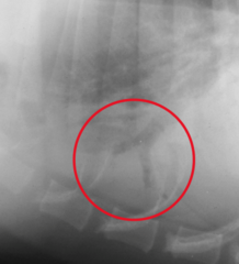
ID sign
Air bronchogram

ID sign
Lobar sign (sharp difference between cranial and caudal lobes)

Is there border effacement present?
Yes

ID sign
Air bronchogram
Point out the lobar sign
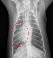
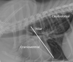
What disease is indicated by cranioventral alveolar pattern?
Bronchopneumonia
What disease is indicated by Caudodorsal alveolar pattern?
Pulmonary edema
What is the classic sign of bronchiolar pattern?
Rings and lines

You should see vessels at the ___th rib on lateral and the ___th on VD
4; 9
Heartworm disease will affect arteries, veins, or both?
Arteries
Left sided heart disease will affect arteries, veins, or both?
Veins
Left to right shunt will affect arteries, veins, or both?
Both
Pulmonary hypertension will affect arteries, veins, or both?
Arteries
Fluid overload and CHF will affect arteries, veins, or both?
Both
ID vessels

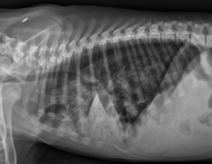
ID the pattern
Alveolar (lobar sign)
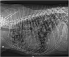
ID the pattern
Structured interstitial

ID the pattern
Bronchiole (rings and lines)

ID the pattern
Alveolar (Caudodorsal)

Diagnosis
Pneumothorax
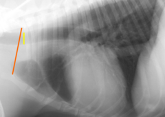
What percentage of the thoracic inlet should the trachea be in a non-brachycephalic?
>20%
If the trachea is easy to see on inspiration but disappears on expiration, you should be suspicious of what?
Collapsing trachea
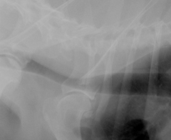
Diagnosis?
Redundant dorsal trachealis muscle
T/F: A normal trachea can deviate to the right, but not the left
True

Diagnosis?
Pneumothorax

What disease can cause ventral deviation of the trachea?
Esophageal disease
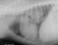
What can cause ventral displacement of the carina and caudal lobar bronchi?
Tracheobronchial lymphadenopathy

Diagnosis?
Diaphragmatic hernia

Diagnosis?
Normal
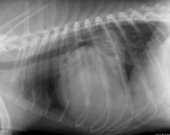
What disease can cause dorsal tracheal deviation?
Cardiac (LA) enlargement
What ICS should the carina be at?
5th
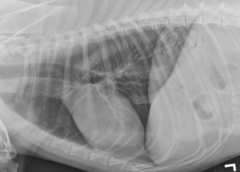
ID lesion
None, normal

Diagnosis?
Normal

Diagnosis?
Diaphragmatic hernia

Diagnosis?
Pneumomediastinum

Diagnosis?
Peritoneopericardial Diaphragmatic Hernia

Diagnosis?
Normal

ID lesion
Pleural fissure lines (seen due to pleural effusion)
What view is best for visualization of the heart if fluid is present?
VD (fluid is below heart, not surrounding it like in DV)


Diagnosis?
Pneumothorax
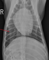
ID the lesion
Skin fold

ID the lesion
Mediastinal fat

Is the trachea dorsally deviated?
Yes, because it is parallel to the spine

ID structure
Descending aorta

What diseases can result in an aortic bulge?
PDA or systemic hypertension

Enlargement of what structure can compress the main stem bronchus?
Left atrium
What is the earliest sign of left atrial enlargement?
Straightening of the cardiac waist


Severe left atrial enlargement can bulge between the main stem bronchi, known as what?
Backpack sign
What sign from VD will allow you to ID left atrial enlargement?
Double opacity
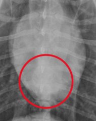
What occurs with the main stem bronchi in left atrial enlargment?
Left bronchus deviation (bow legged cowboy)

With more advanced left atrial enlargement, what other structure will enlarge?
Left auricle

At what clock position is the left atrium in a cat?
1:00 - 2:00

The left set of cranial vessels is more (cranial/caudal) than the right set
Cranial

Normal pulmonary vessels should be what size compared to the 4th rib?
3/4th

Are these vessels enlarged?
Yes, due to overcirculation to the lung (left to right shunt)
What sign on VD is indicative of right heart enlargement?
Reverse D

You would expect the CVC to be (bigger than/equal to/smaller than) the aorta in right heart failure
Bigger than

How can you ID a normal stomach axis?
Parallel to the ribs

Enlargement of what organ can shift the stomach axis caudally?
Liver


Ascites?
No

Ascites?
Yes
Abdominal effusion due to right heart failure can often be identified due to the lack of visualization of what structure?
Falciform fat pad

Long standing left heart disease leading to right heart changes is often due to what disease?
Mitral regurgitation
What are the most common causes of systemic hypertension in cats?
Hyperthyroidism and renal disease
What lung pattern does pulmonary edema present as in cats?
Patchy interstitial to alveolar


What sign is classic for feline HCM?
Valentine heart
What signs can you expect to see with canine DCM?
Left sided enlargement


Diagnosis?
Pericardial effusion (globoid)
Pericardial effusion will present with what side heart failure?
Right (easier to compress)
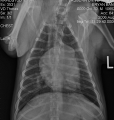
Diagnosis?
Heartworms
What lung pattern can you expect with heartworms?
Bronchiole

PDA connects what two vessels?
Aorta to pulmonary artery (Left to right shunt, overcirculation of left side)
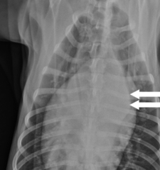
What radiographic sign indicates PDA?
Ductus bump
What side of the heart is affected by pulmonic stenosis?
Right side (blood can't flow easily, right pumps harder)
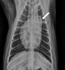
ID the lesion associated with pulmonic stenosis
Enlarged main pulmonary artery
Ventricular septal defects will lead to an overcirculation of what?
Pulmonary vasculature (enlarged A/V)
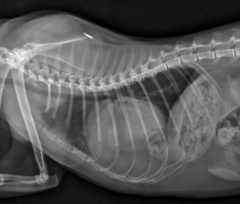
Diagnosis?
Peritoneopericardial Diaphragmatic Hernia
What are the four features of tetralogy of fallot?
Pulmonic stenosis
Right ventricular hypertrophy
Subaortic VSD
Overriding aorta
What grade are you going to make on this test?
55 (out of 55)