Biology Lab 4: Infraglottic Cavity, Renal Pelvis, and Related Structures
1/52
There's no tags or description
Looks like no tags are added yet.
Name | Mastery | Learn | Test | Matching | Spaced |
|---|
No study sessions yet.
53 Terms
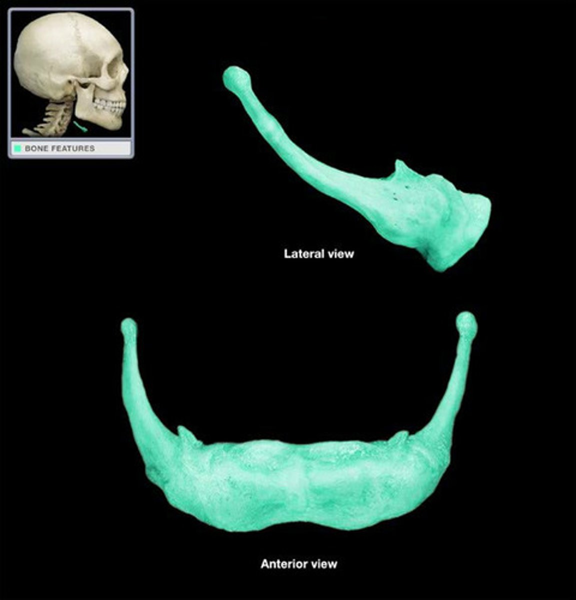
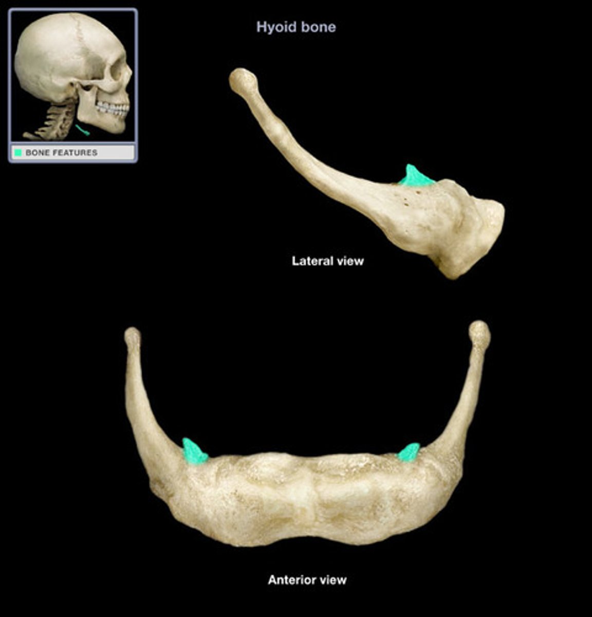
Hyoid
Note: superior to the thyroid cartilage, has three parts (the body forms the central segment of the hyoid; bilaterally the greater horns project backwards and are larger and longer than the conical shaped lesser horns)
Name the Bone
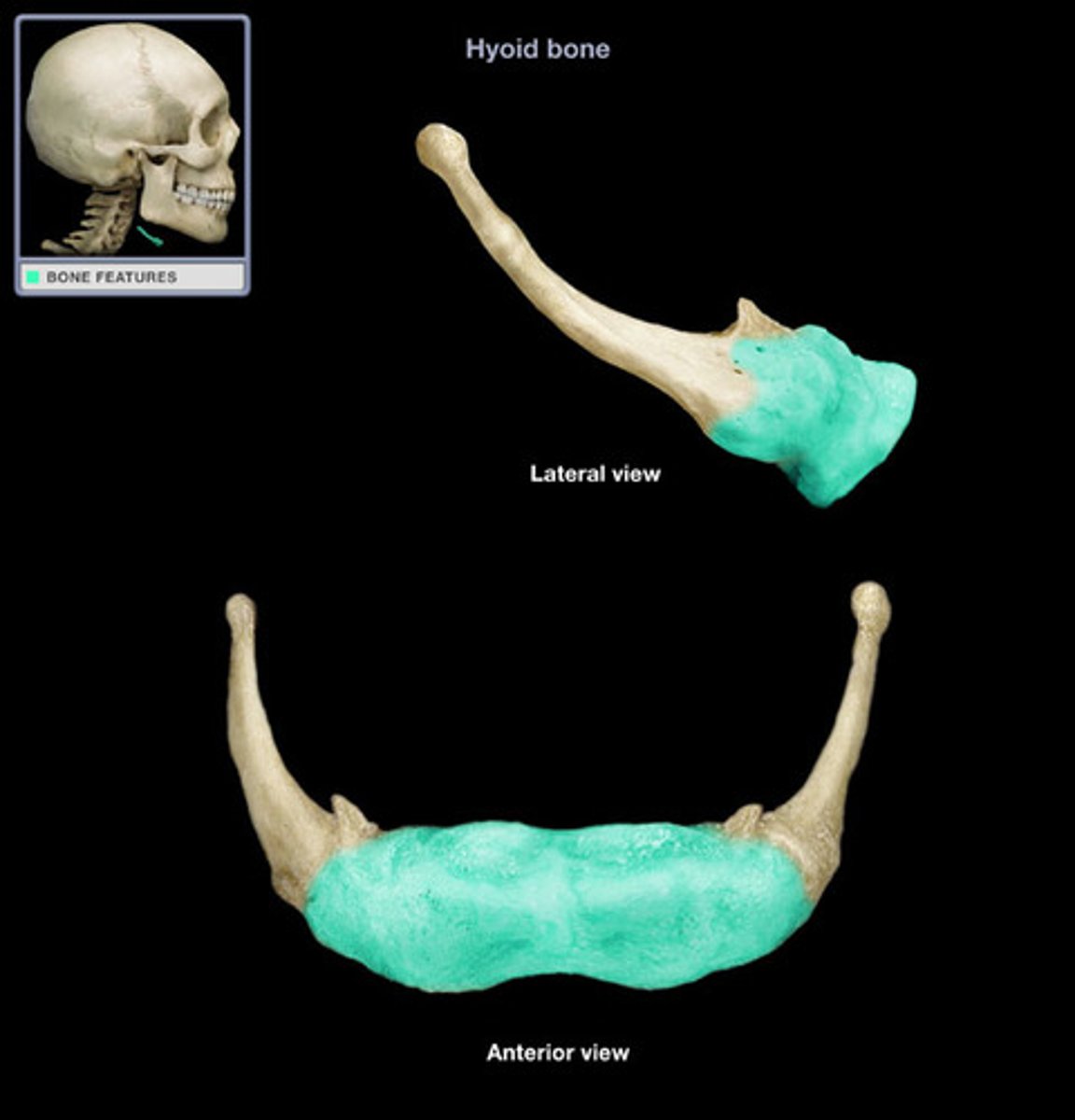
Body of Hyoid
Name the Specific Part of the Hyoid
Greater Horns of Hyoid
Name the Specific Part of the Hyoid
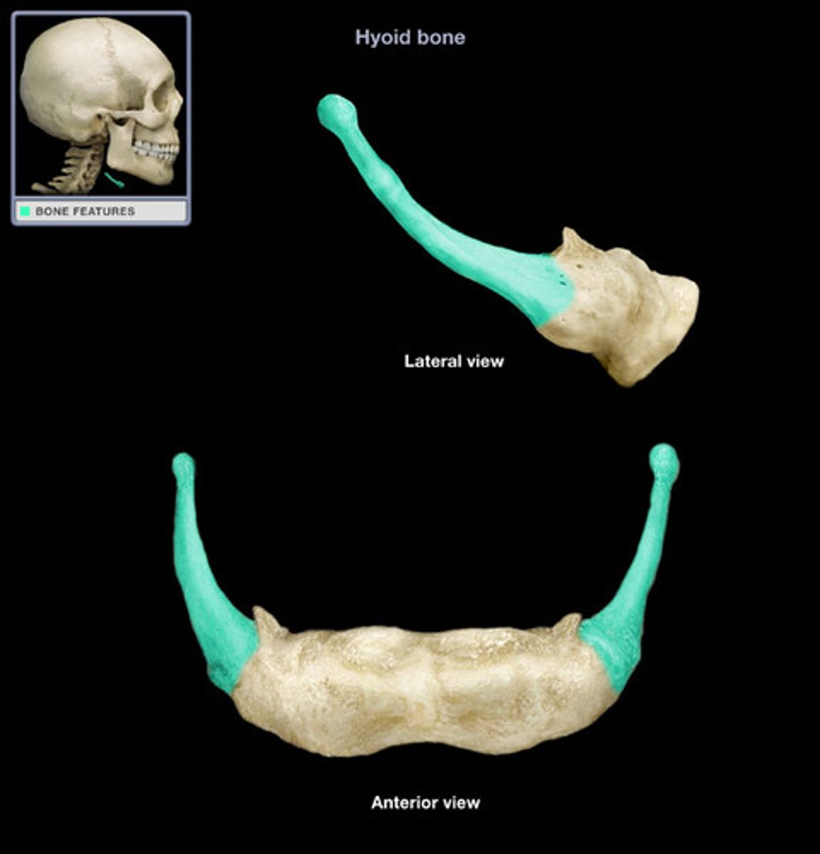
Lesser Horns of Hyoid
Name the Specific Part of the Hyoid
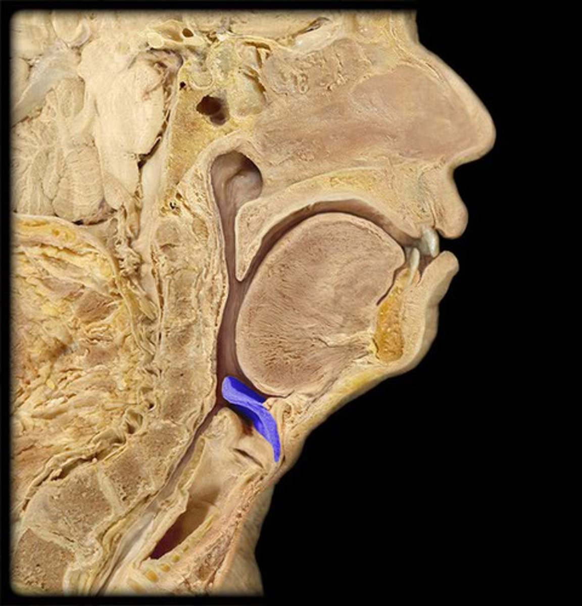
Epiglottis (Lateral View)
Name the Structure
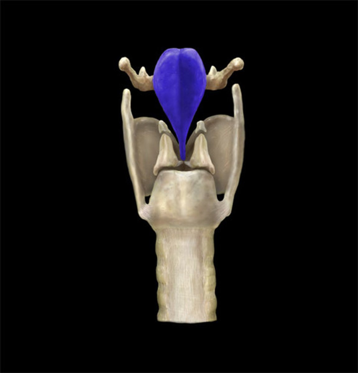
Epiglottis (Posterior View)
Name the Structure
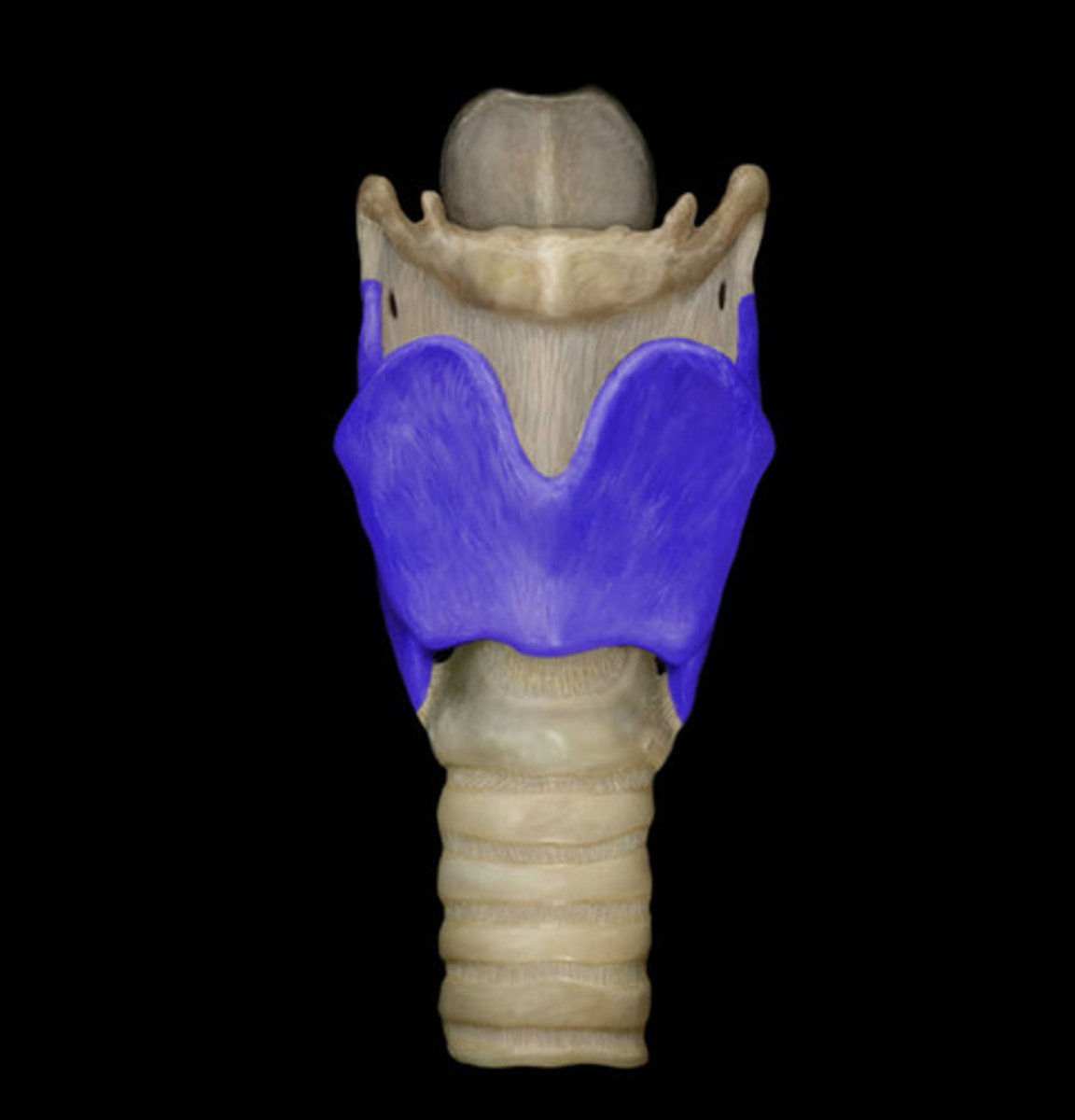
Thyrohyoid Membrane (Anterior View)
Note: attached to superior border of thyroid cartilage and inferior border of hyoid bone, pierced by internal laryngeal nerve and superior laryngeal artery and vein
Name the Structure

Thyroid Cartilage (Anterior View)
Note: composed of two, plate-like laminae that fuse to form a protrusion called the laryngeal prominence (Adam's Apple), posterior border of each lamina has a superior horn and an inferior horn
Name the Structure
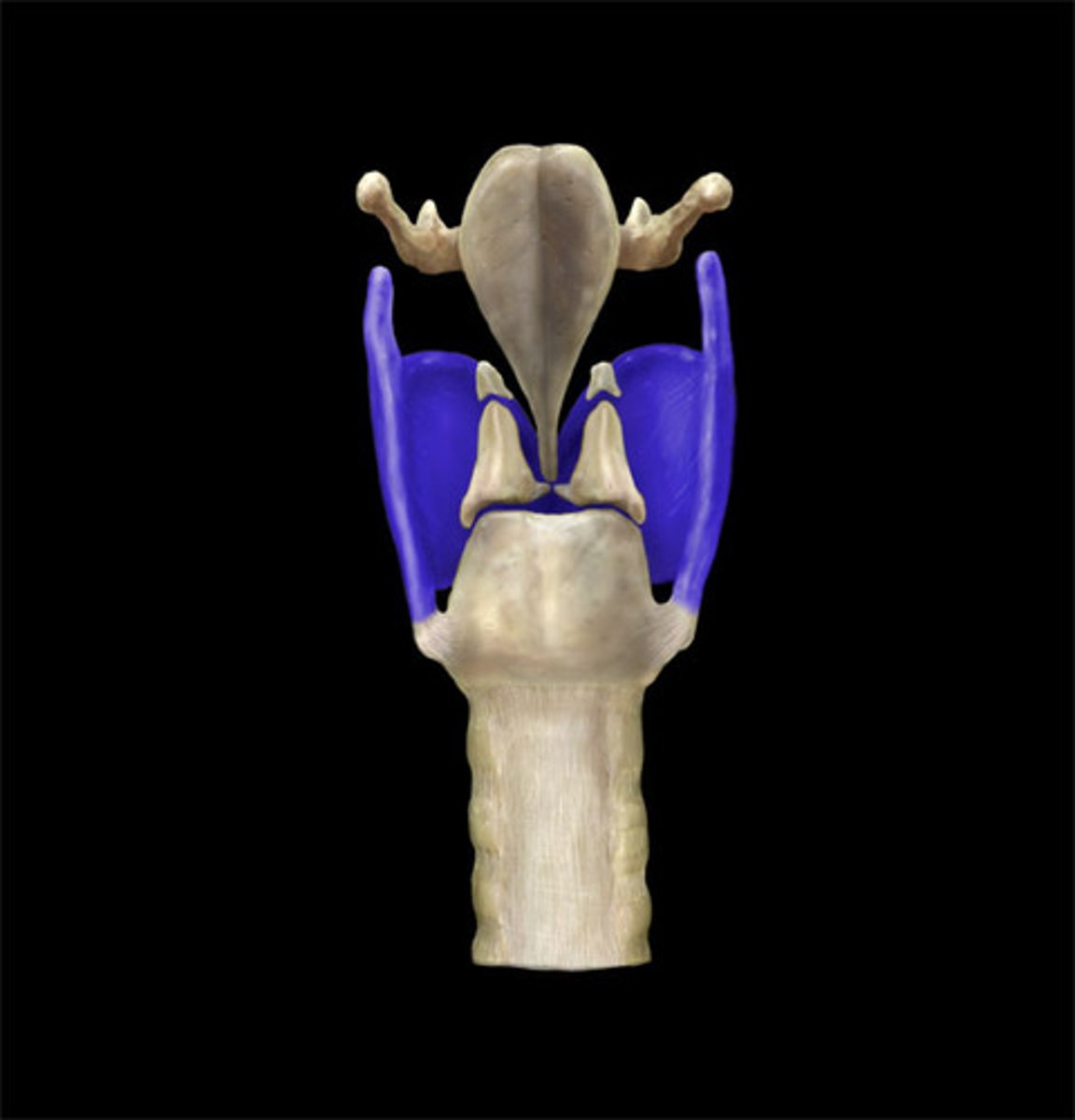
Thyroid Cartilage (Posterior View)
Name the Structure
Laryngeal Prominence (Anterior View)
Name the Part of the Thyroid Cartilage
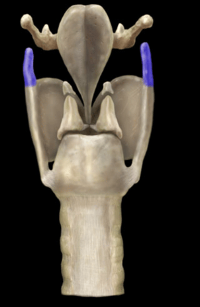
Superior Horn (Posterior View)
Note: this is inferior to greater horn of hyoid when viewed posteriorly
Name the Part of the Thyroid Cartilage
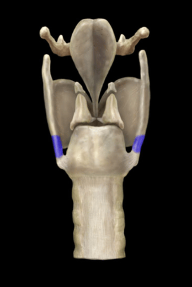
Inferior Horn (Posterior View)
Name the Part of the Thyroid Cartilage
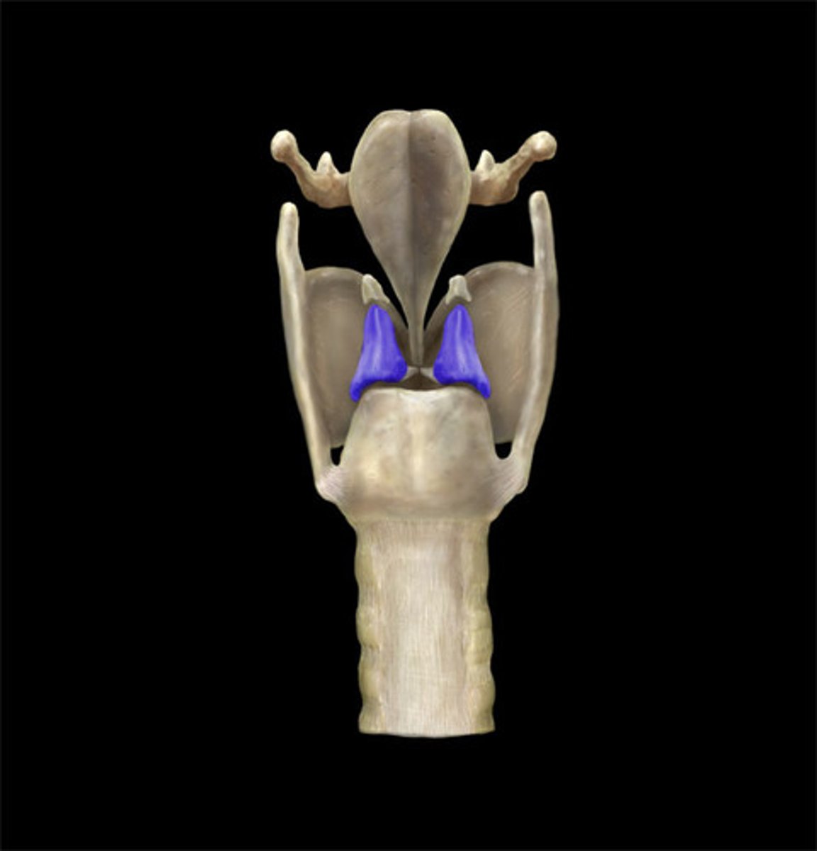
Arytenoid Cartilages (Posterior View)
Note: paired pyramid-shaped structures superior to the cricoid cartilages, attach to vocal folds/ligaments (vocal process of each arytenoid cartilage is directed anteriorly and attaches to vocal ligament), muscular process of each arytenoid cartilage projects posterolaterally from the base of the arytenoid cartilage and attaches to posterior cricoarytenoid muscle and lateral cricoarytenoid muscle
Name the Structure
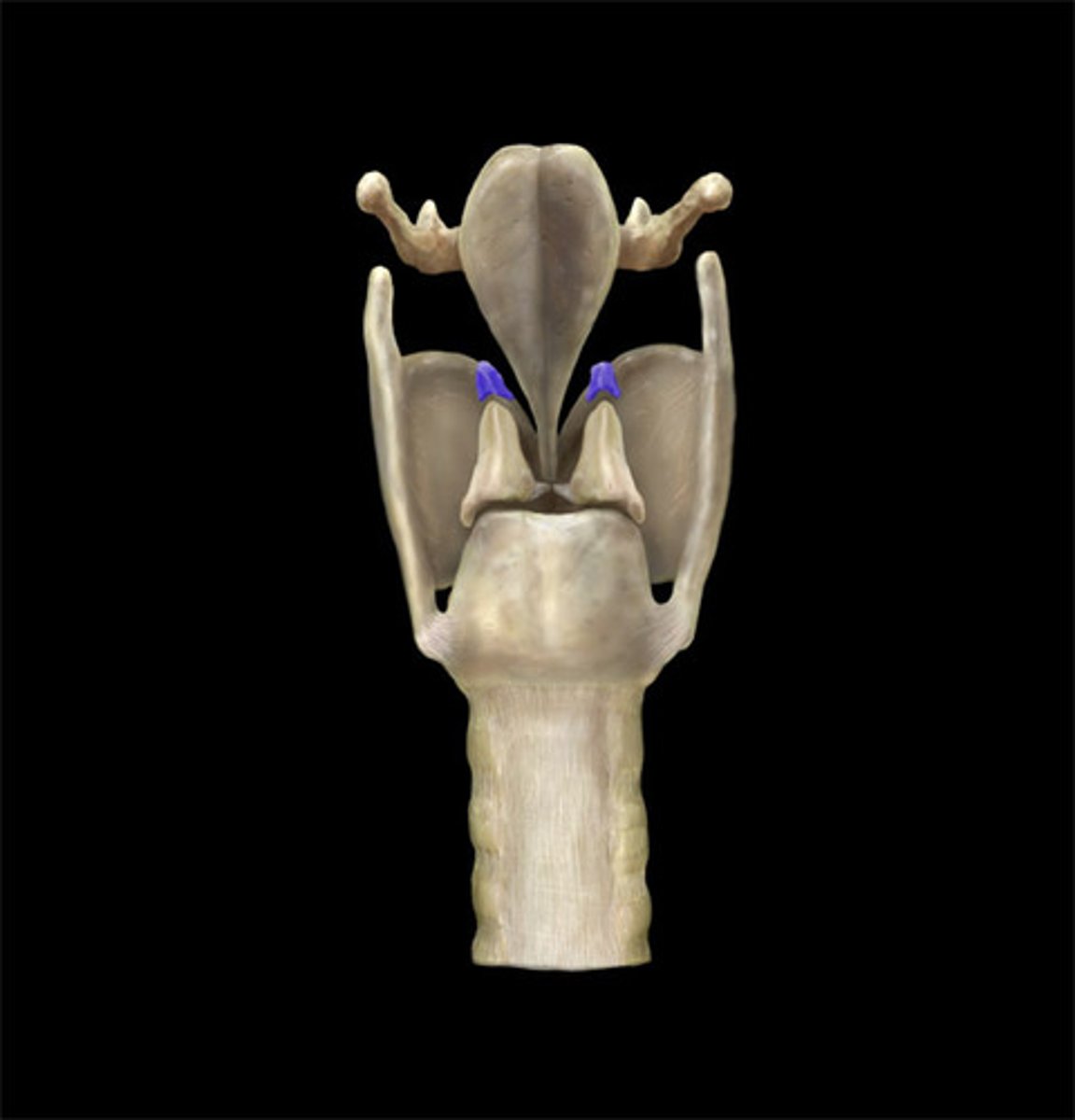
Corniculate Cartilage (Posterior View)
Name the Structure
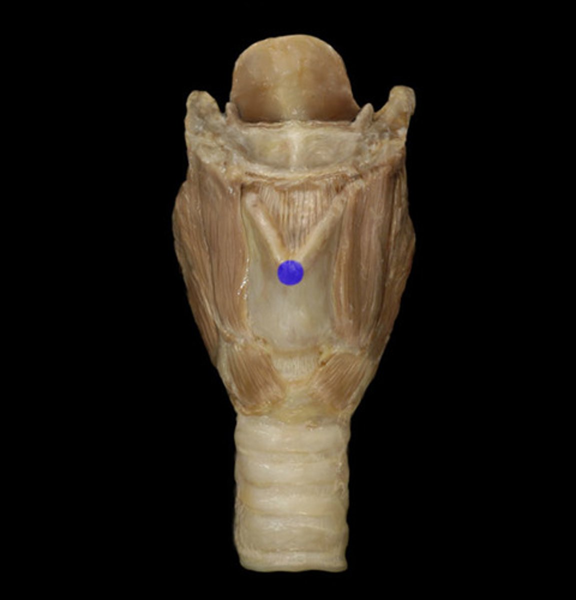
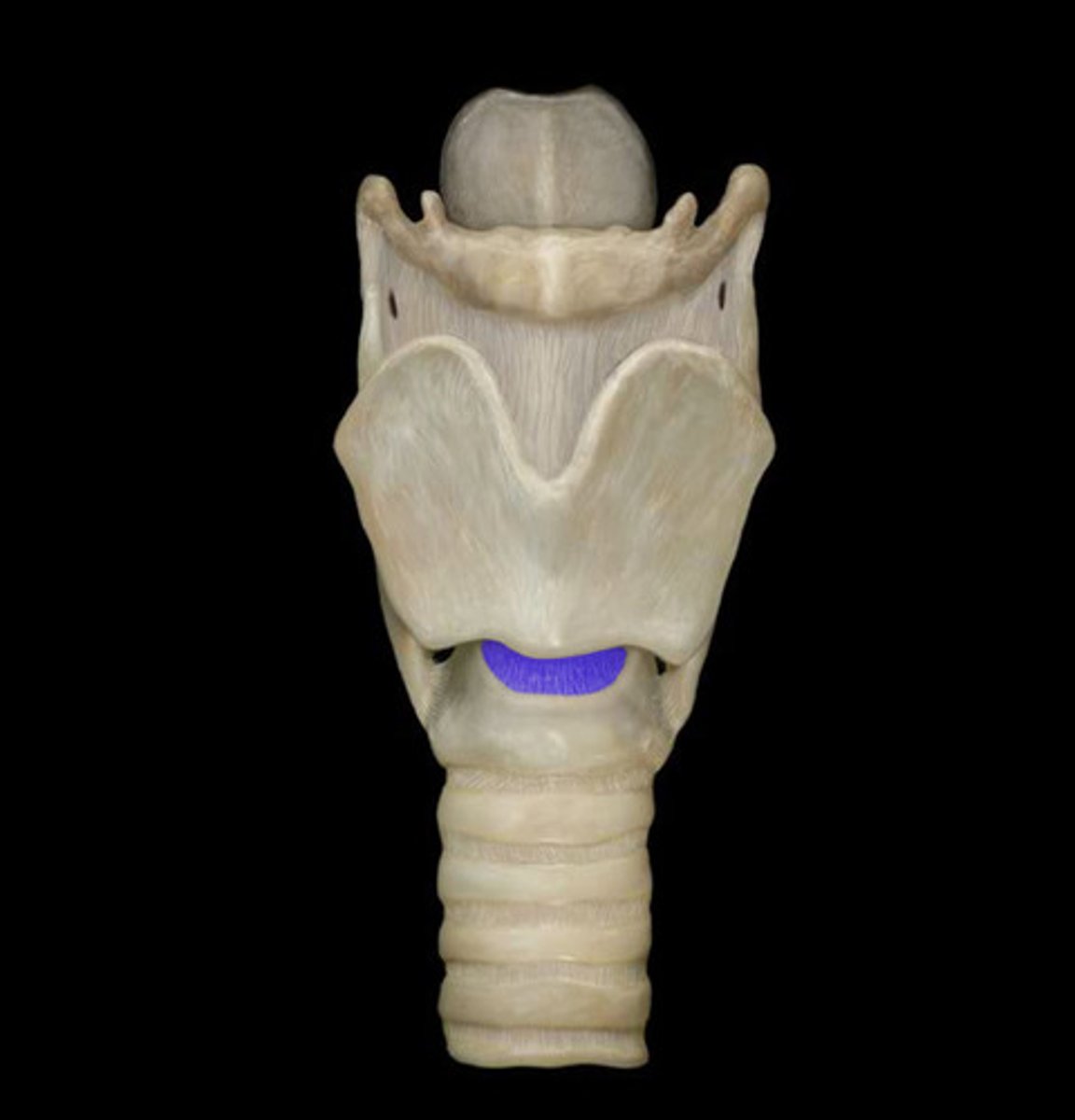
Cricothyroid Ligament (Anterior View)
Name the Structure
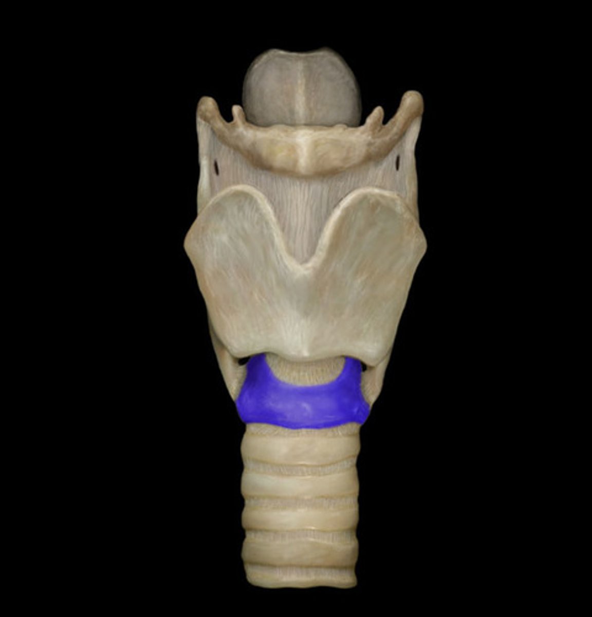
Cricoid Cartilage (Posterior View)
Note: wider posteriorly compared to anteriorly
Name the Structure
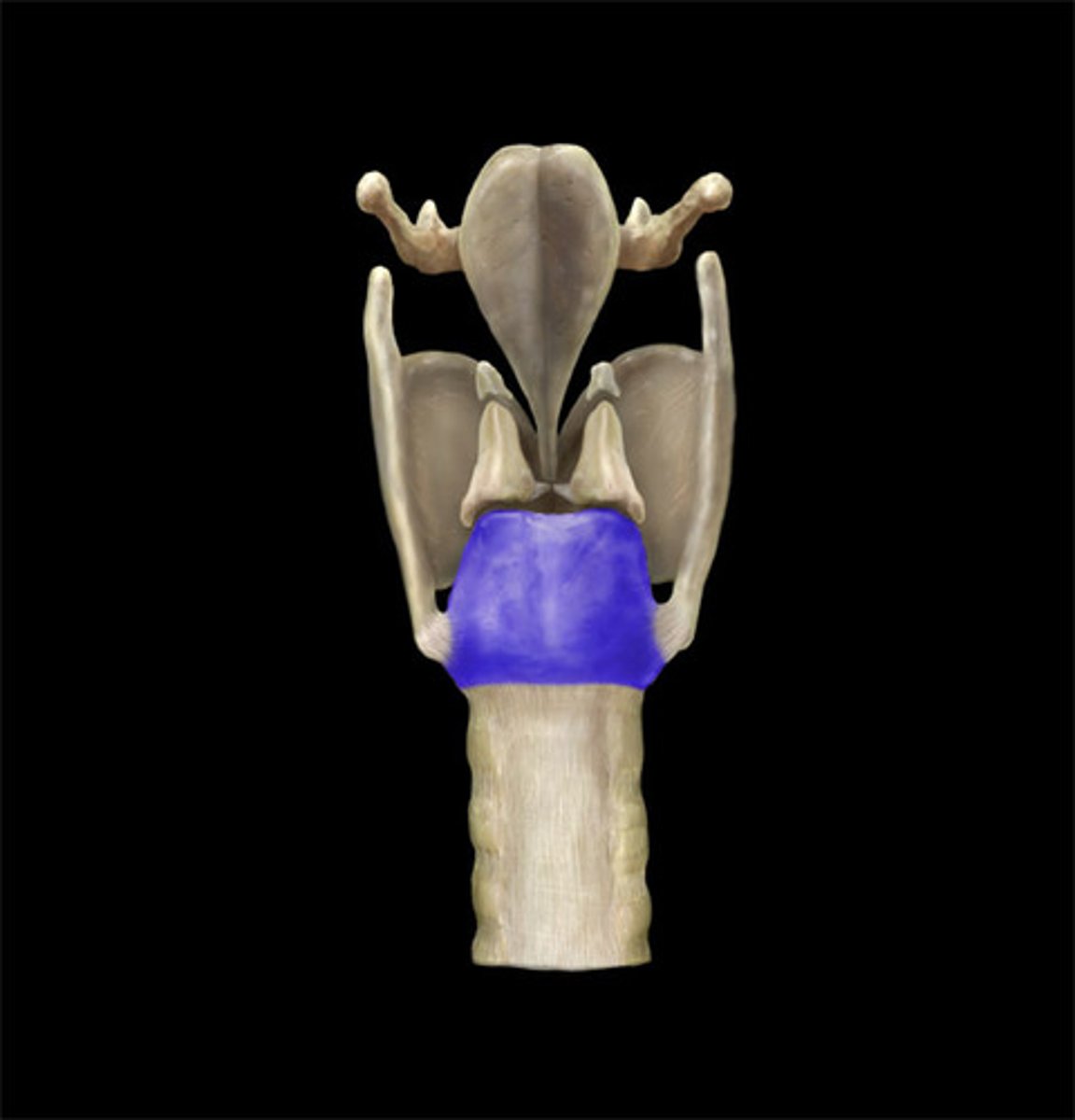
Cricoid Cartilage (Anterior View)
Name the Structure
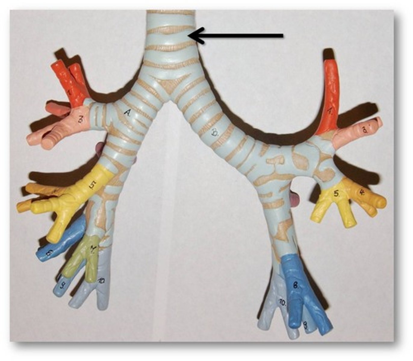
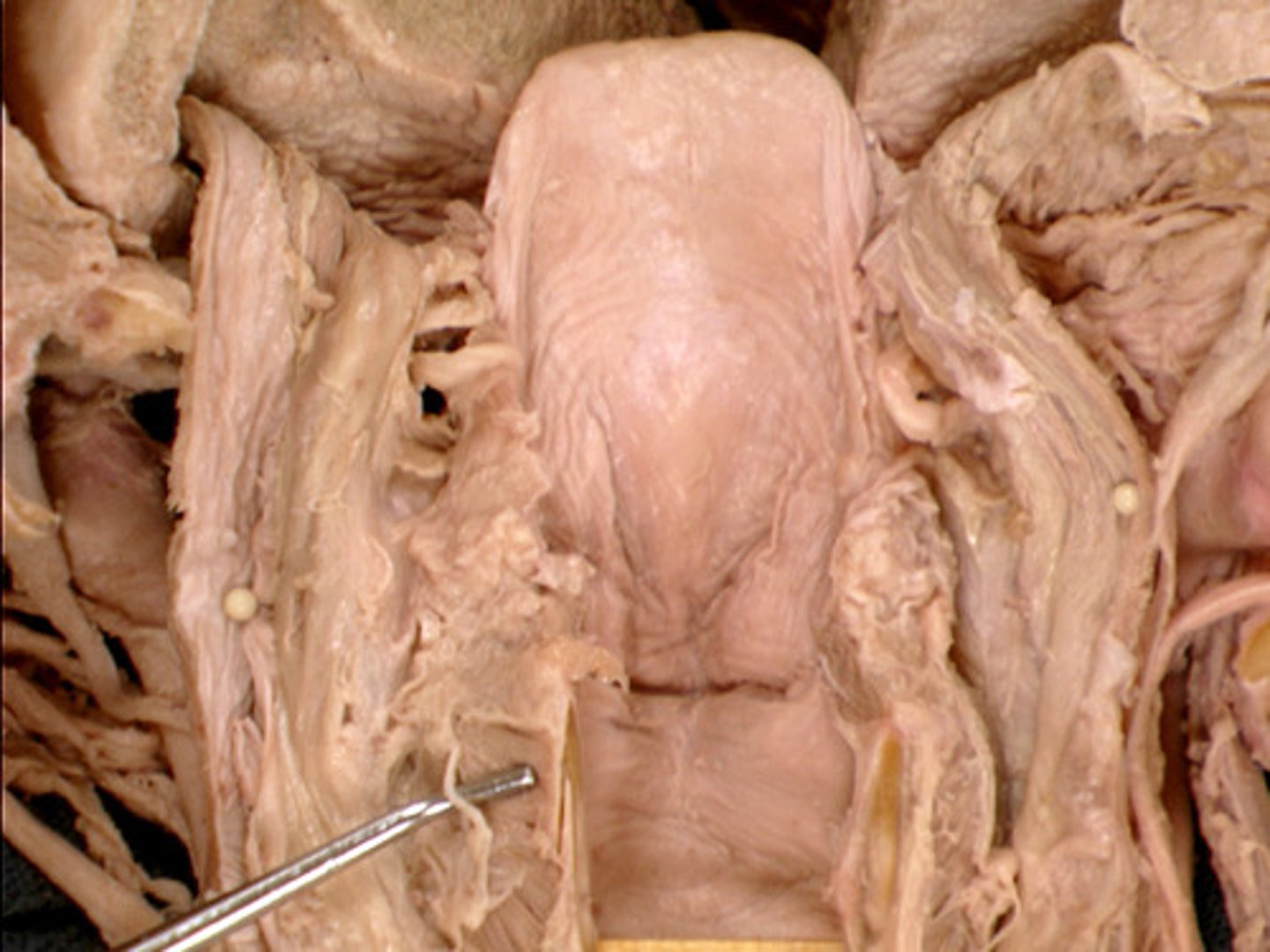
Trachea
Note: numerous C-shaped cartilaginous "rings", located inferior to the larynx
Name the Structure
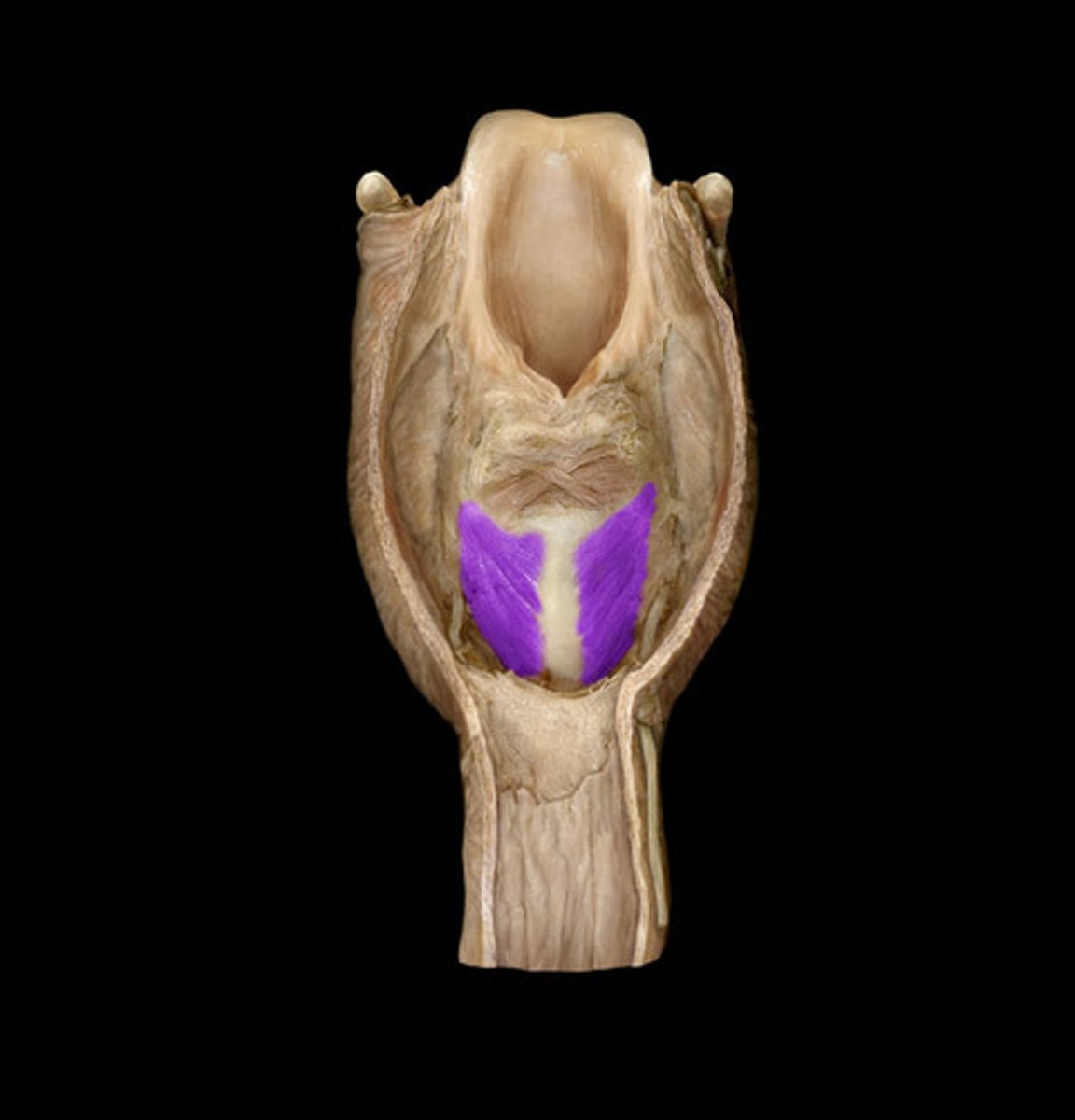
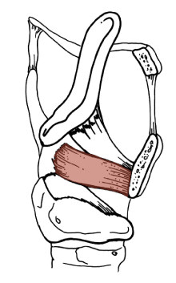
Cricothyroid (Anterior View)
Note: cricothyroid LIGAMENT is MEDIAL while cricothyroid MUSCLE is LATERAL (remember that L goes with M), located in between cricoid and thyroid cartilages, tenses the vocal ligament (makes it more taut, thus raises pitch), only larynx muscle with motor innervation by the external laryngeal nerve (branch of superior laryngeal branch of vagus nerve, while everything else is innervated by inferior branch of recurrent laryngeal branch of vagus nerve)
Name the Muscle

Posterior Cricoarytenoid (Posterior View)
Note: abduct the vocal folds, thus opening the rima glottidis (only muscle that opens vocal cords)
Name the Muscle
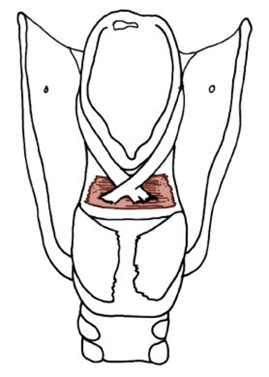
Arytenoid (Posterior View)
Note: has oblique and transverse heads, oblique is continuous with aryepiglottal fold
Name the "X" Shaped Muscle Above the Purple Muscle

Oblique Head of Arytenoid
Name the Part of the Arytenoid (#6)
Transverse Head of Arytenoid
Name the Part of the Arytenoid
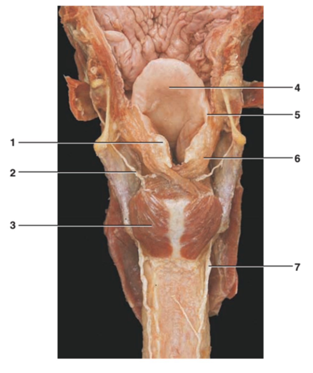
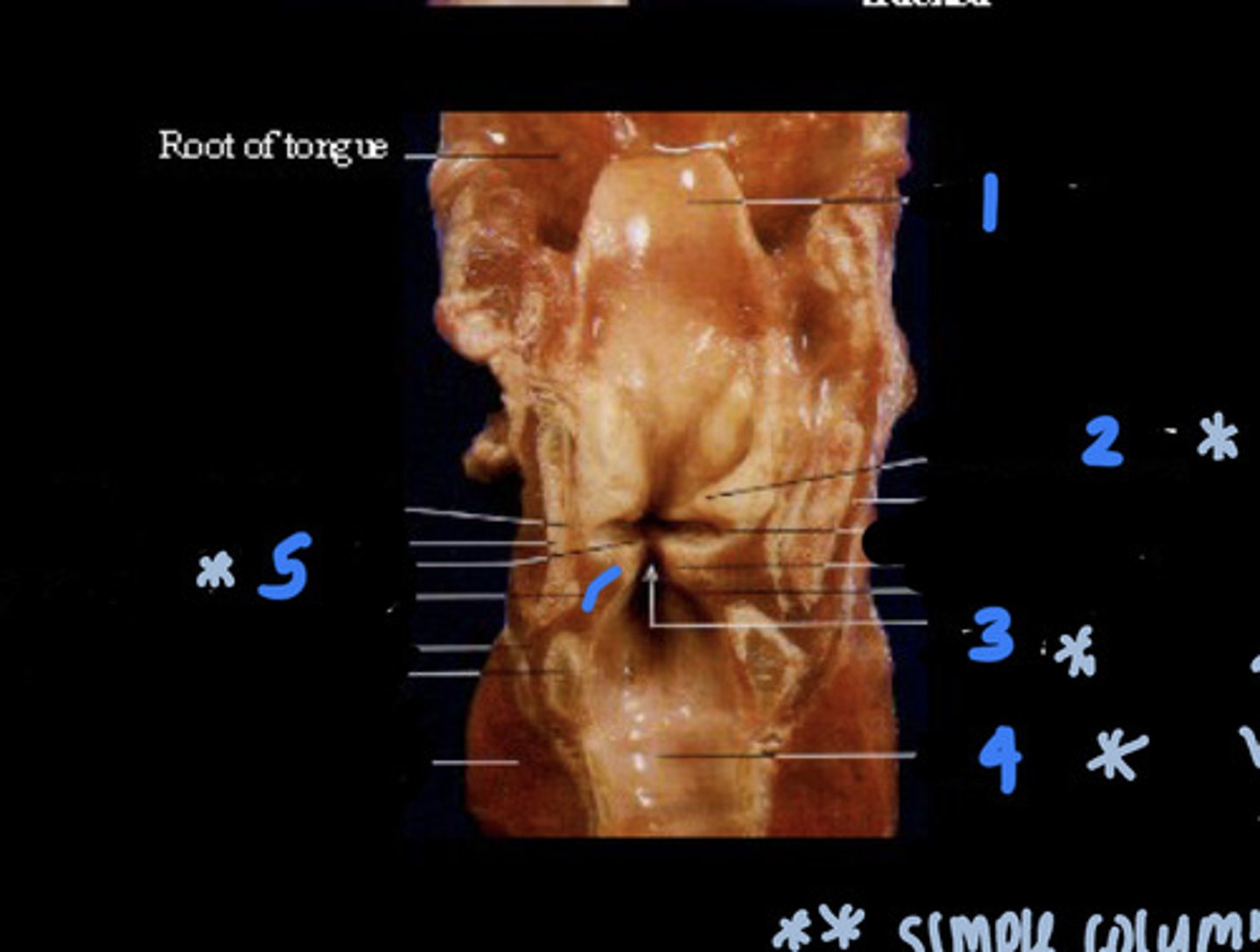
Vestibular Folds (Coronal View)
Note: superior to vocal cords
Name Structure #2

Vestibular Folds (Lateral/Saggital View)
Name the Structure
Vestibular Folds (Superior/Axial View)
Name the Structure
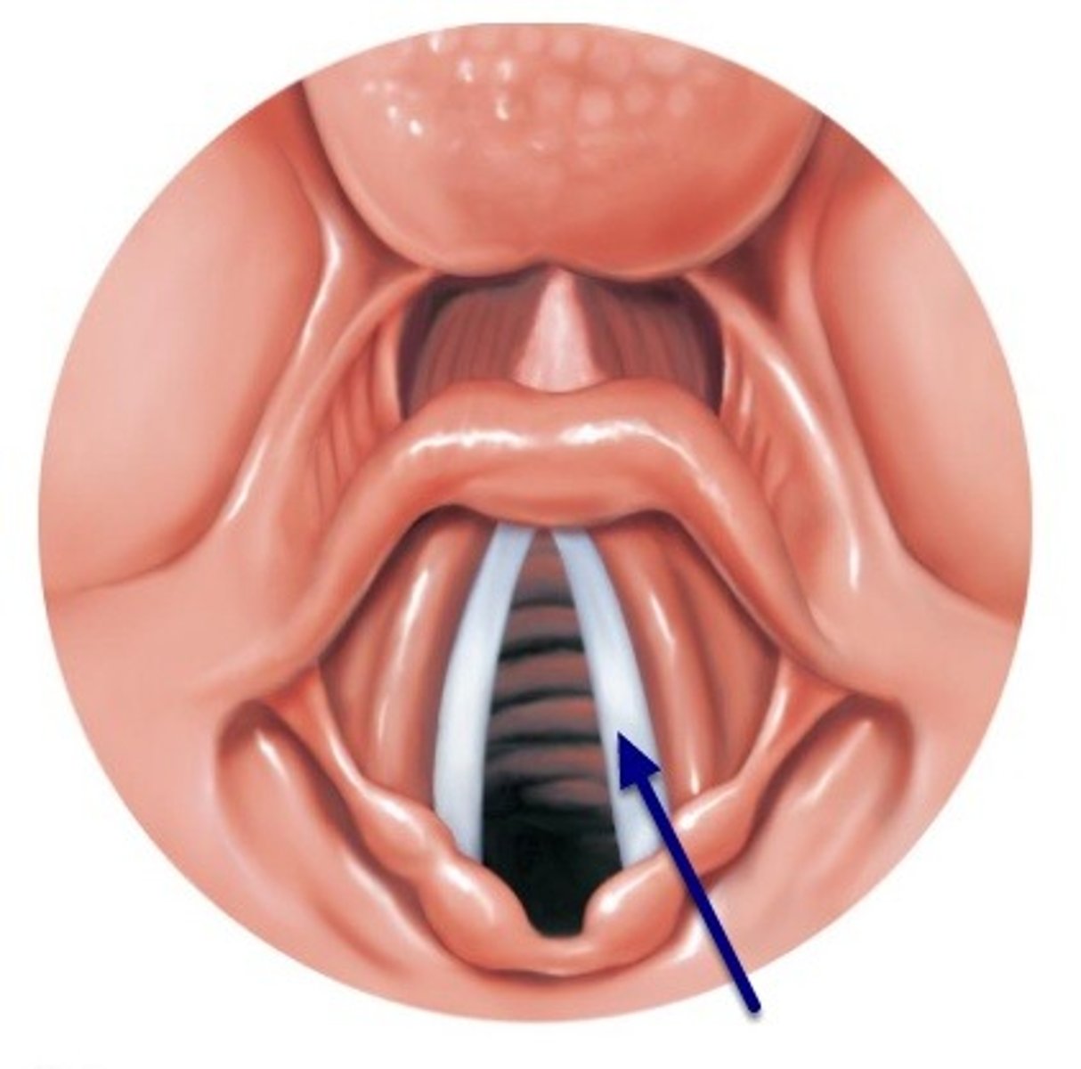
Vocal Folds (Coronal View)
Note: inferior to vestibular cords
Name Structure #5

Vocal Folds (Lateral/Saggital View)
Name the Structure
Vocal Folds (Superior/Axial View)
Name the Structure
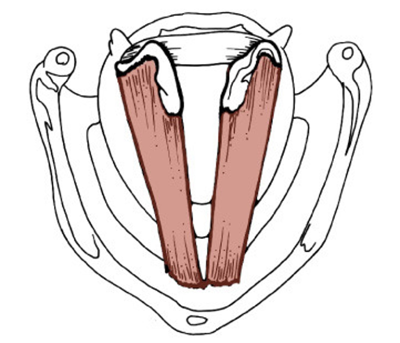
Thyroarytenoid (Superior/Axial View)
Note: this is a muscle WITHIN the vocal fold (if asked for a muscle within the vocal fold, THIS is the answer), attaches anteriorly to thyroid cartilage, sometimes the medial segment is called the vocalis muscle (though we don't need to ID that for this exam), relaxes vocal ligament and produces lower pitch
Name the Muscle
Thyroarytenoid (Lateral/Saggital View)
Name the Muscle
Aryepiglottic Folds (Posterior View)
Note: mucosa in between the arytenoid cartilages and the epiglottis
Name the Structure
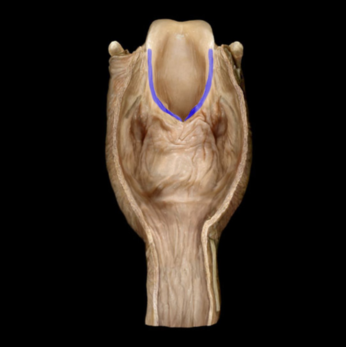
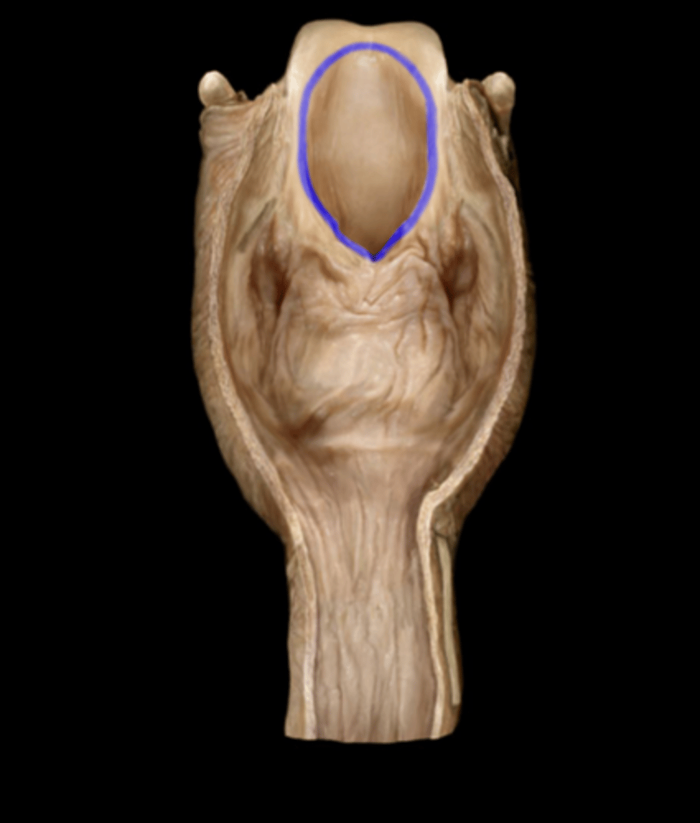
Laryngeal Inlet (Posterior View)
Name the Opening (NOT the Structure)
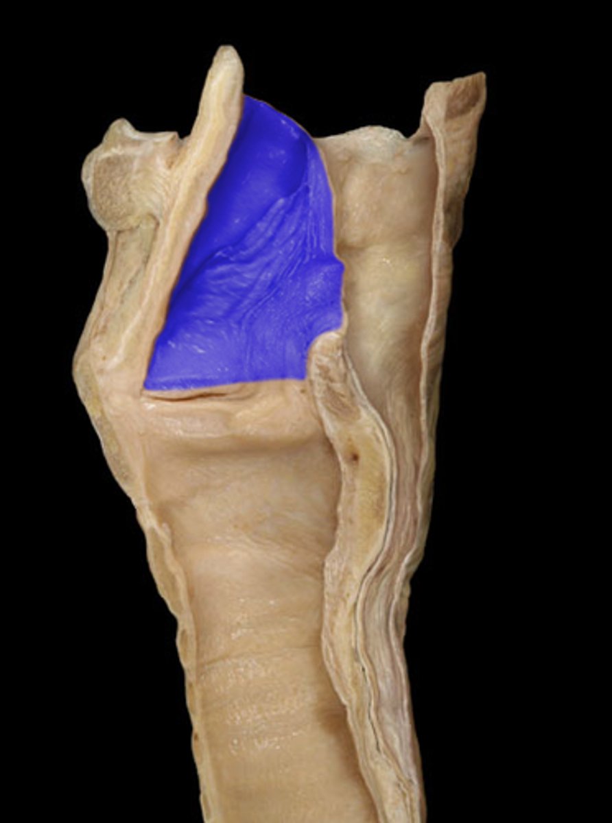
Vestibule (Posterior View)
Note: space between laryngeal inlet and the vestibular folds
Name the Opening Between #1 & #2

Vestibule (Lateral View)
Name the Opening
Ventricle (Posterior View)
Note: space between vestibular folds and vocal folds
Name the Opening Between #2 & #5
Ventricle (Lateral View)
Name the Opening
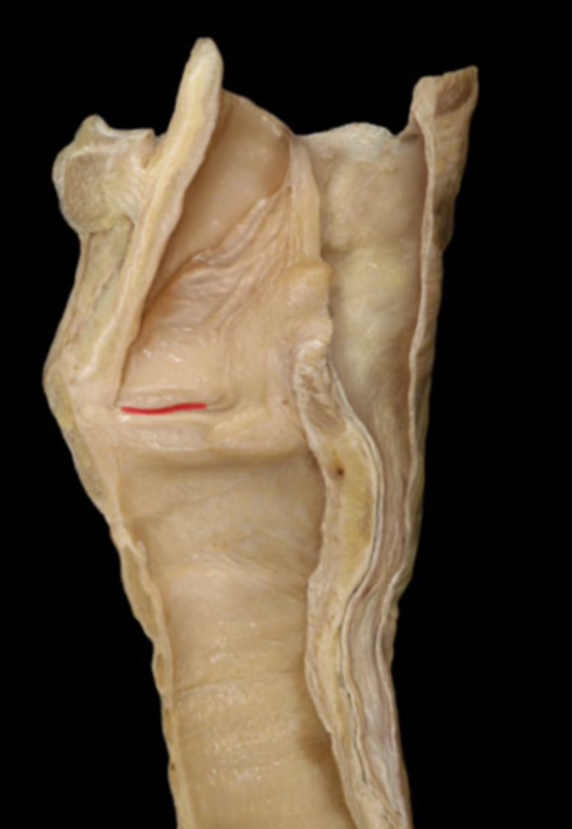
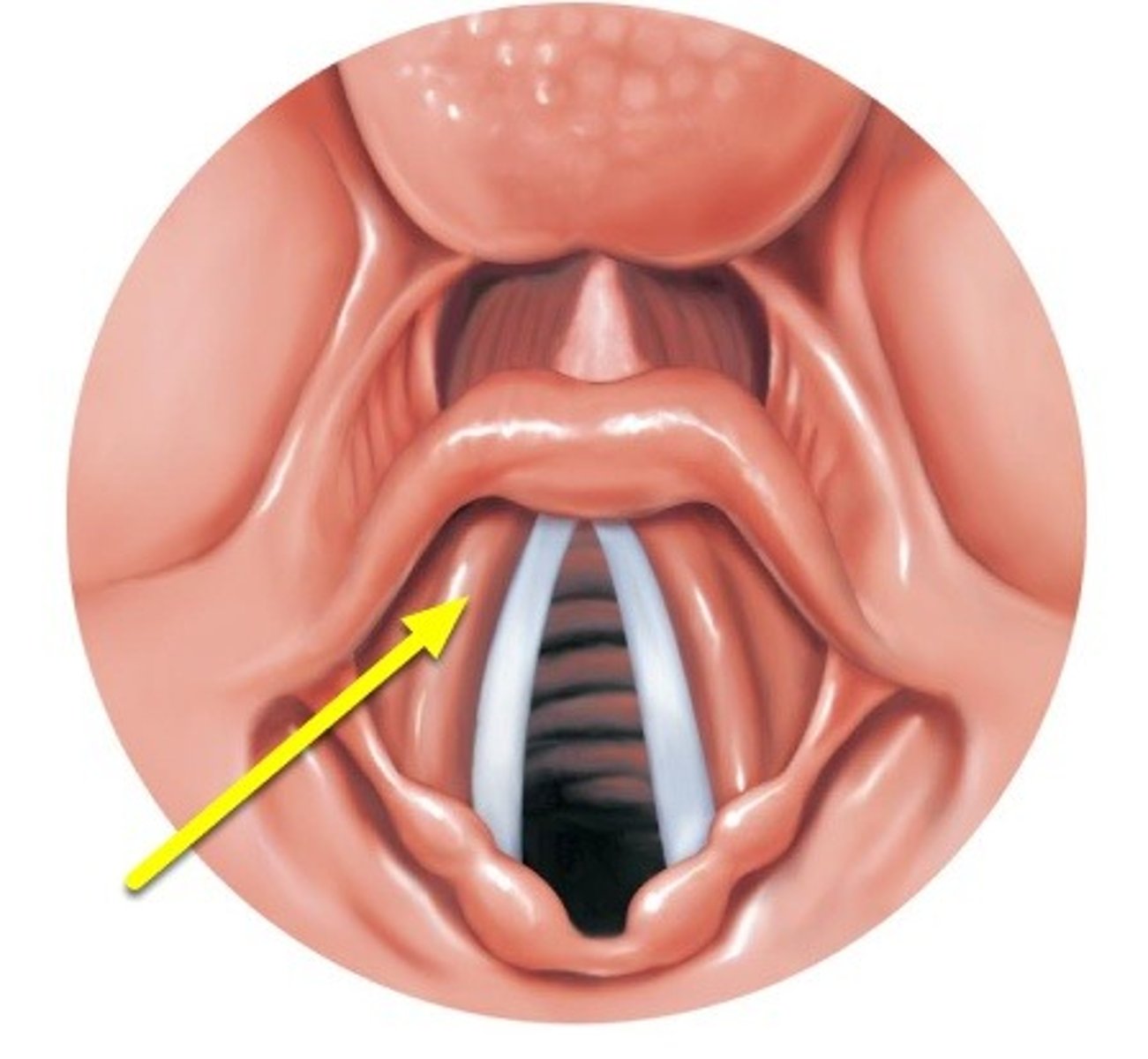
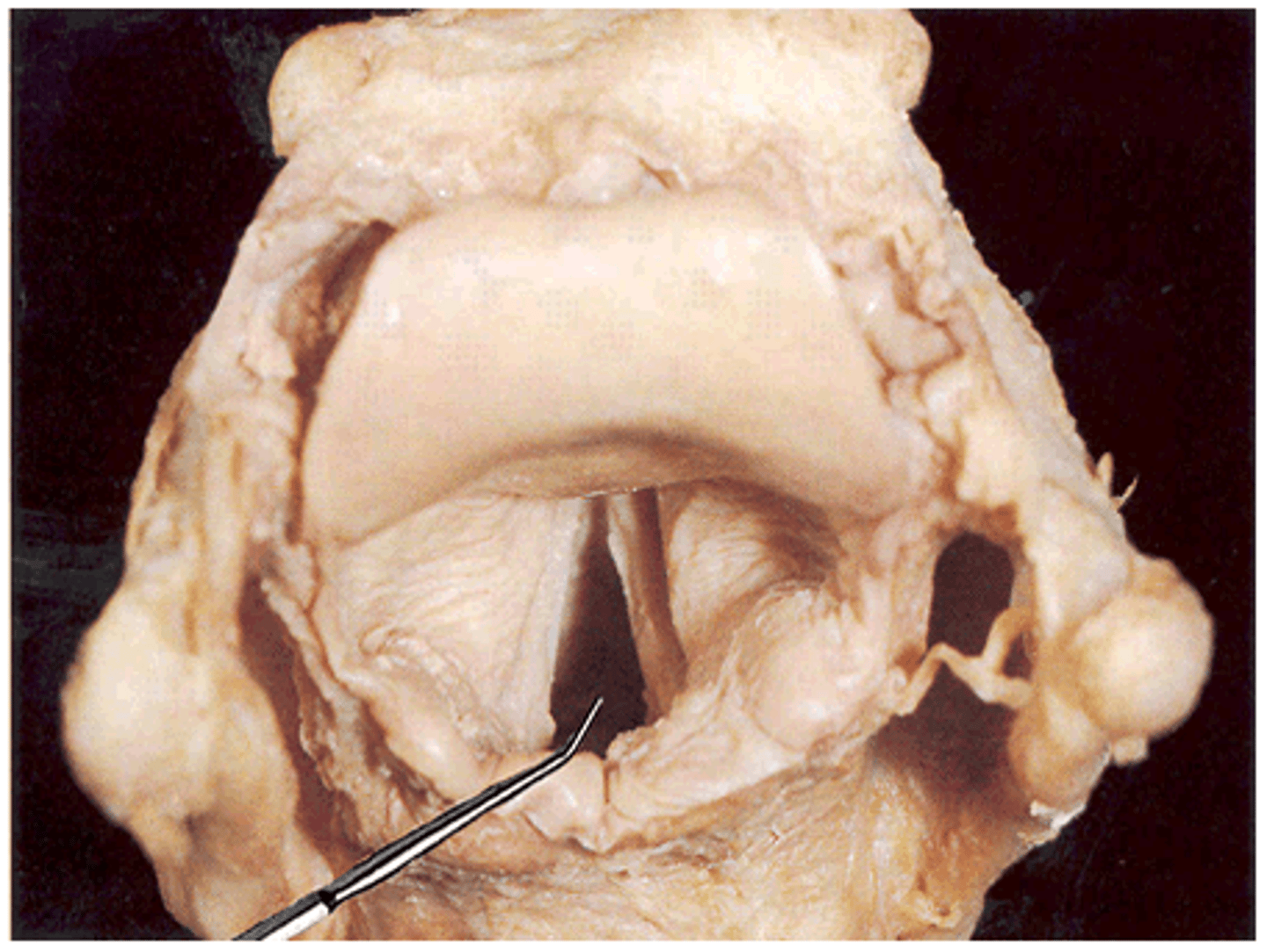
Rima Glottidis (Posterior View)
Note: slit between vocal folds where speech is produced
Name Opening #3

Rima Glottidis (Superior/Axial View)
Name the Opening
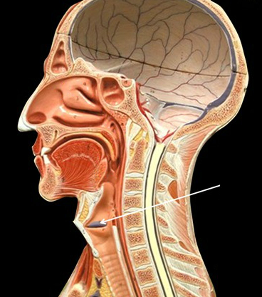
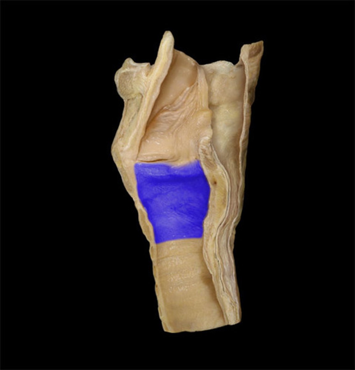
Infraglottic Cavity (Posterior View)
Note: space from vocal folds to cricoid cartilages (inferior is trachea)
Name Opening #4

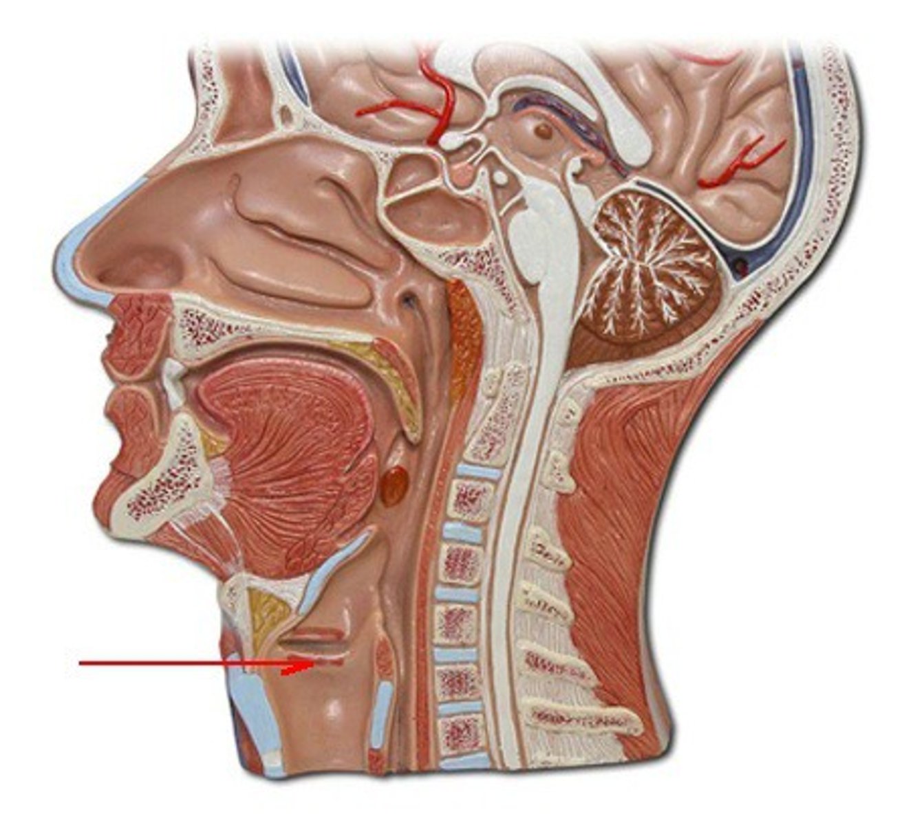
Infraglottic Cavity (Lateral View)
Name the Opening
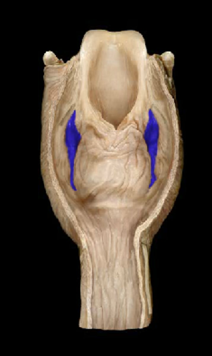
Piriform Recess
Note: lateral to laryngeal inlet & medial to thyroid cartilage
Name the Space (food can get caught here)
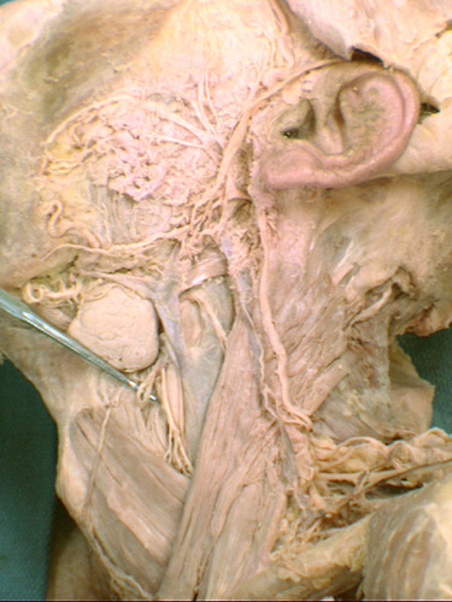
Internal Laryngeal Nerve
Note: lateral to the aryepiglottic folds, branch of superior laryngeal branch of vagus nerve, supplies all sensory innervation superior to the vocal folds
IMPORTANT: if you see a posterior view of the larynx with the epligottis visualized and there is a nerve running laterally, this is the internal laryngeal
Name the Nerve
Inferior Laryngeal Nerve
Note: medial to cricothyroid joint, supplies sensory innervation of the vocal folds and sensory innervation of the mucosa inferior to the vocal folds, supplies motor innervation to whole larynx, except the cricothyroid muscle
Name the Nerve
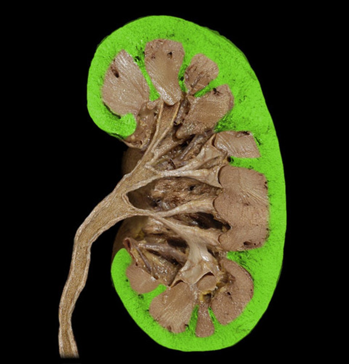
Renal Cortex
Name the Kidney Region
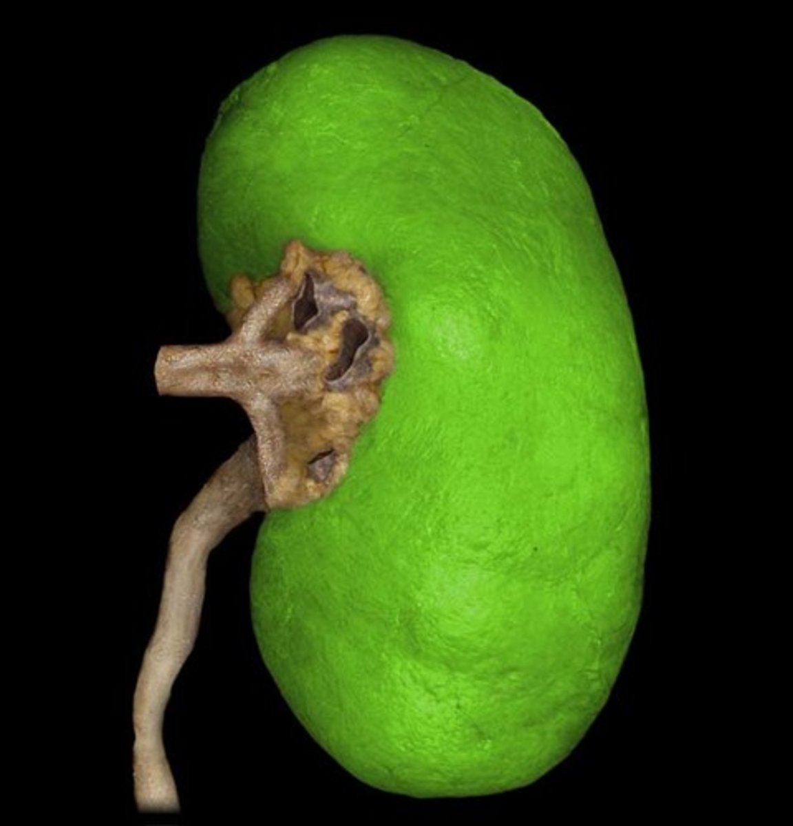
Fibrous Capsule
Name the Kidney Region
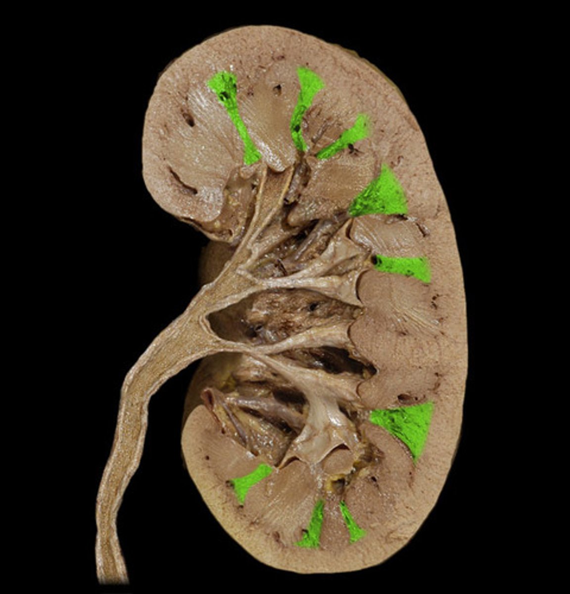
Renal Medulla (AKA Renal Pyramids)
Name the Kidney Region

Renal Columns
Note: these are cortical tissue invading inwards
Name the Kidney Region
Renal Papilla
Note: papilla are the apex of a pyramid
Name the Kidney Region
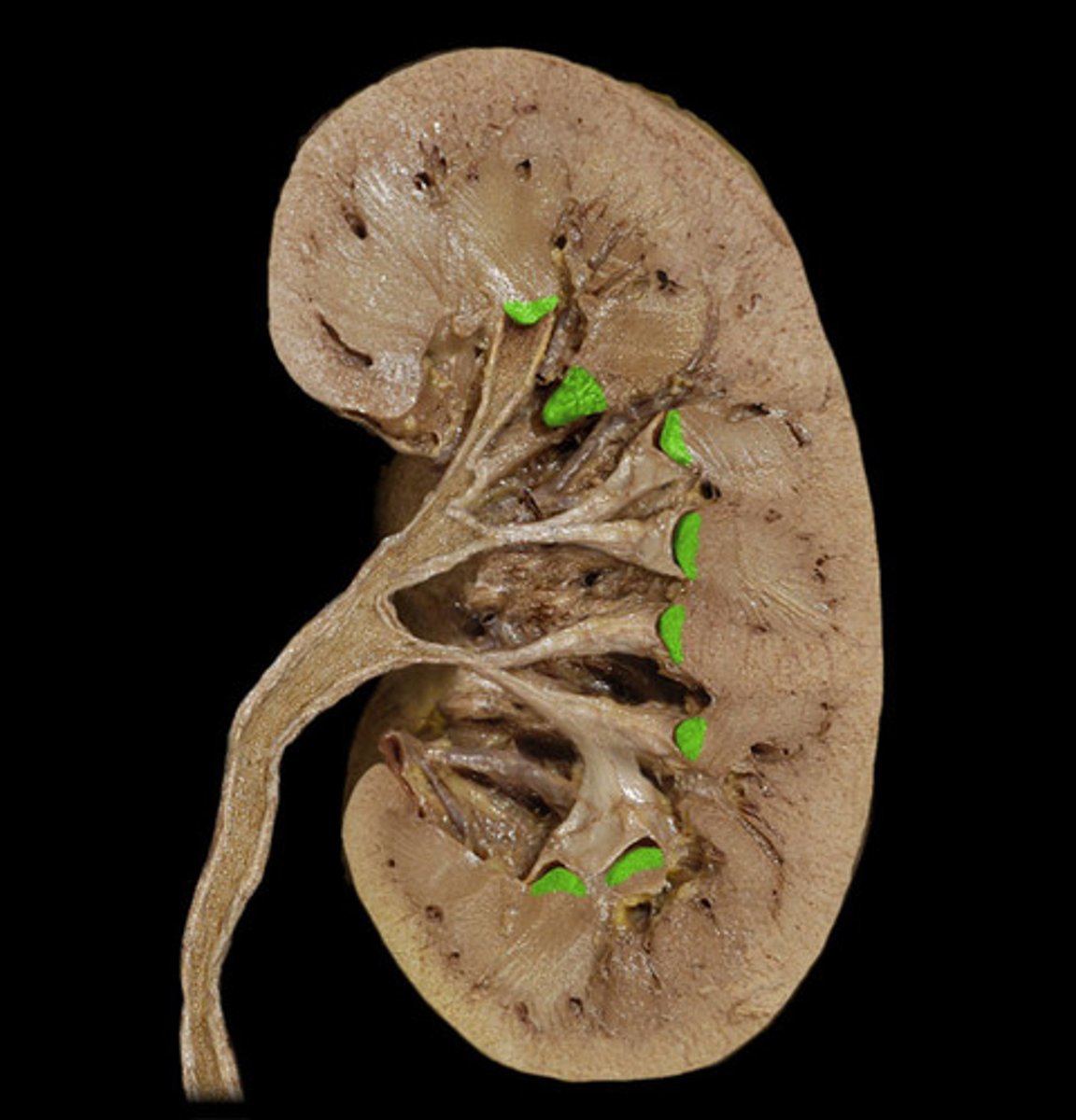
Minor Calyx
Note: miniscule space immediately distal to a renal papilla
Name the Kidney Region
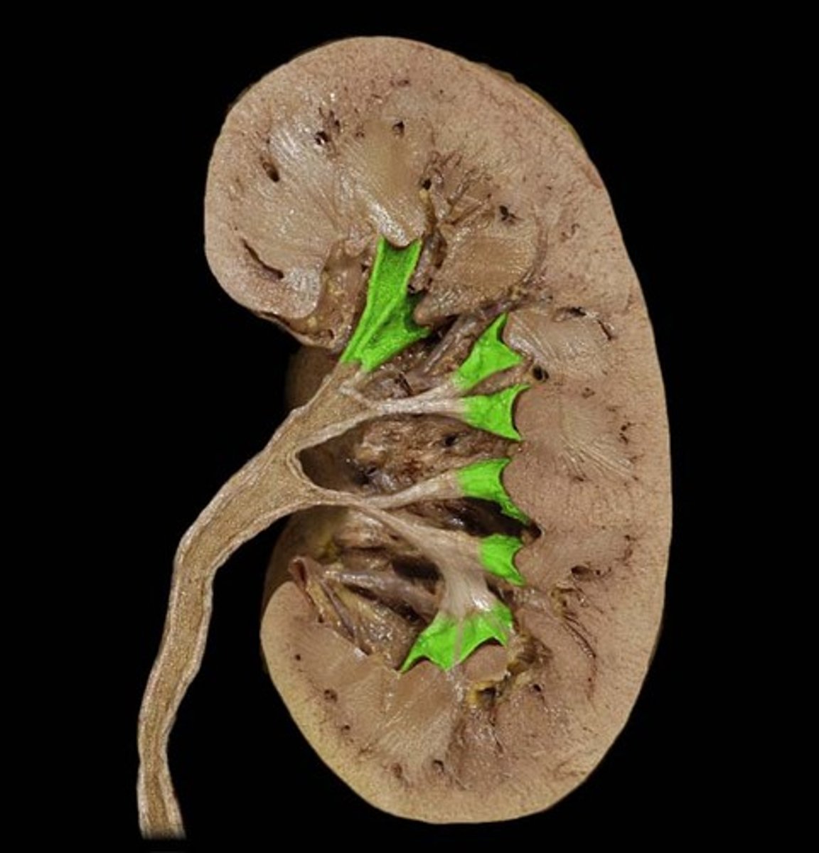
Major Calyx
Note: formed by convergence of minor calyces
Name the Kidney Region
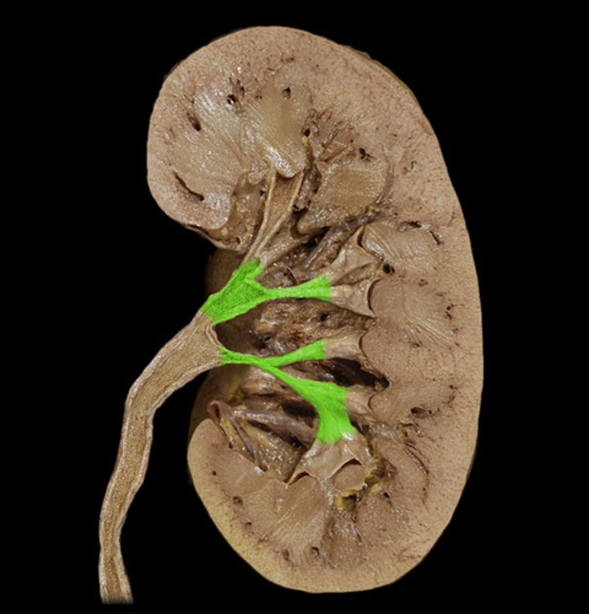
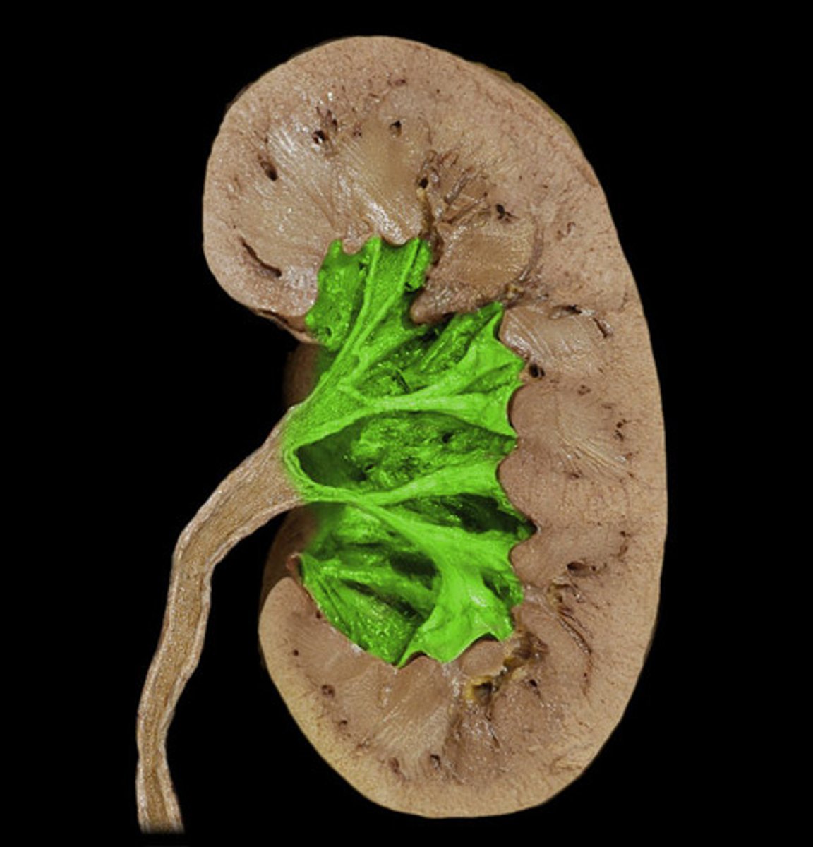
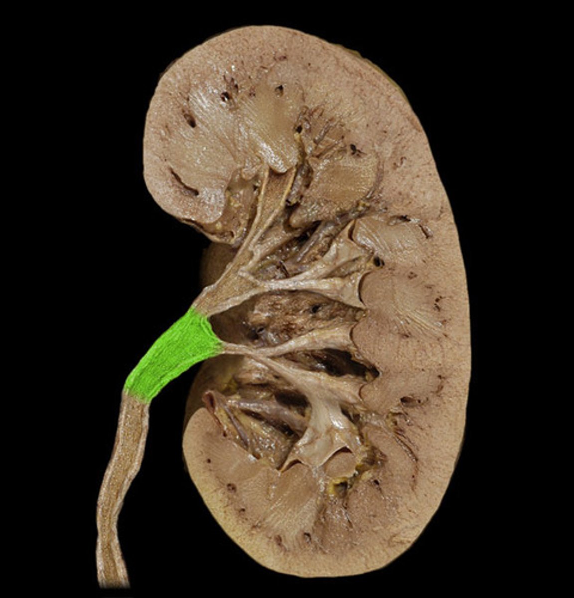
Renal Pelvis
Note: funnel shaped, narrows into ureter, sometimes can see sympathetic ganglion which innervates it
Name the Kidney Region
Renal Sinus
Note: space filed with the renal pelvis, perirenal fat, segmental arteries and segmental veins
Name the Space