Final Practical (Urinary, Respiratory, Digestive, Reproductive)
1/189
There's no tags or description
Looks like no tags are added yet.
Name | Mastery | Learn | Test | Matching | Spaced |
|---|
No study sessions yet.
190 Terms
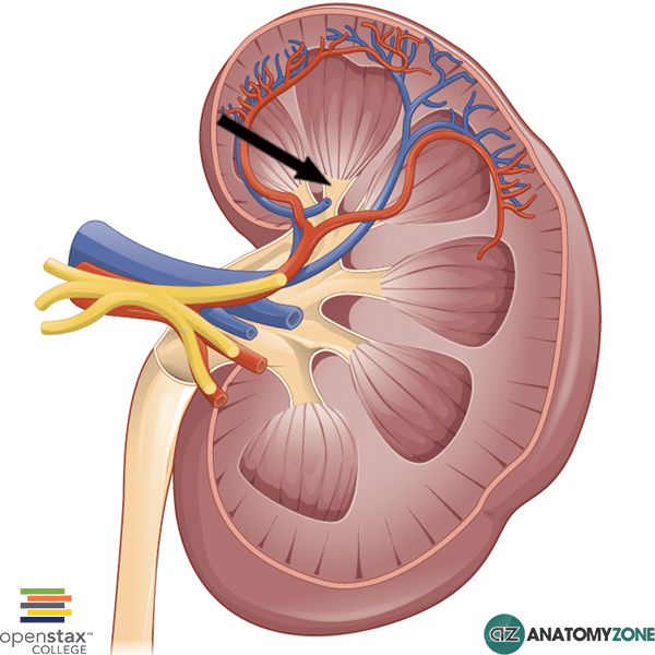
Kidney
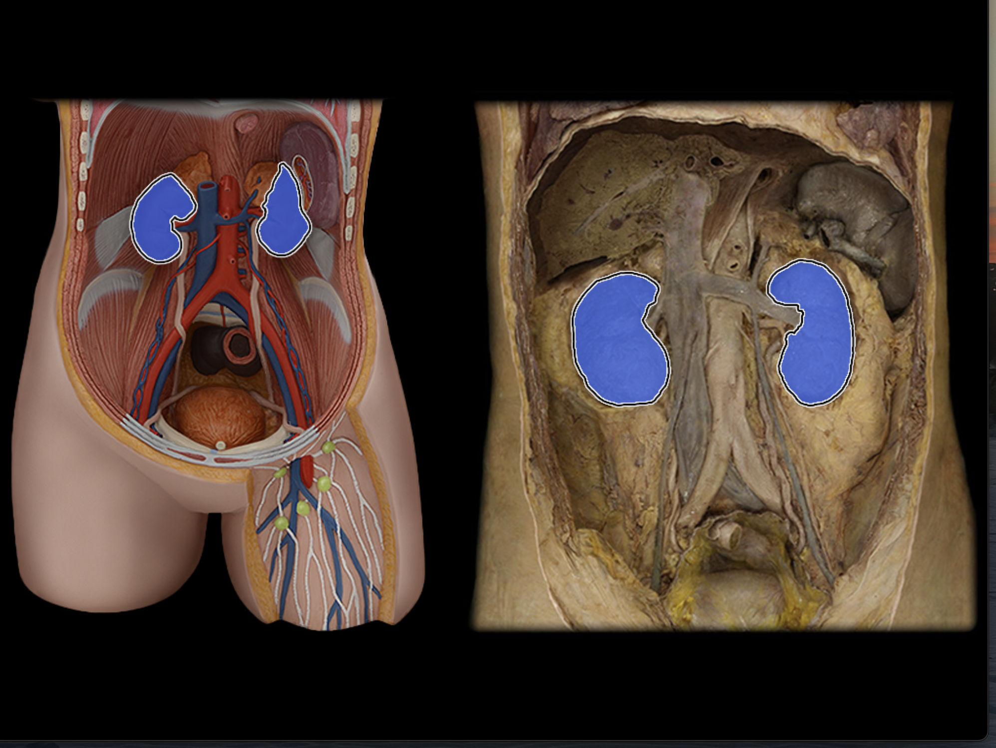
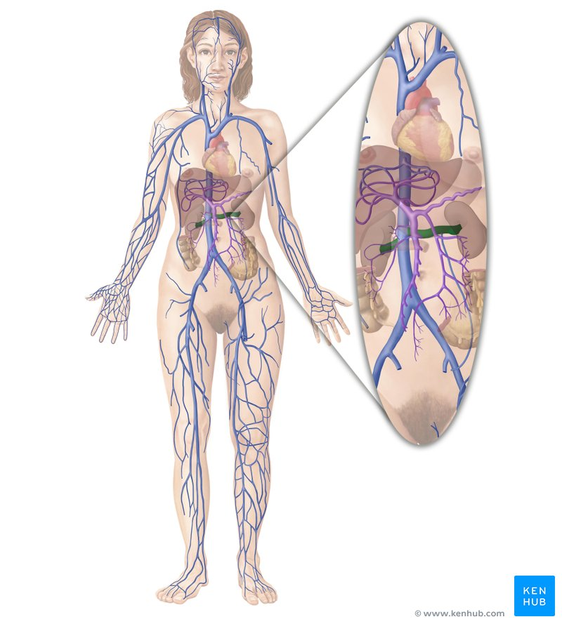
Renal artery
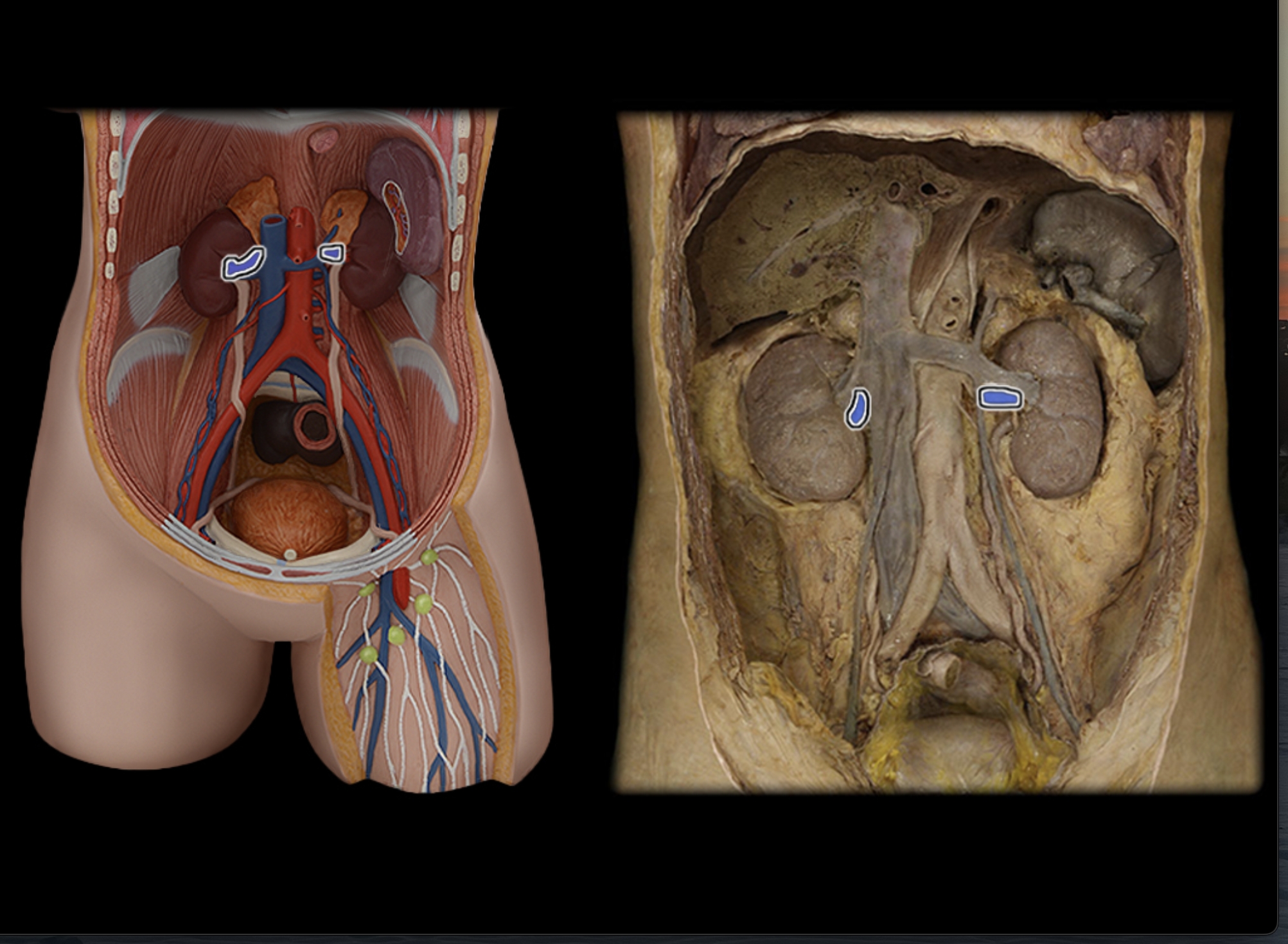
Renal vein
Ureter
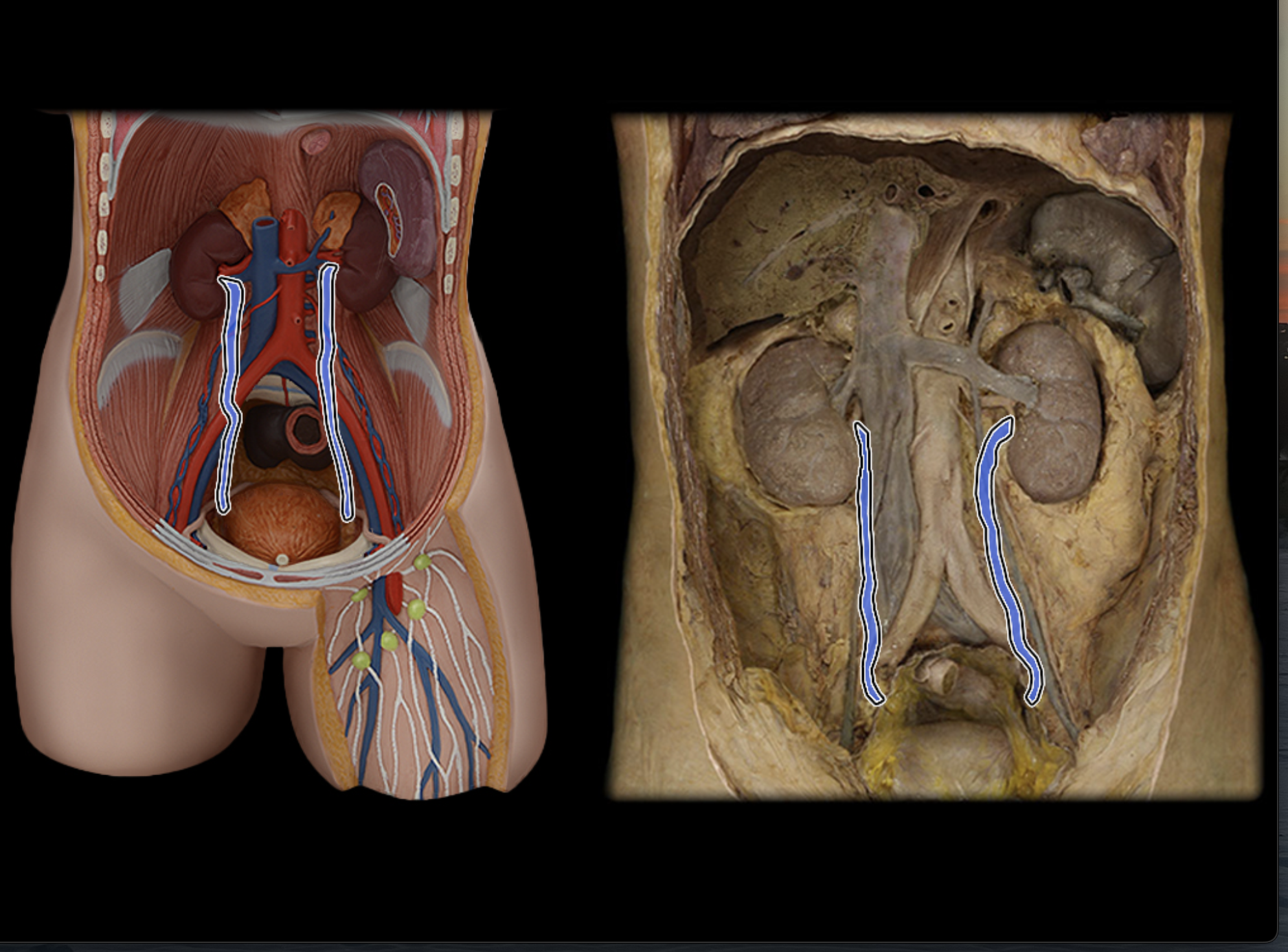
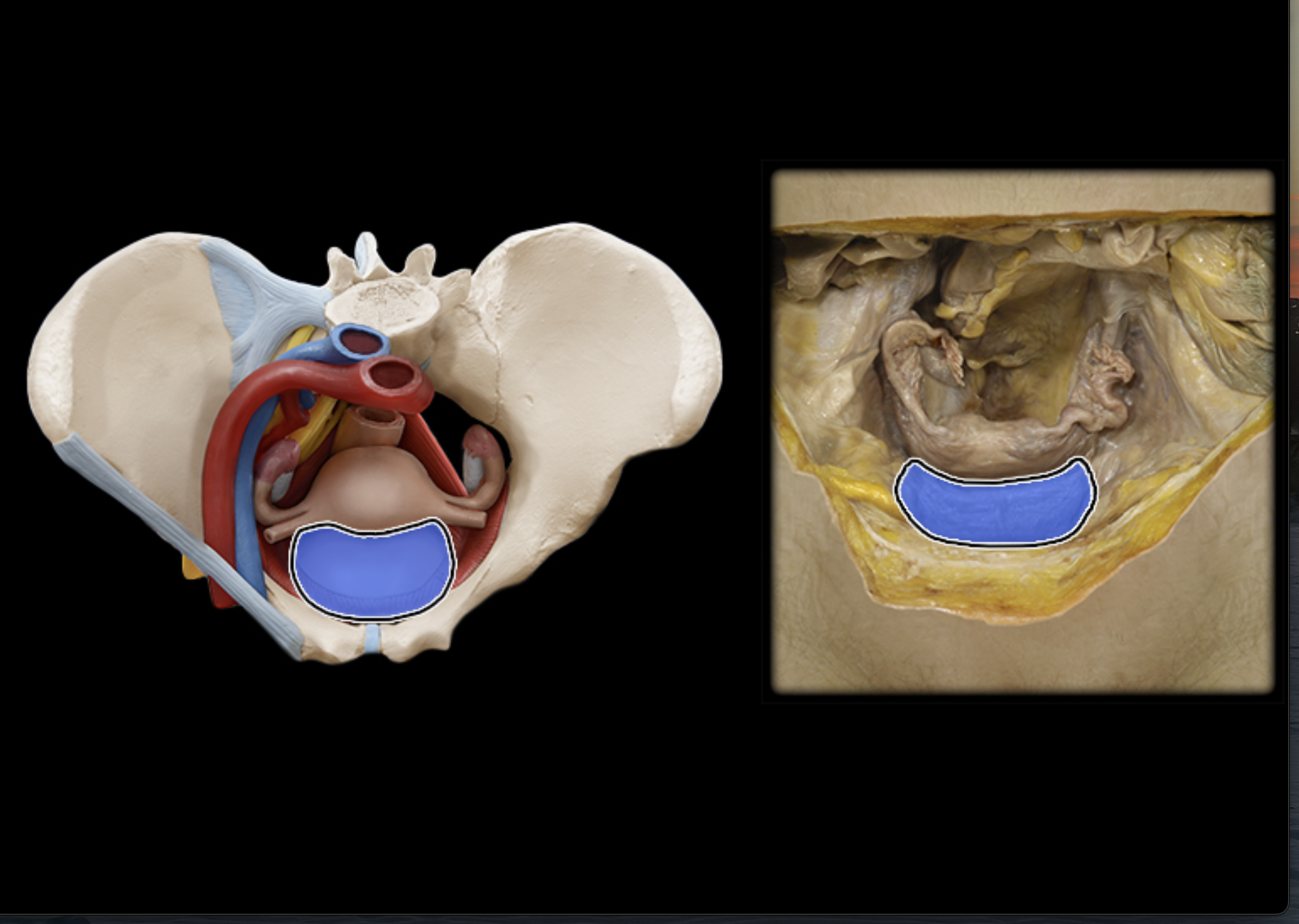
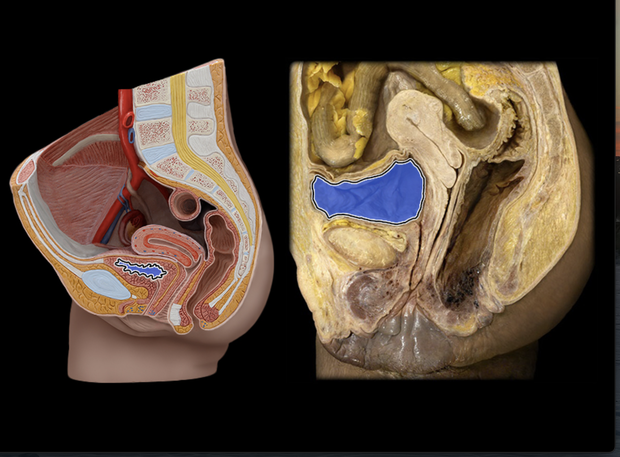
Urinary bladder
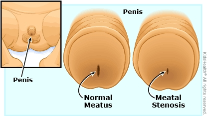
External urethral meatus
The opening at the end of the urethra
Renal capsule
Outer layer surrounding kidney
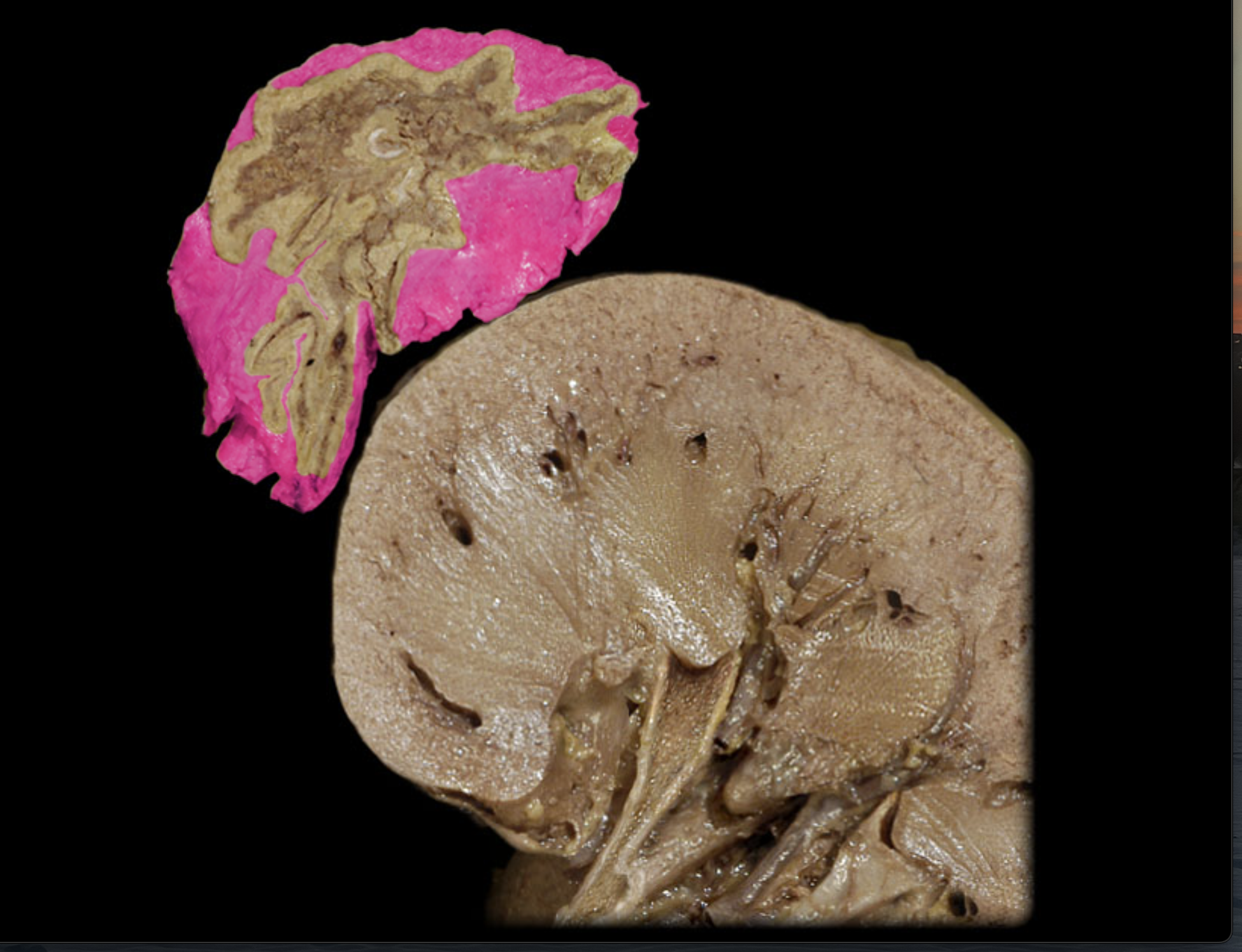
Renal cortex
Middle layer of kidney
Renal medulla
Inner layer of kidney
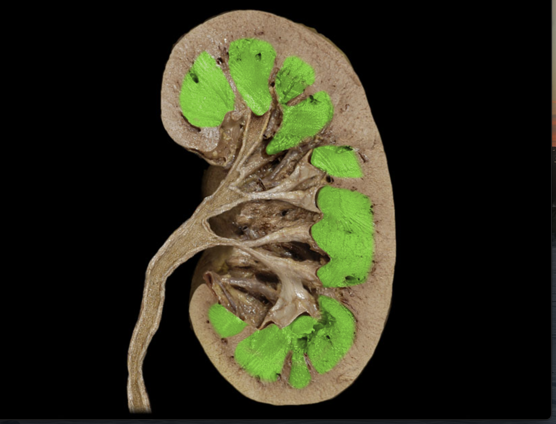
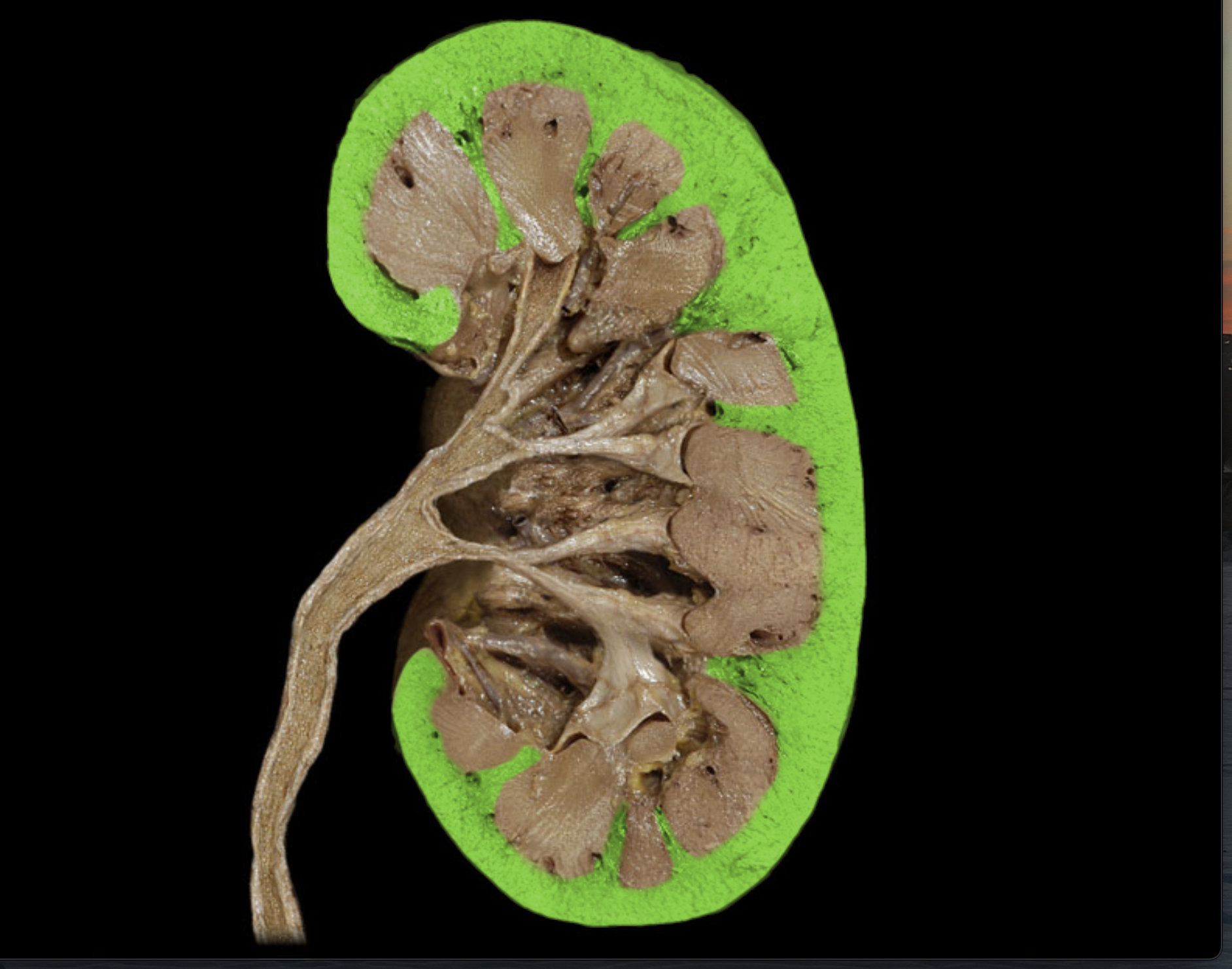
Renal pyramids
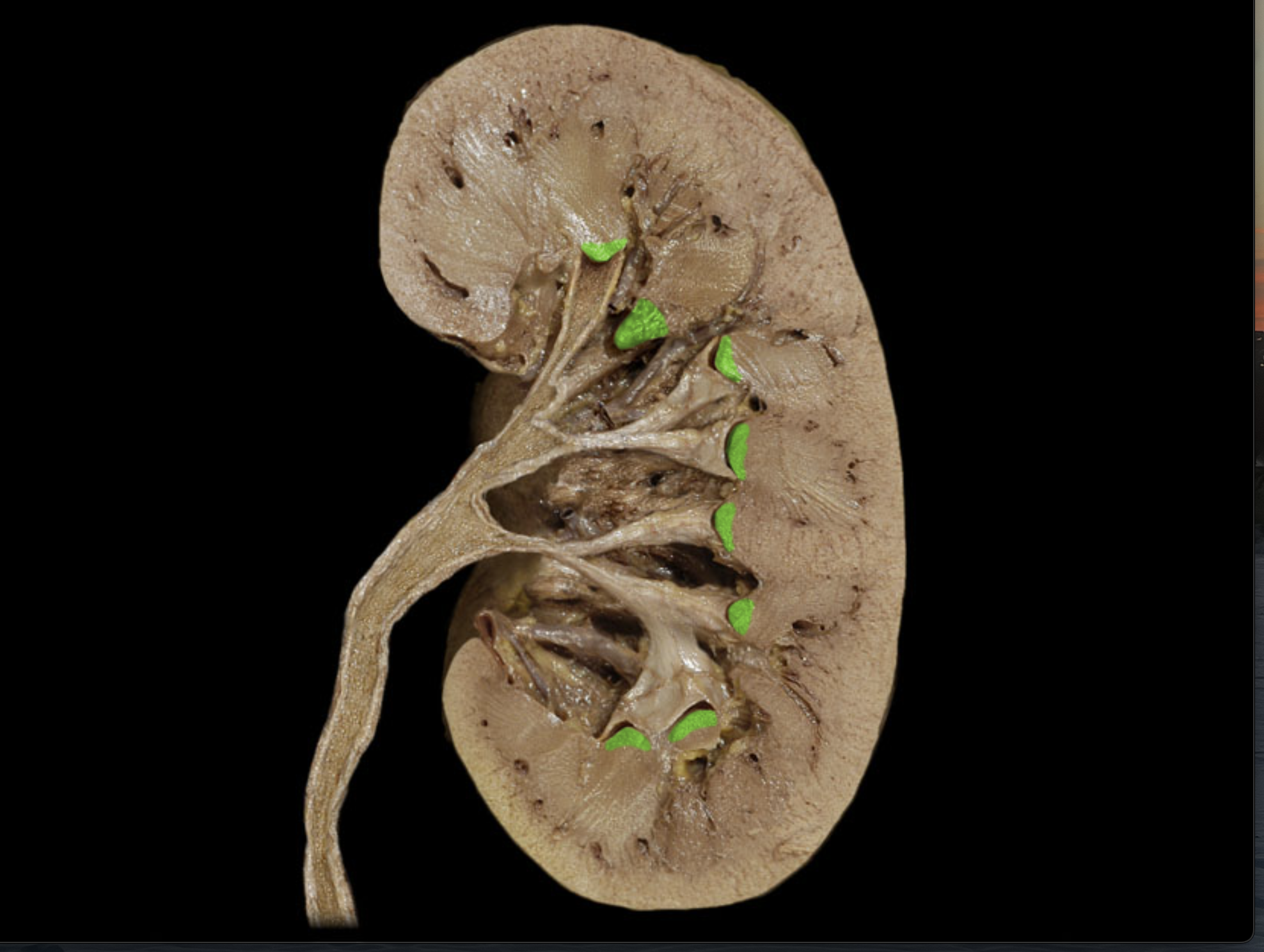
Renal papillae
End of each pyramid
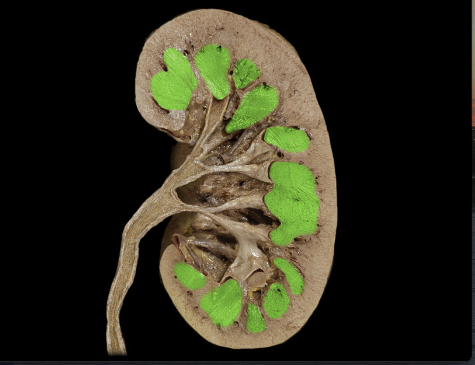
Renal columns
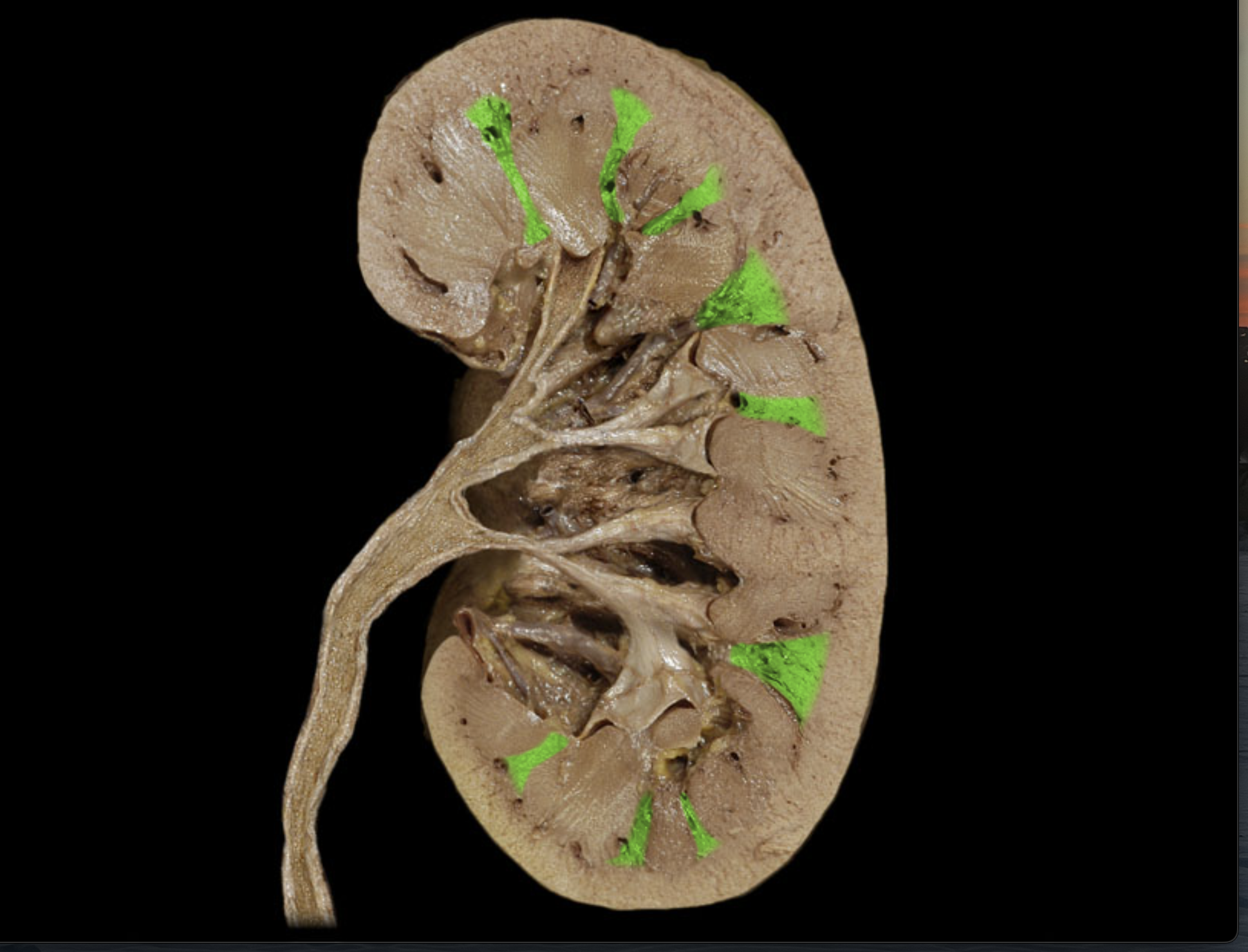
Renal lobes
(Blue box)
Includes one pyramid, cortex above and column on either side
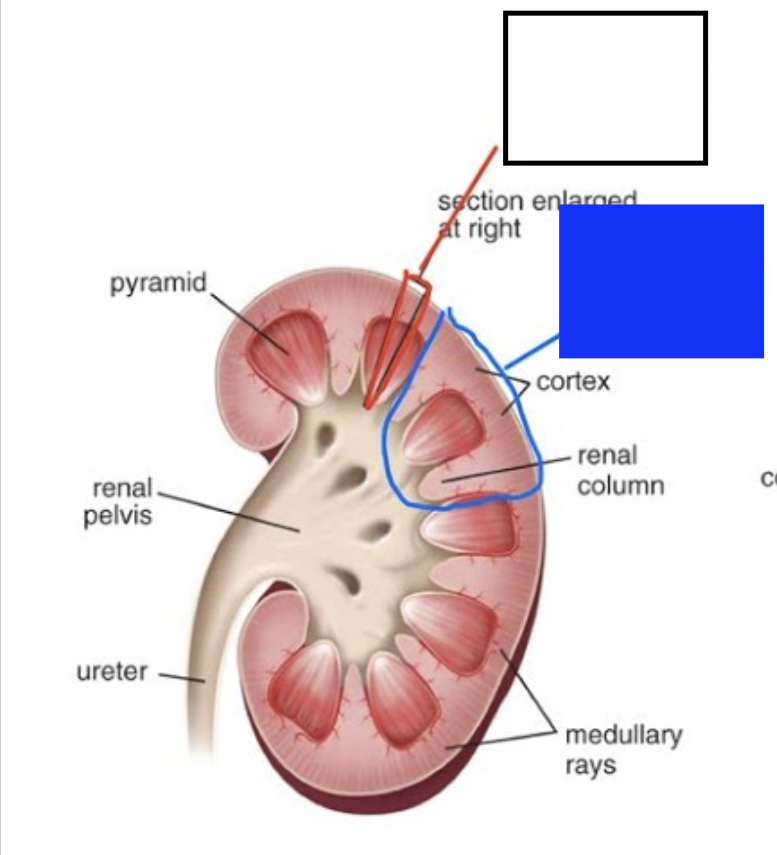
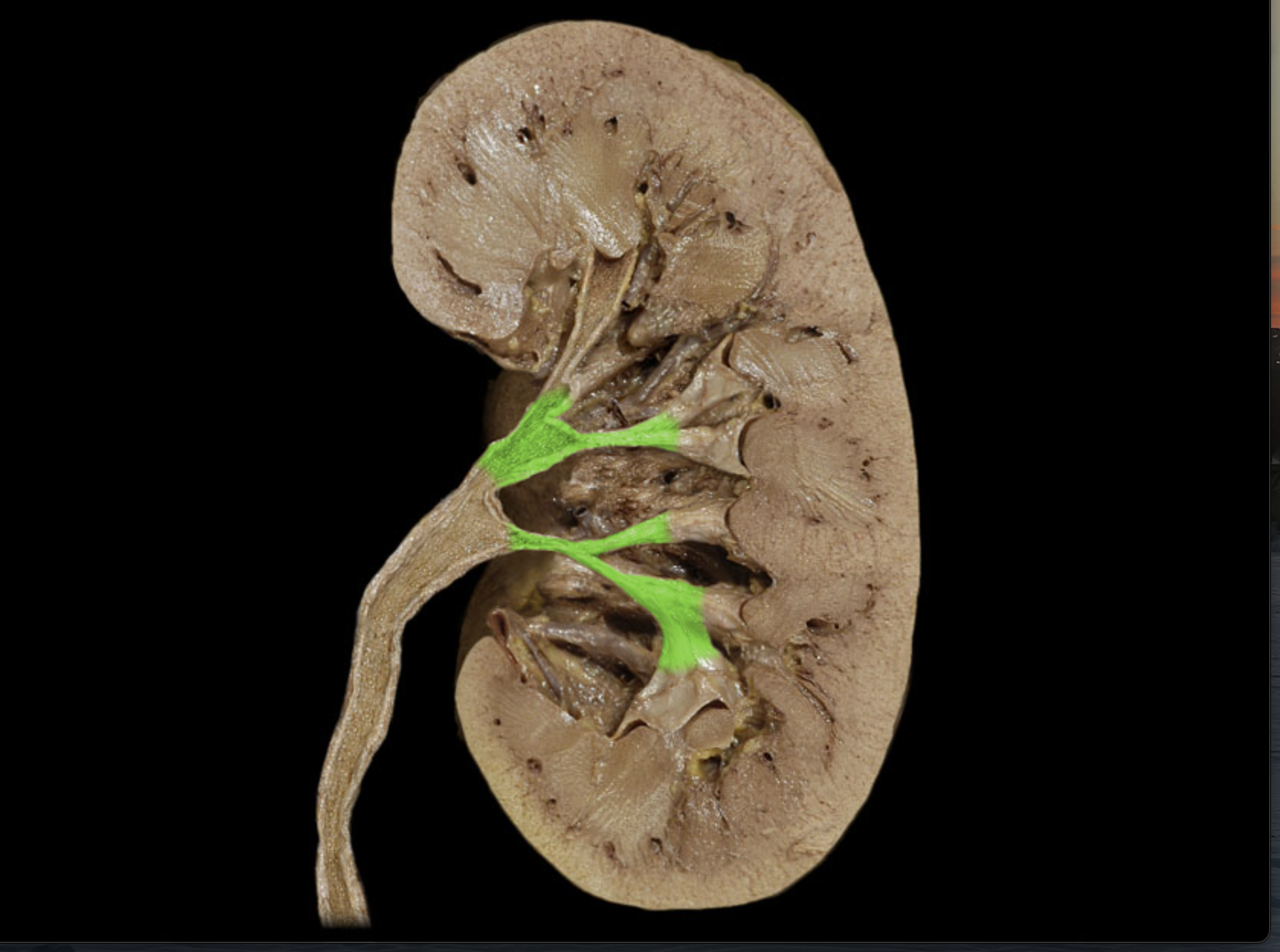
Renal calices
Tan tube going through pyramid
Minor calyx
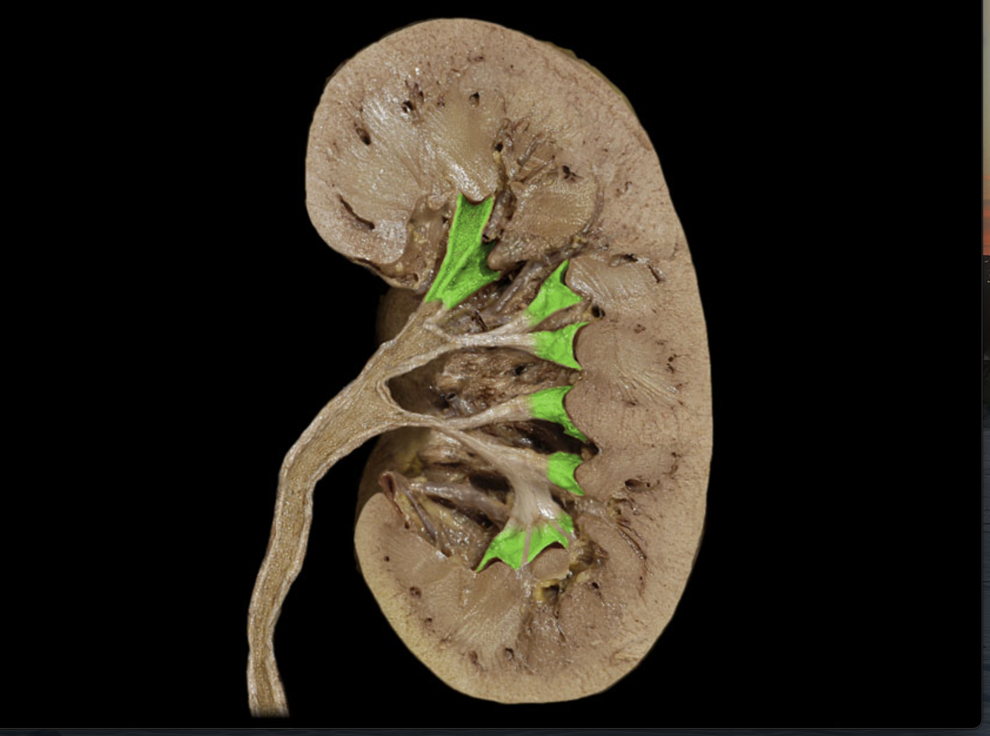
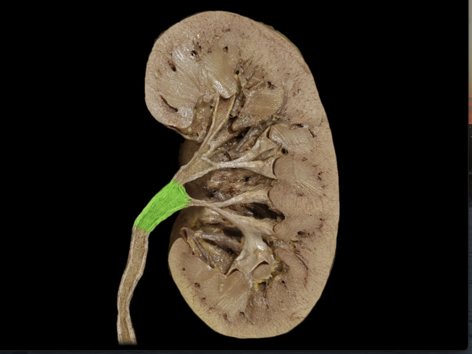
Major calyx
Renal pelvis
Detrusor muscle
Middle layer of urinary bladder
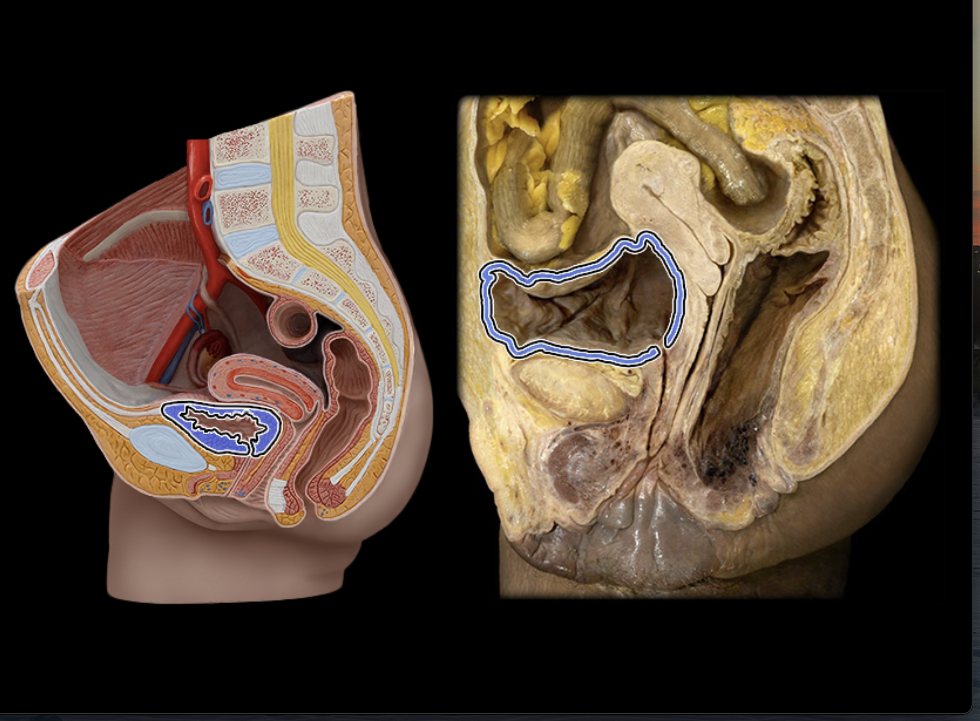
Rugae (urinary bladder)
Fold/ridges in urinary bladder
Urethra
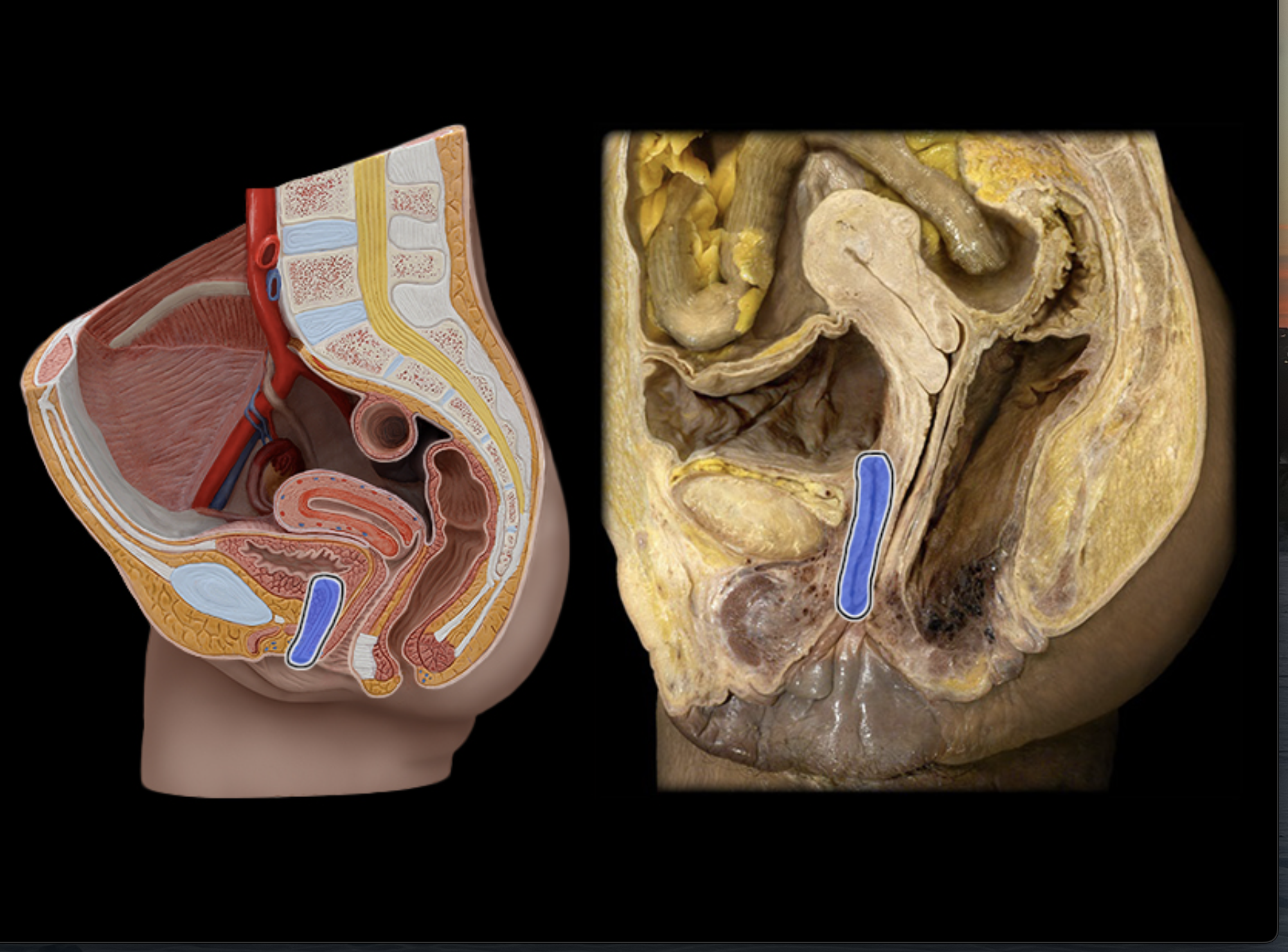
Internal urinary sphincter (male)
External urinary sphincter (female)
Rhinis
Dorsum
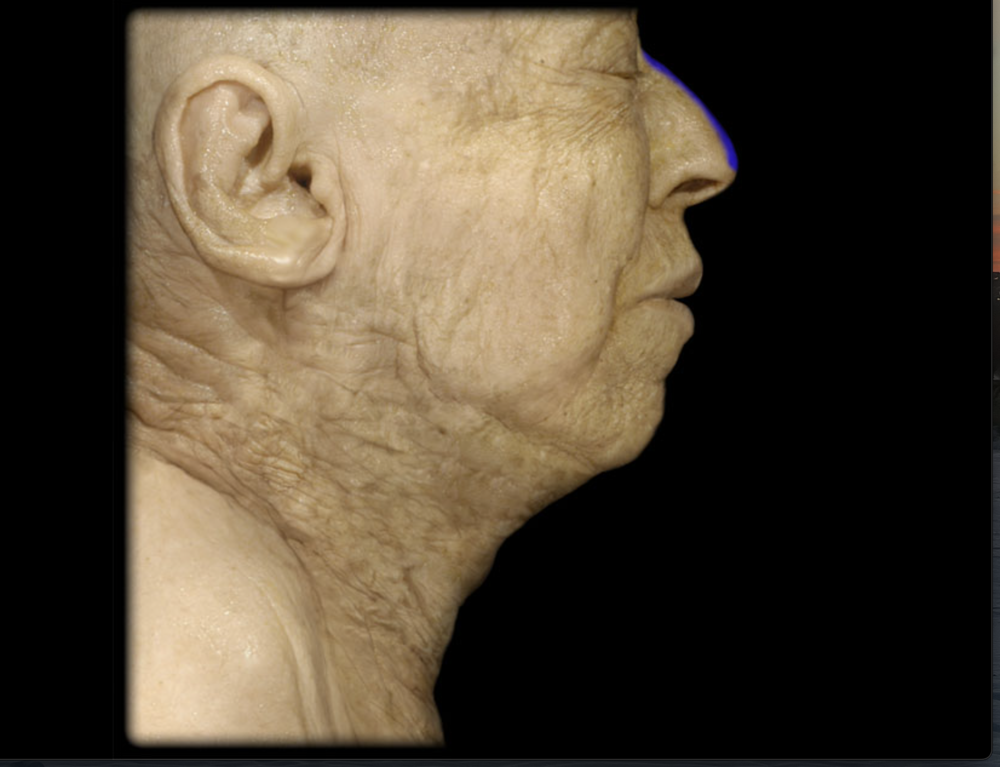
Apex of nose
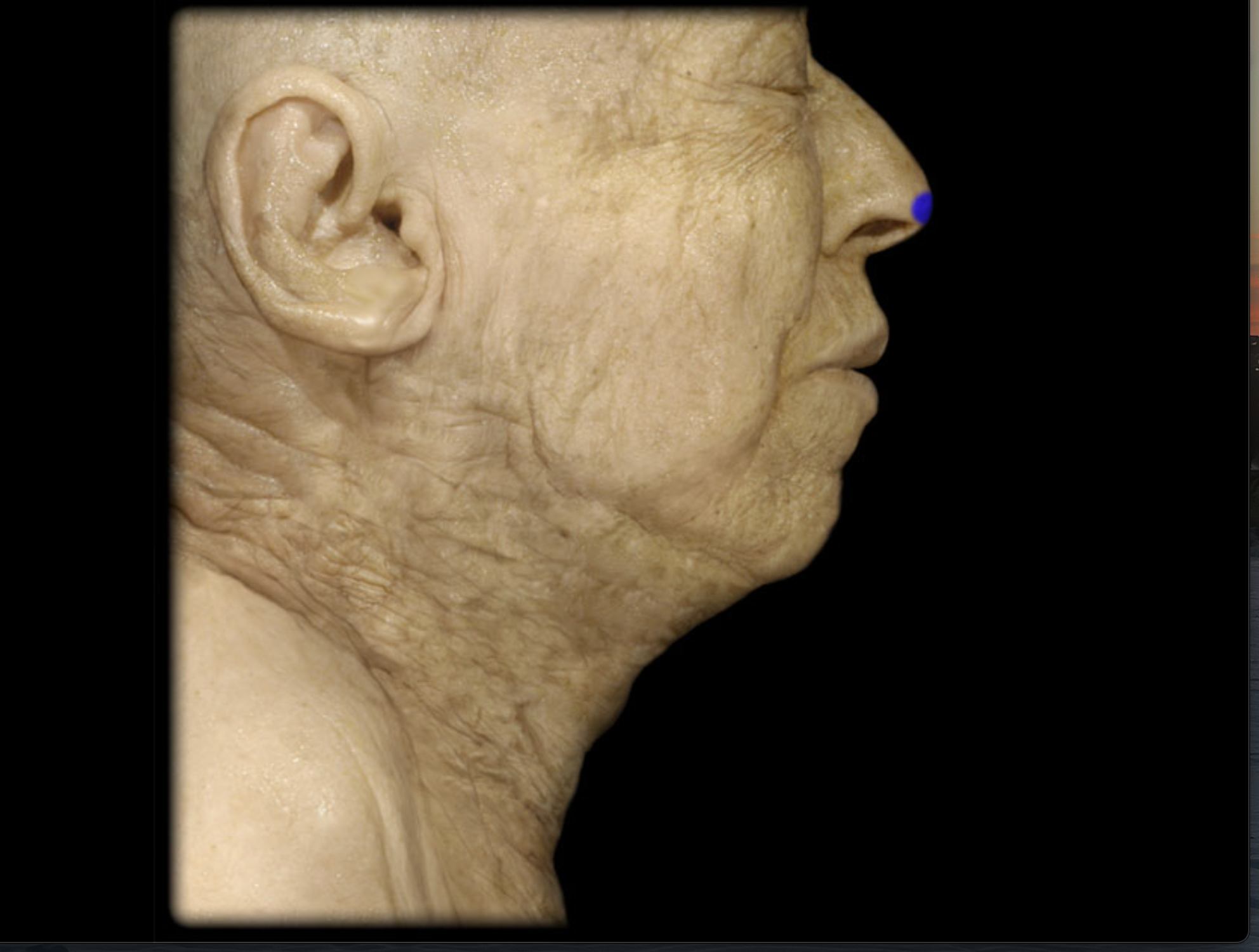
Columella
The tissue structure that separates the nostrils and forms the base of the nasal tip.
Ala nasi
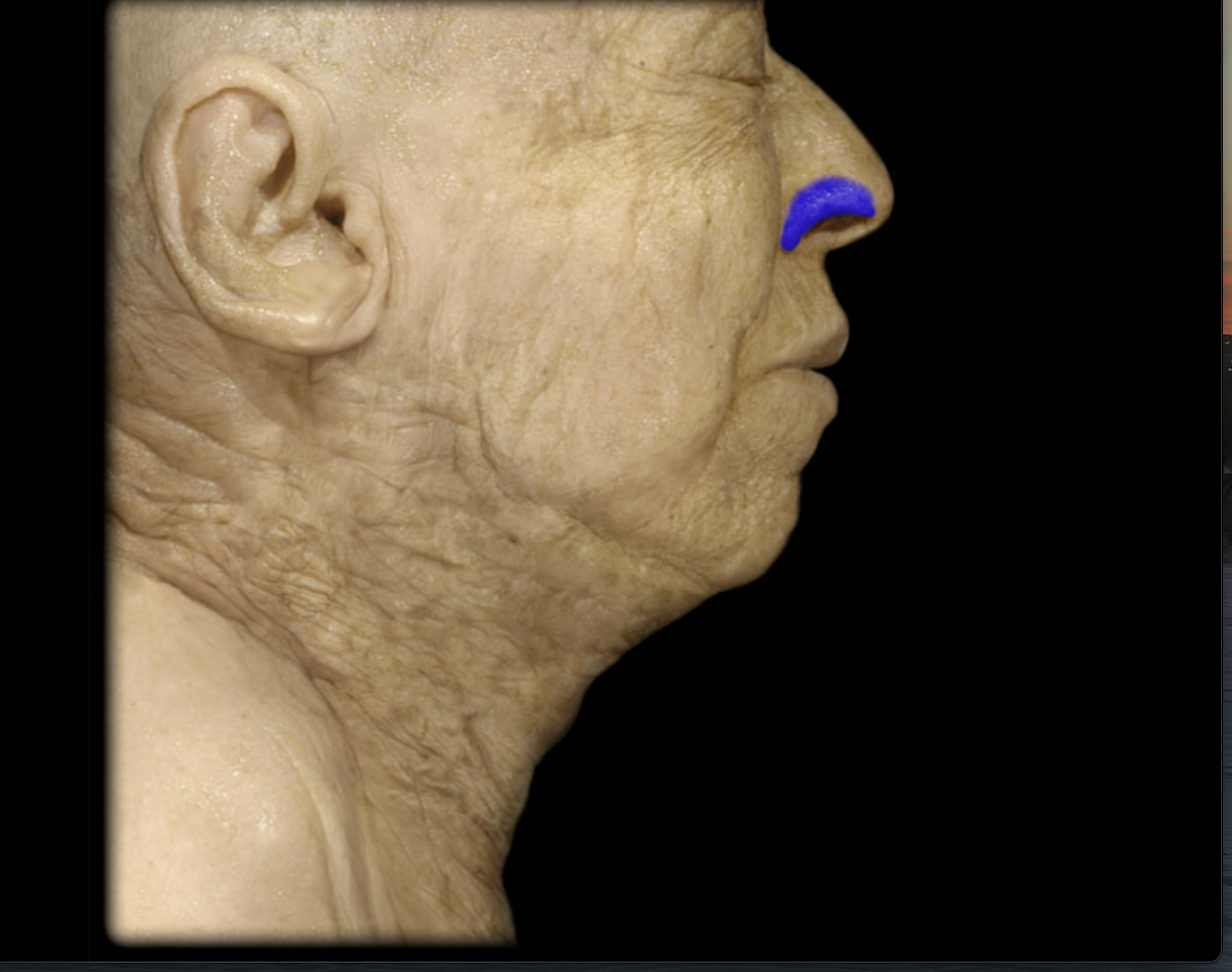
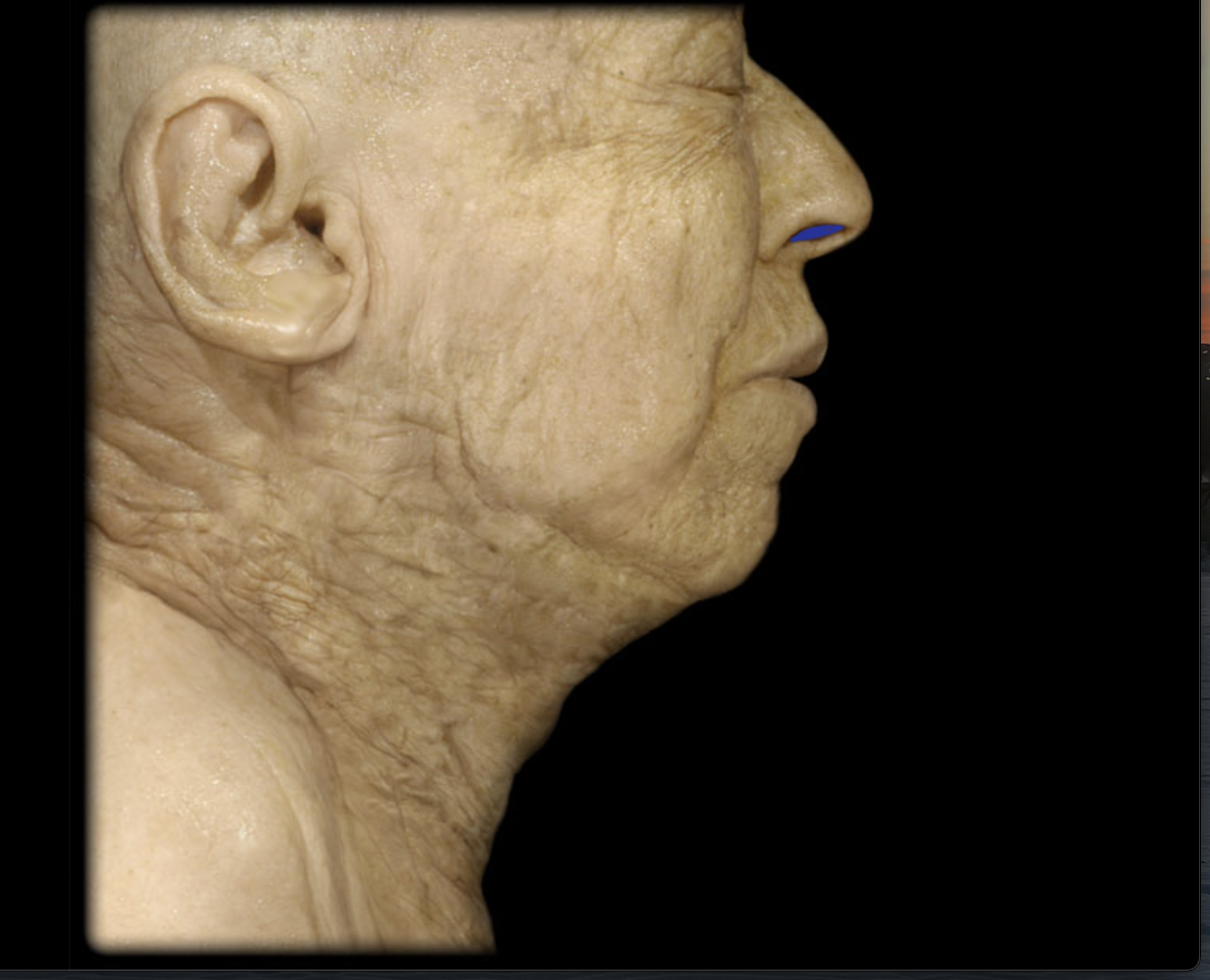
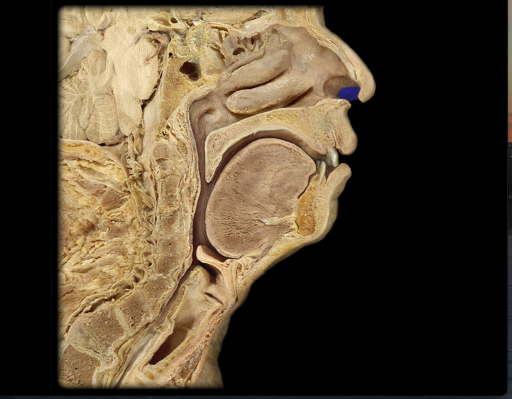
Anterior nares
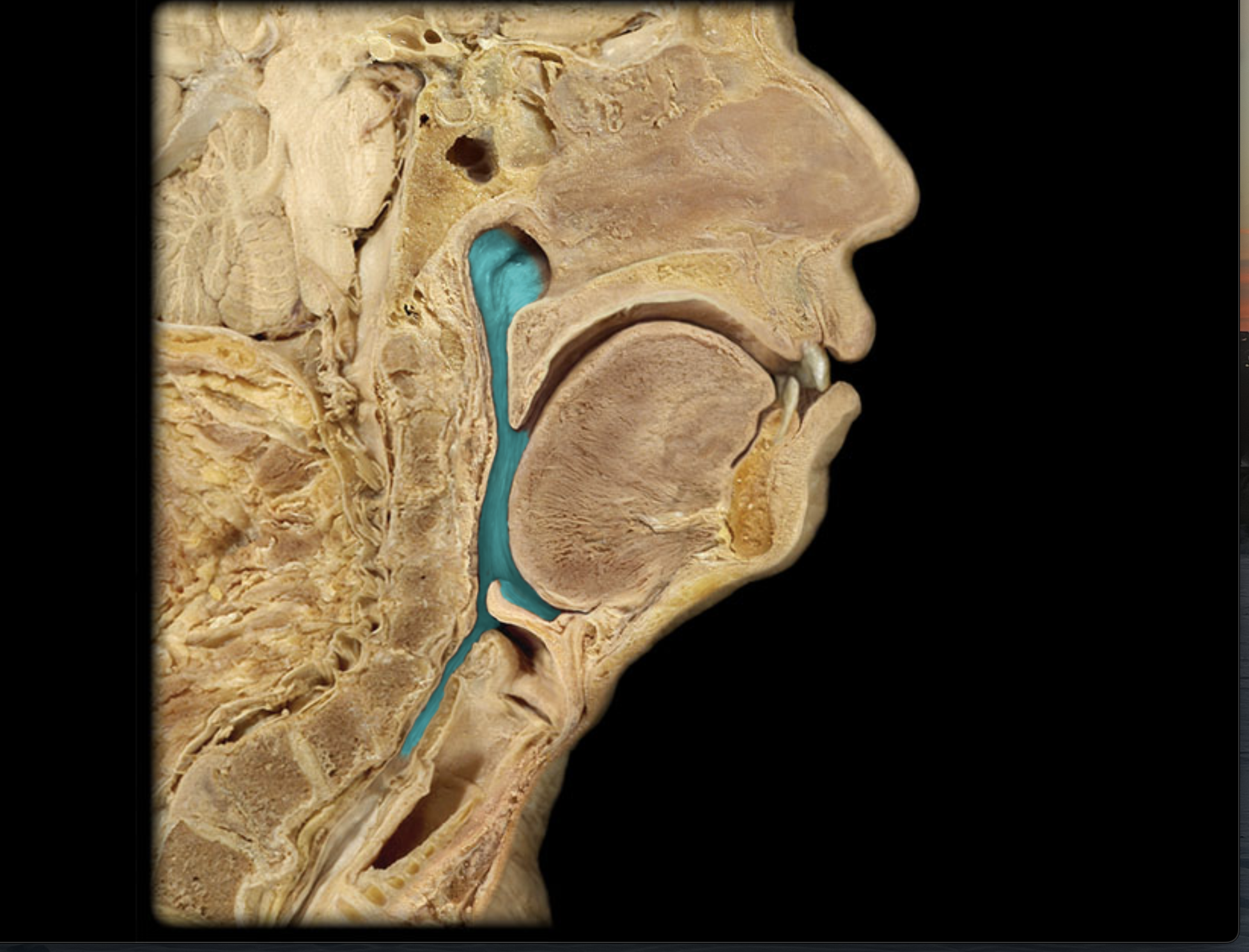
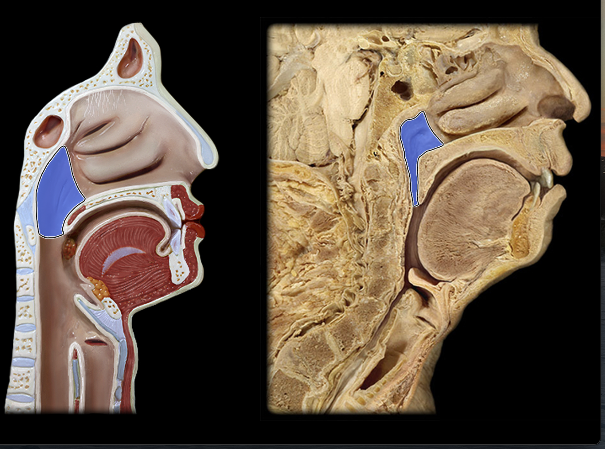
Nasal cavity
Nasal vestibule
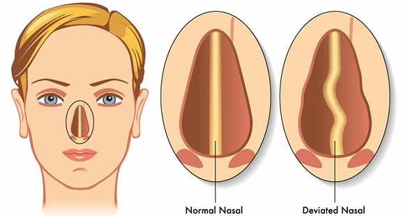
Nasal septum
Middle part inside nose
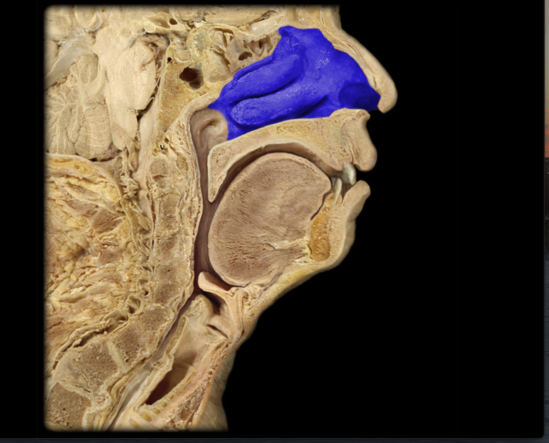
Conchae (superior, middle, inferior)
3 flaps inside nasal cavity
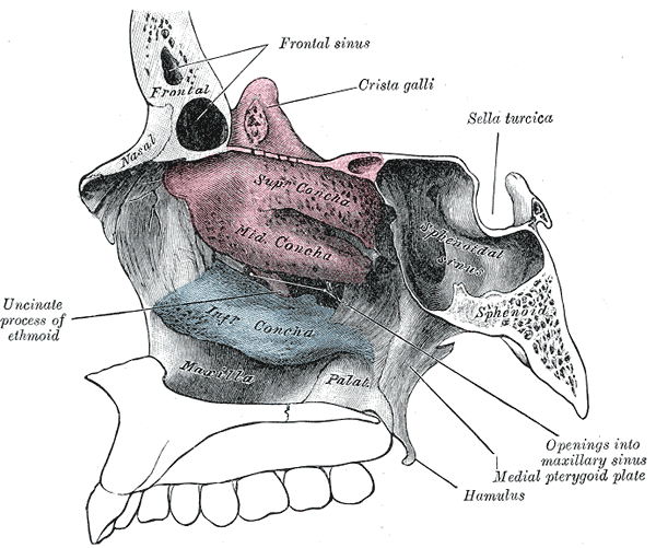
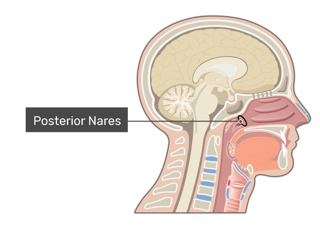
Posterior nares
End of nasal cavity, narrows
Pharynx
Nasopharynx
Oropharynx
Laryngopharynx
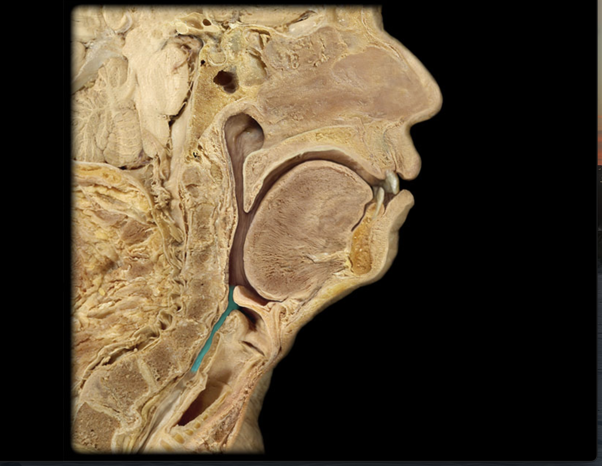
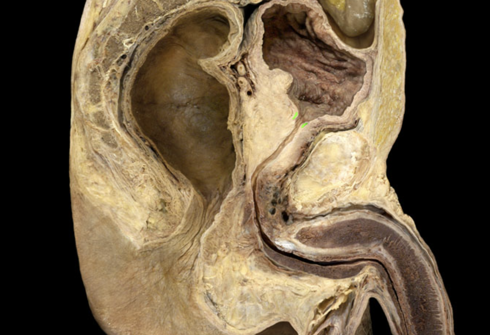
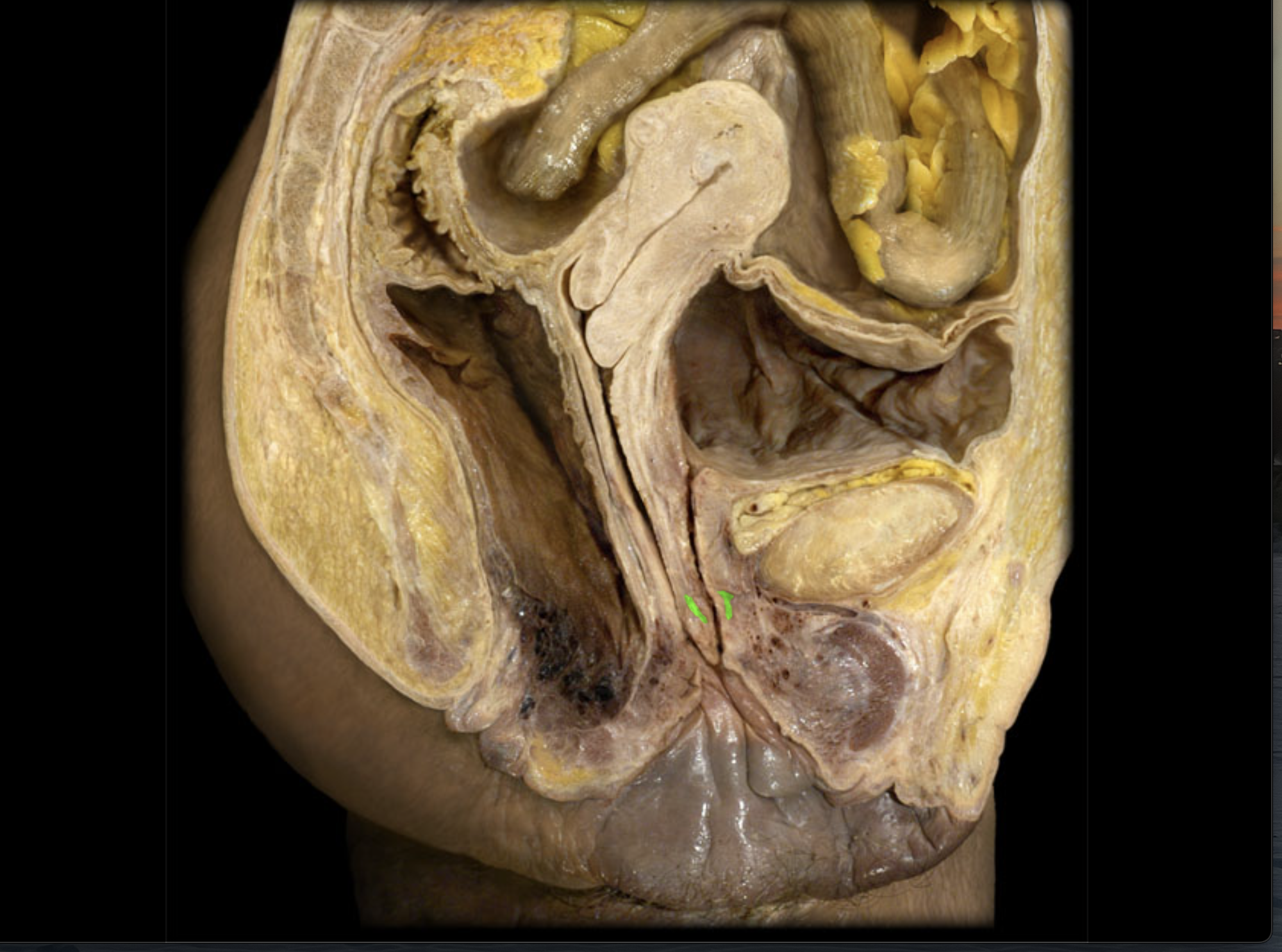
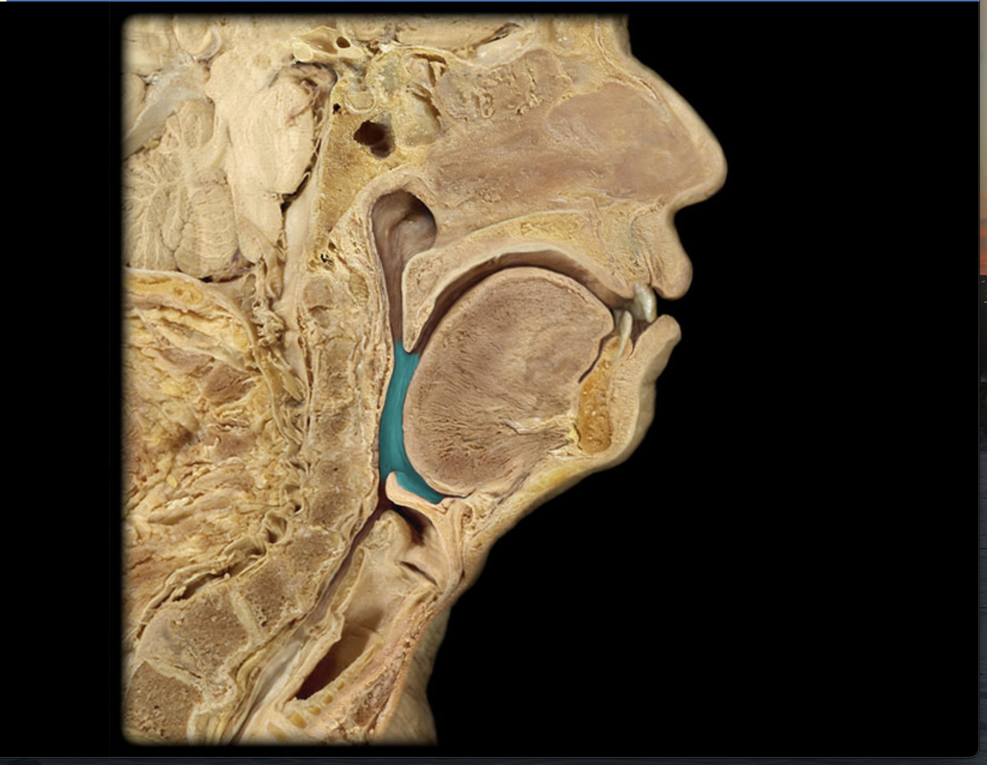
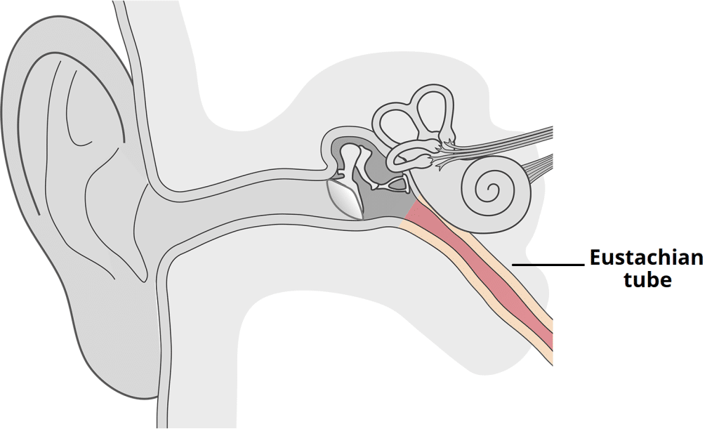
Torus tubarius
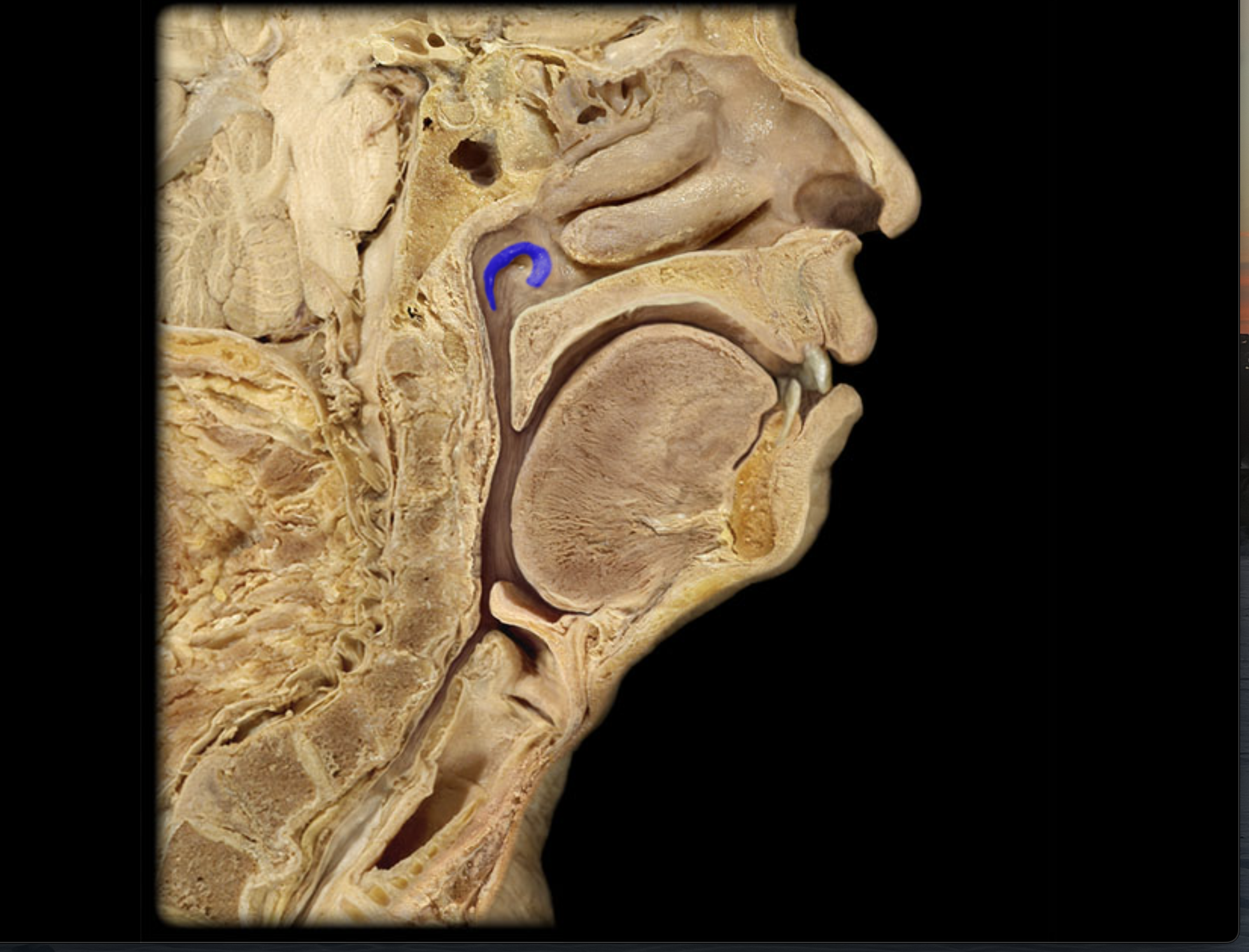
Eustachian tube
A canal that connects the middle ear to the nasopharynx, helping to equalize air pressure.
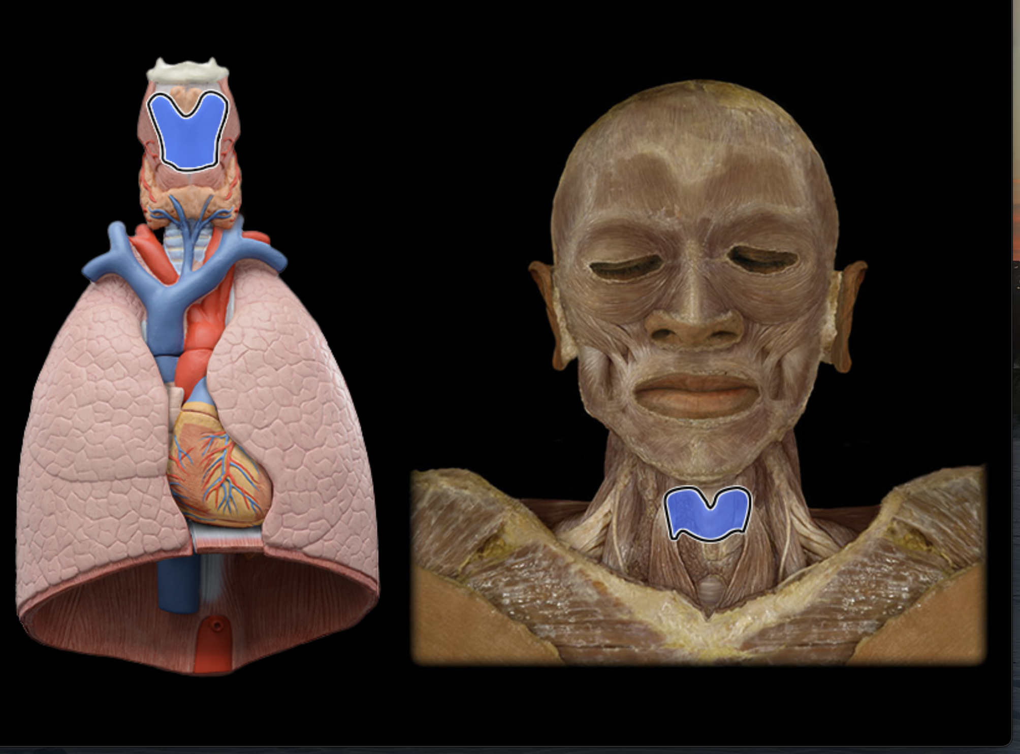
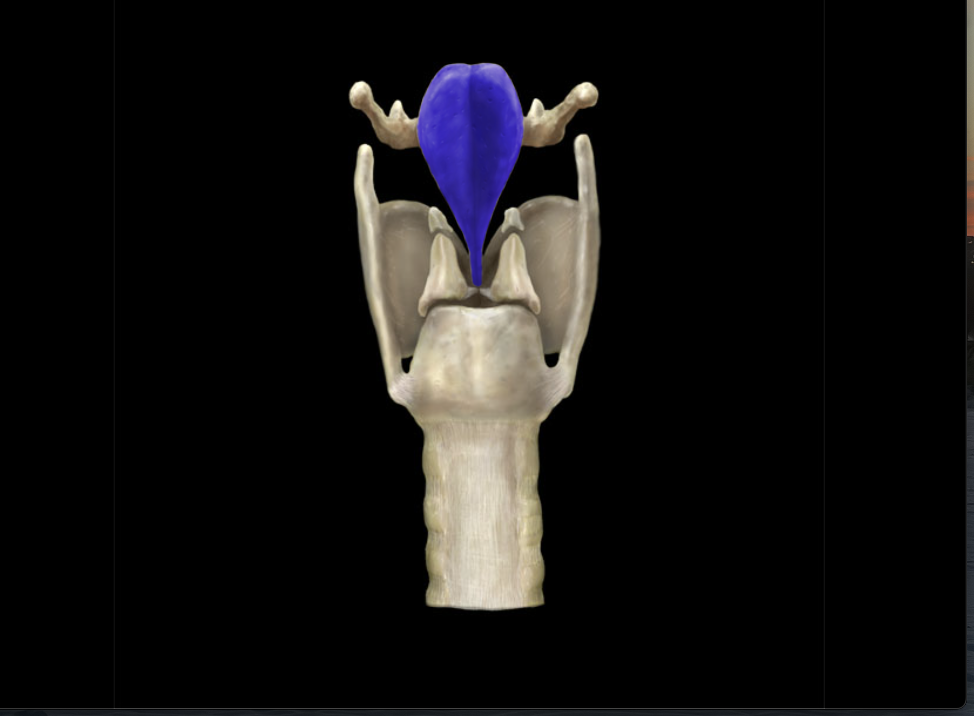
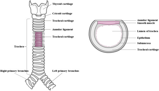
Larynx
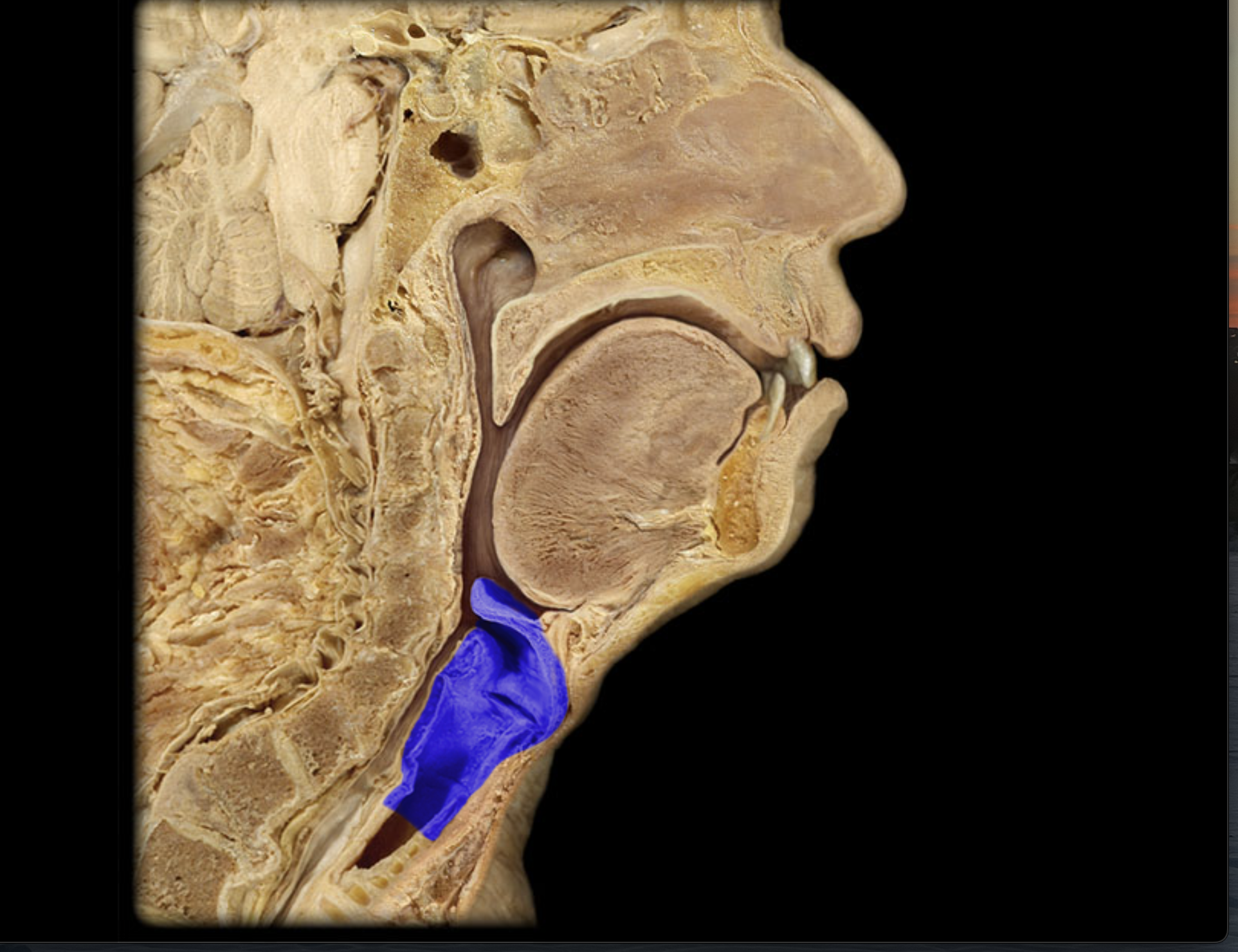
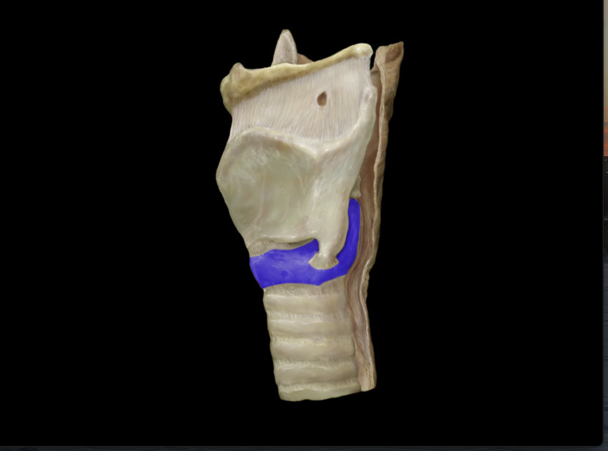
Thyroid cartilage
Laryngeal prominence
Cricoid cartilage
Arytenoid cartilage
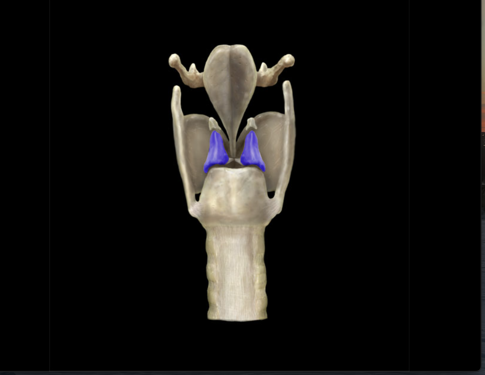
Epiglottis
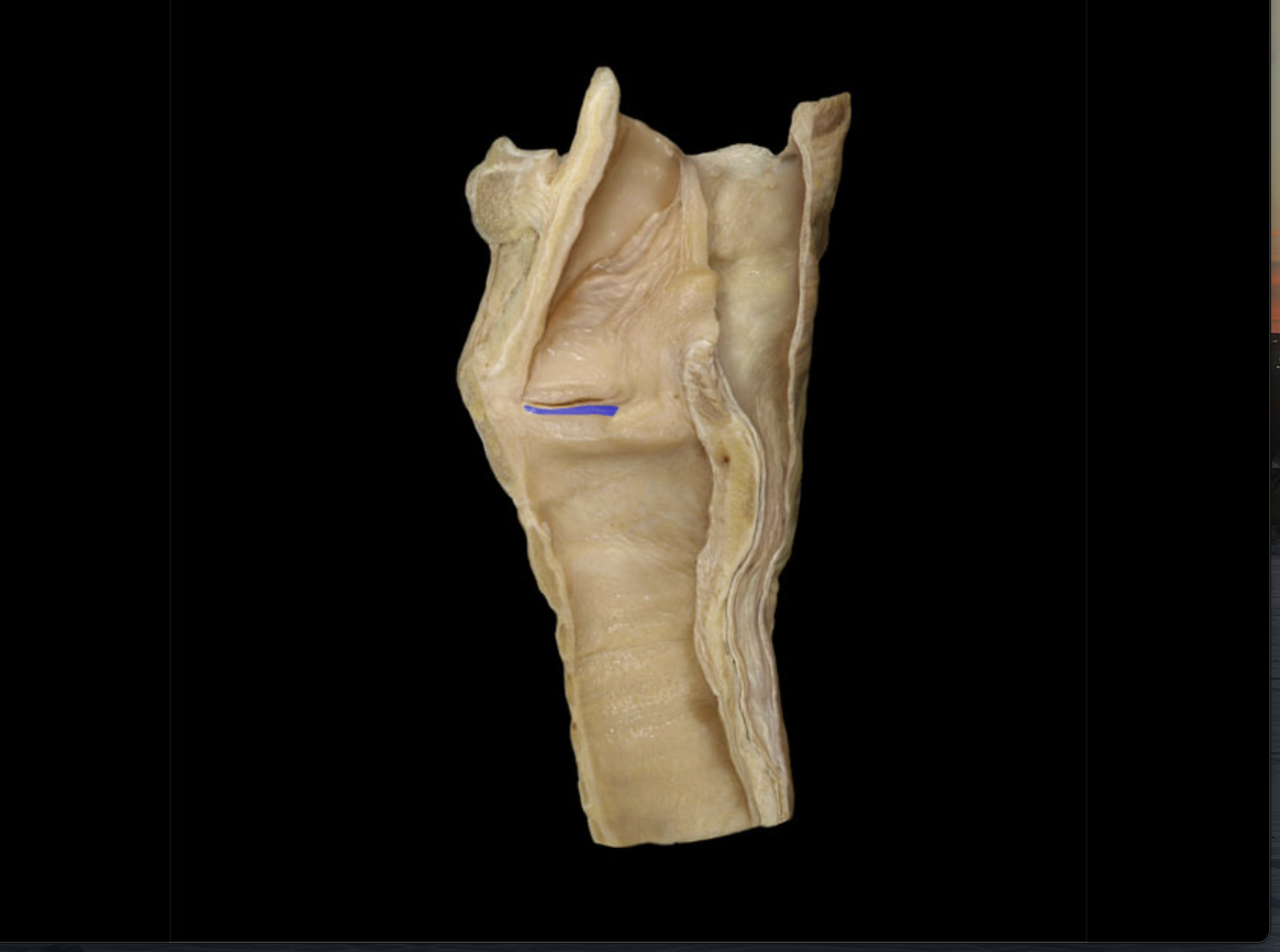
Vestibular folds
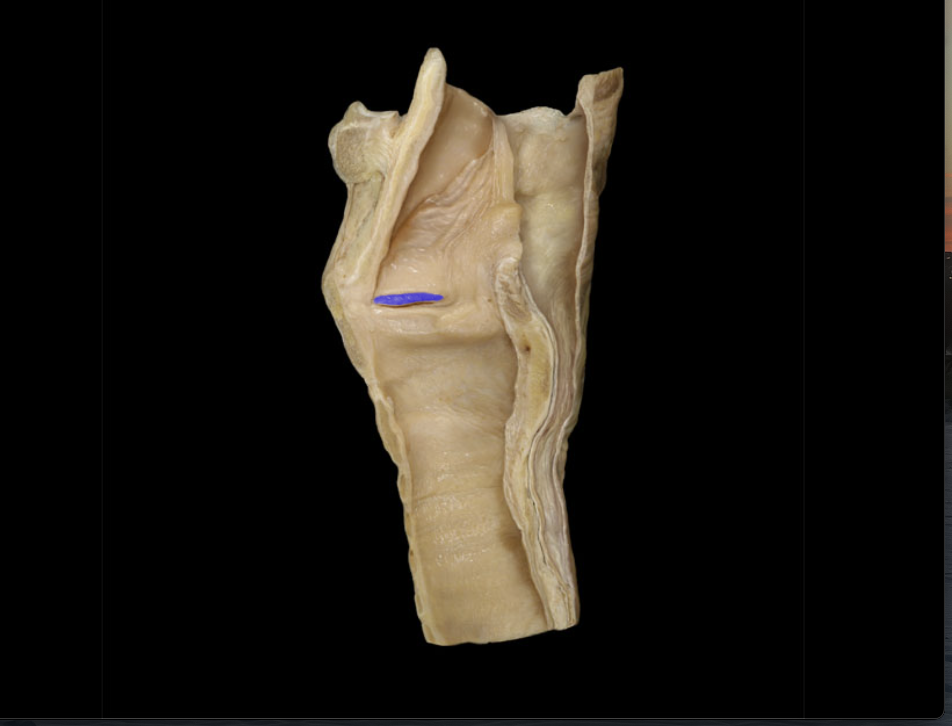
Vocal folds
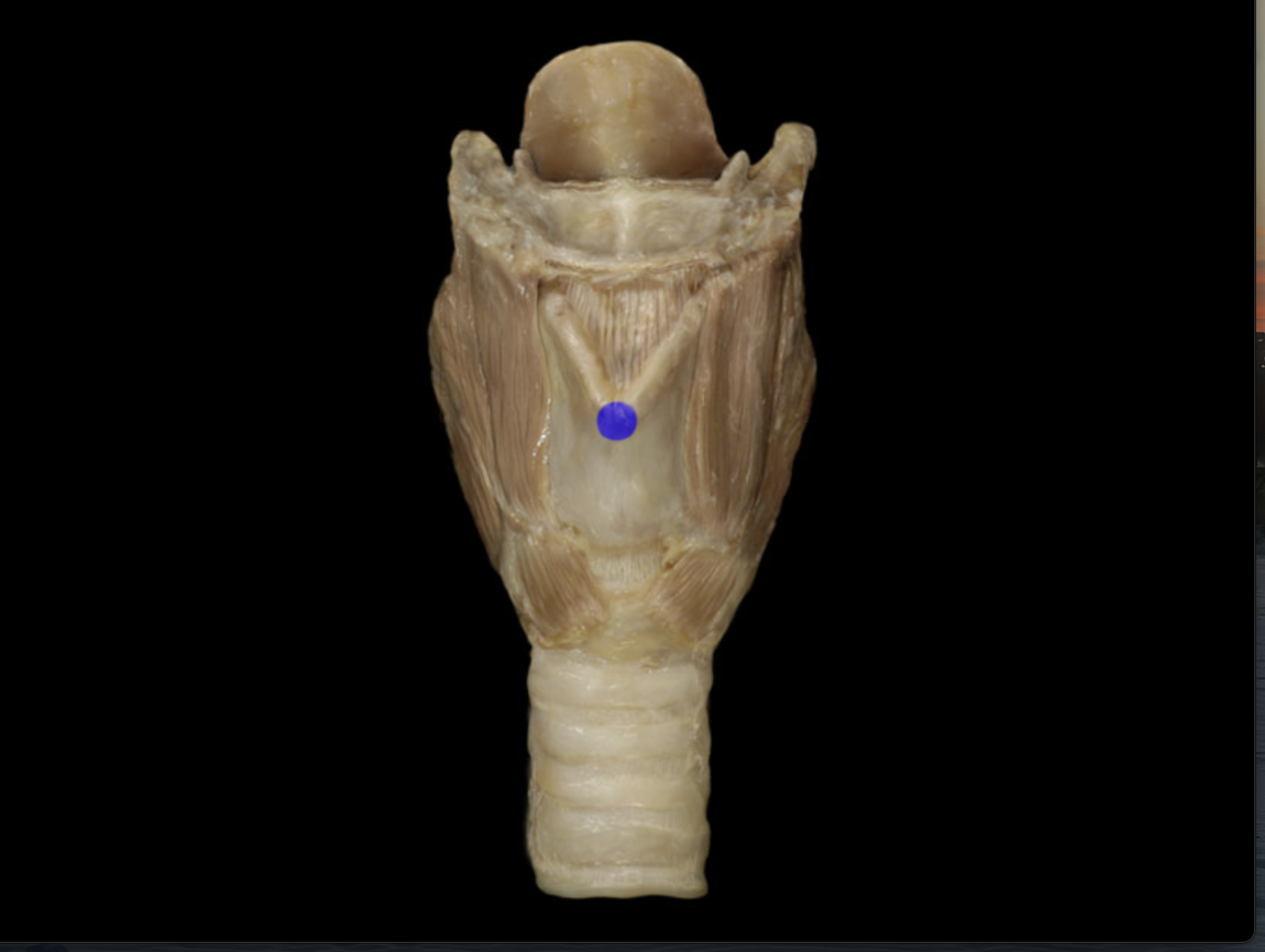
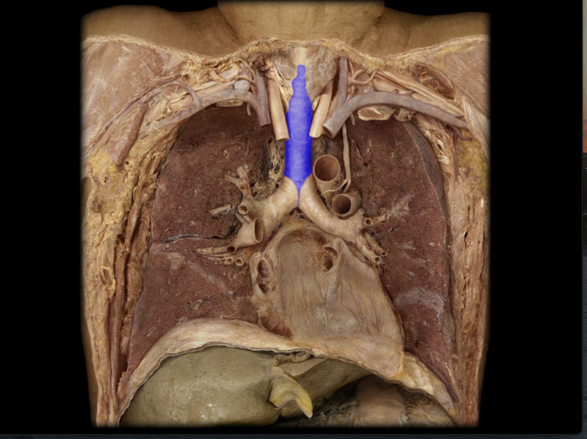
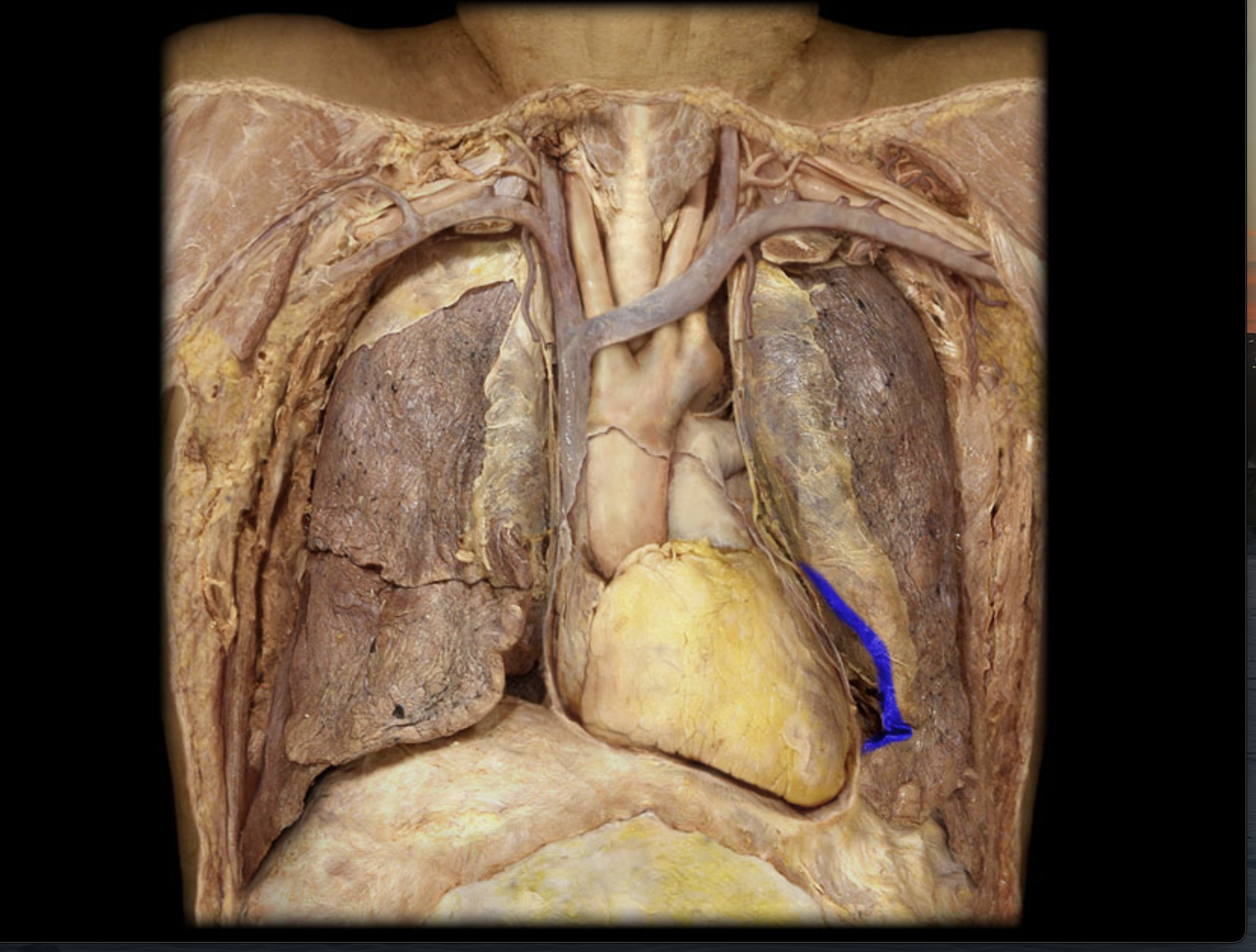
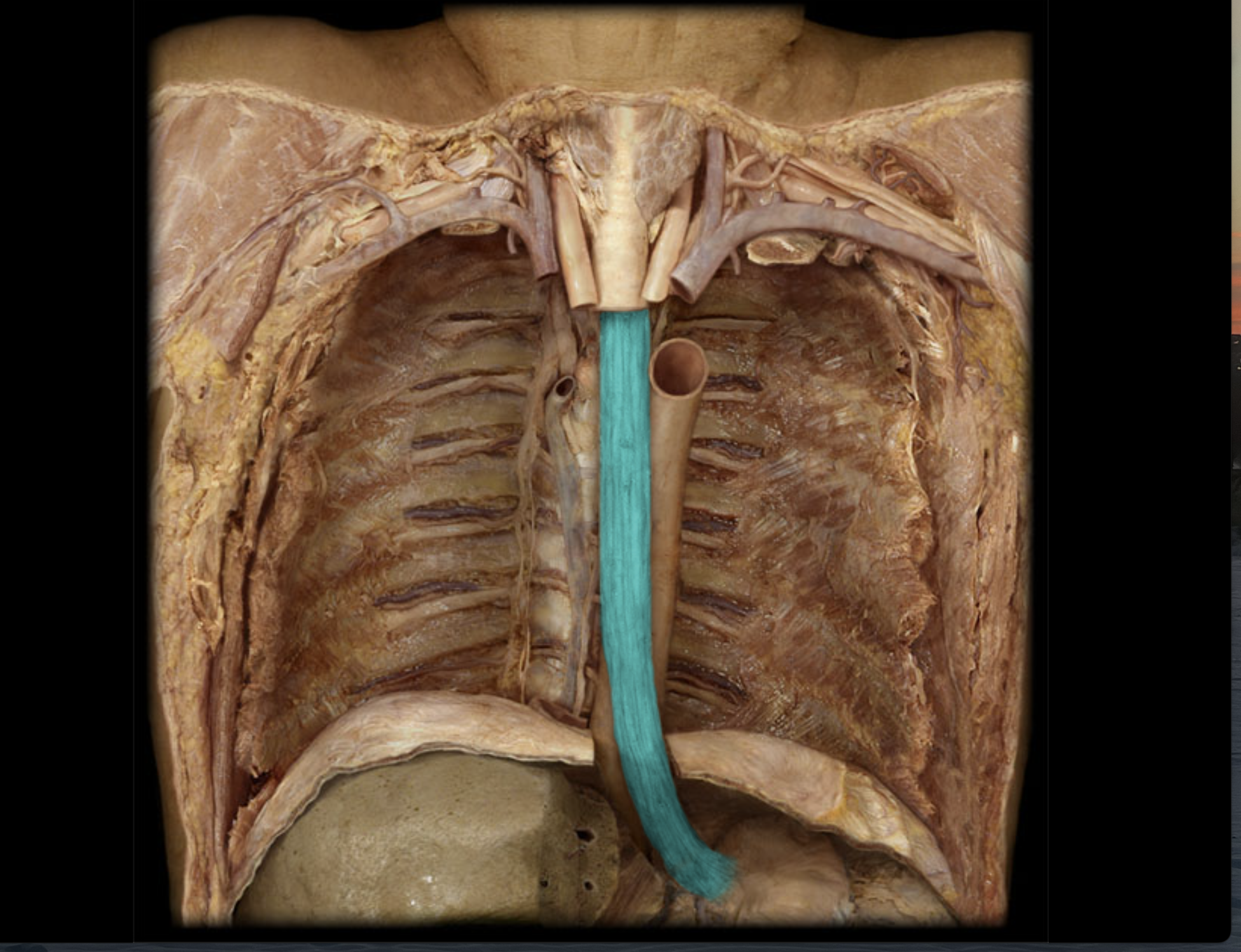
Trachea
Trachealis
Posterior, middle part of ring forming trachea
Carina
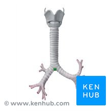
Right/left primary bronchi
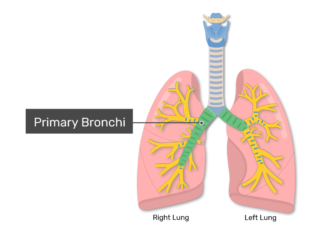
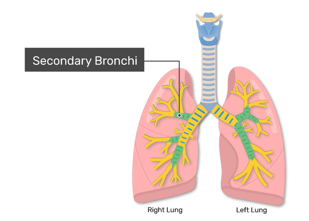
Secondary bronchi
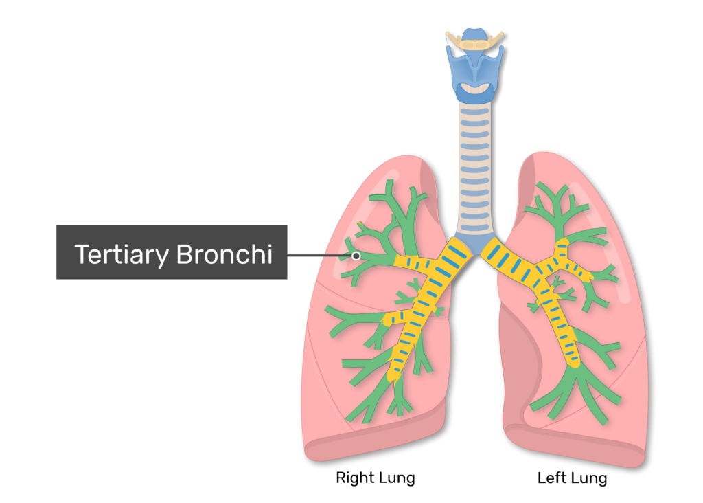
Tertiary bronchi
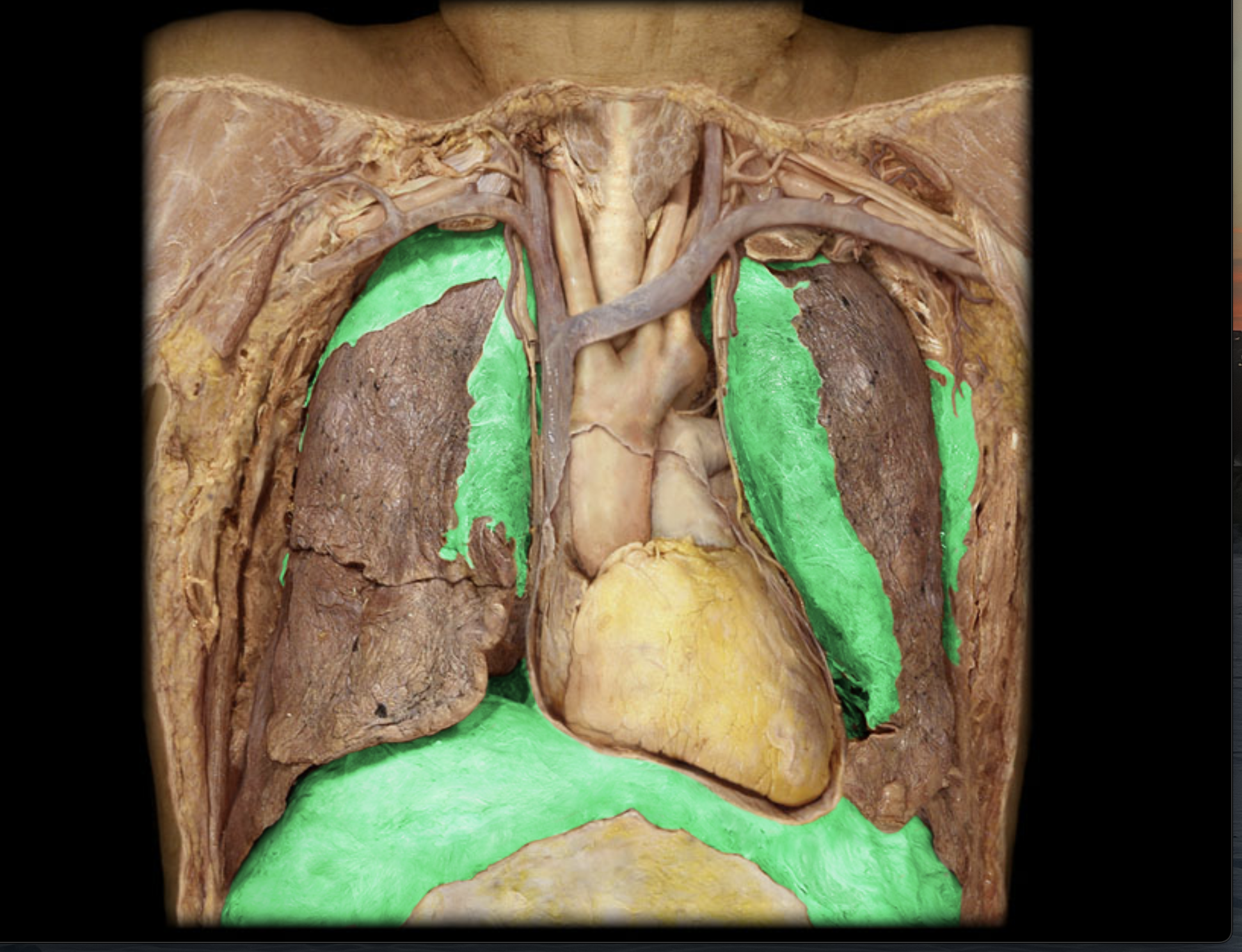
Apex of lung
Top of lung
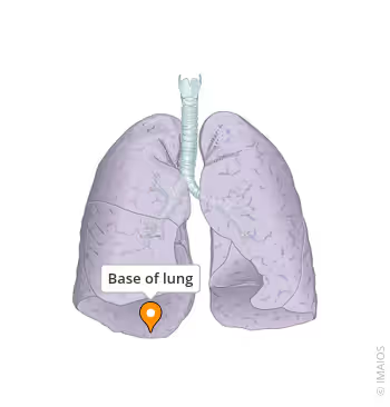
Base of lung
Bottom part of the lung.
Visceral pleura
Membrane covering the lungs
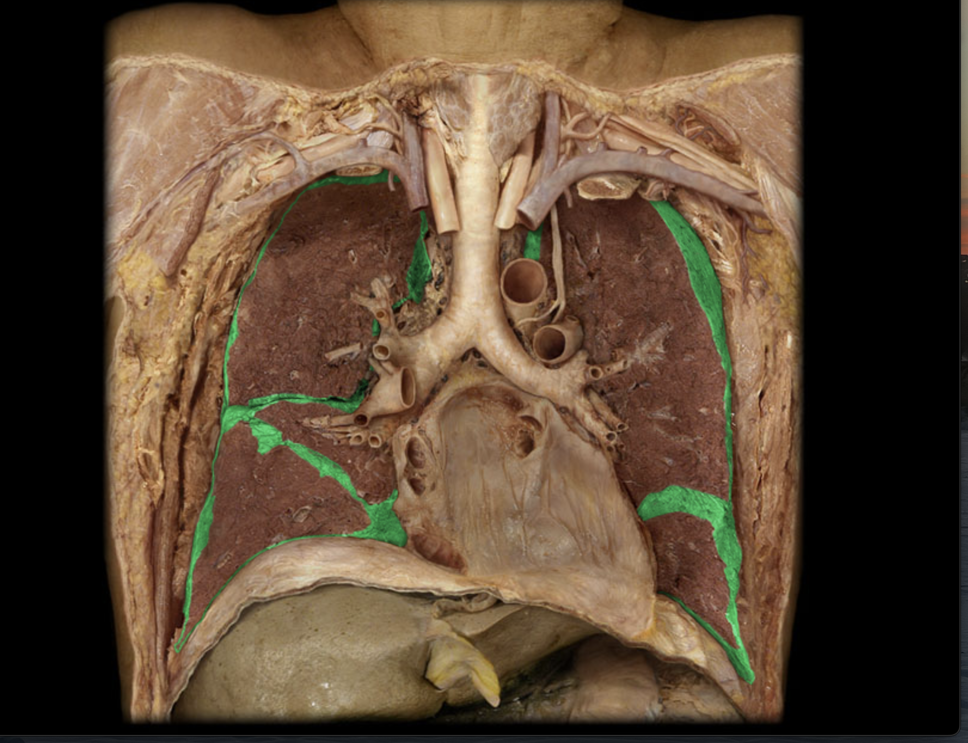
Parietal pleura
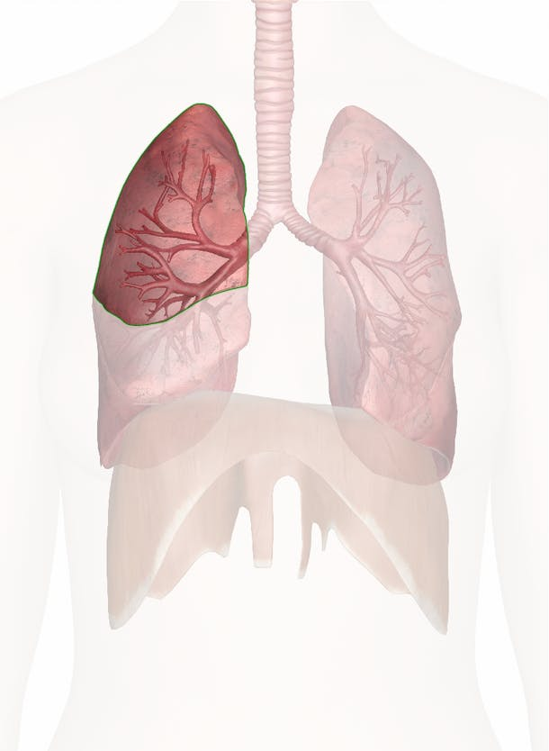
Right lung lobes (superior, middle, inferior)
(right)

Horizontal fissure
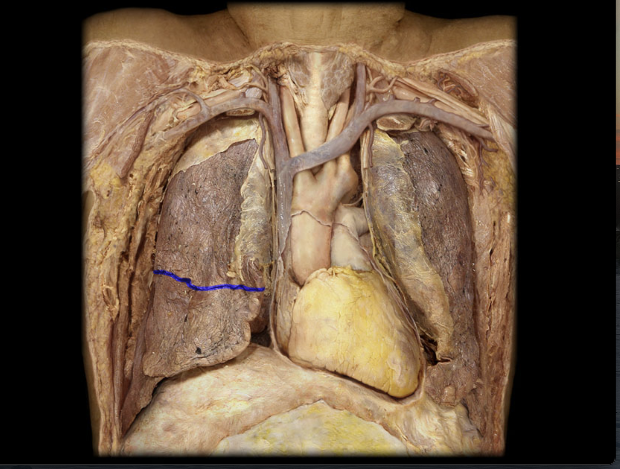
Oblique fissure
Left lung lobes (superior and inferior)
(Left)
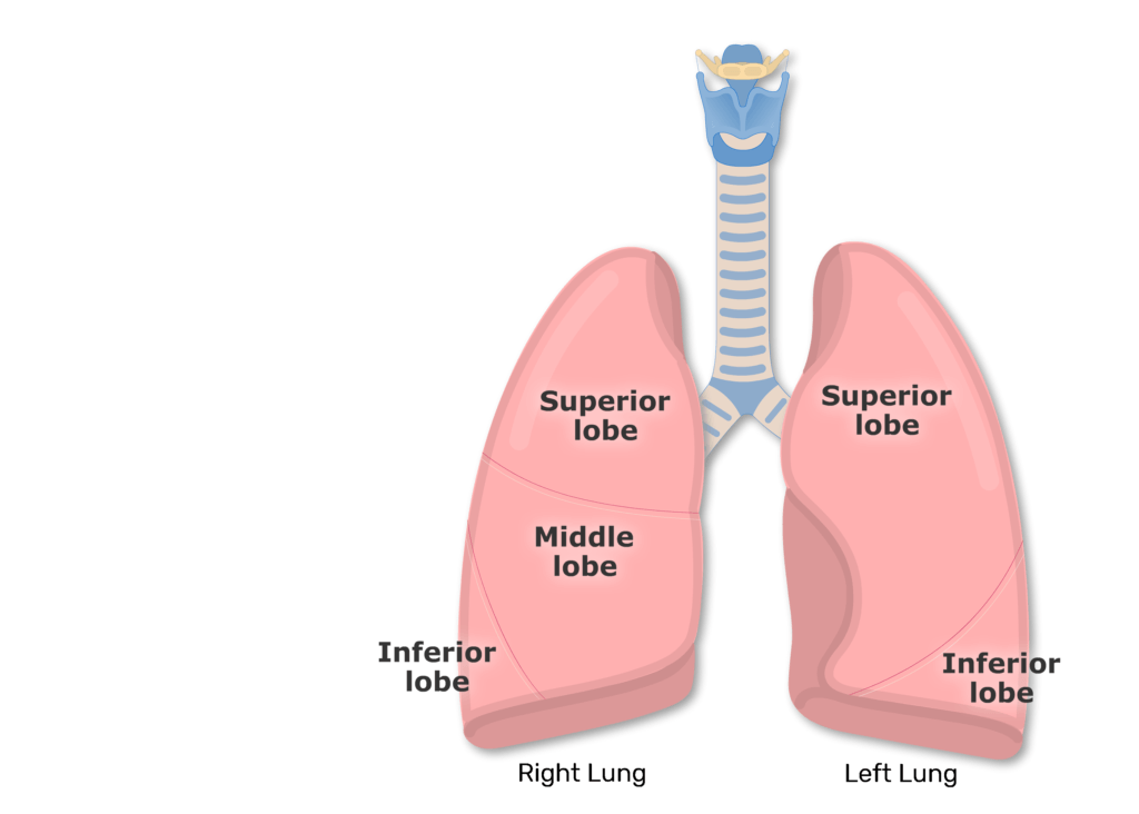
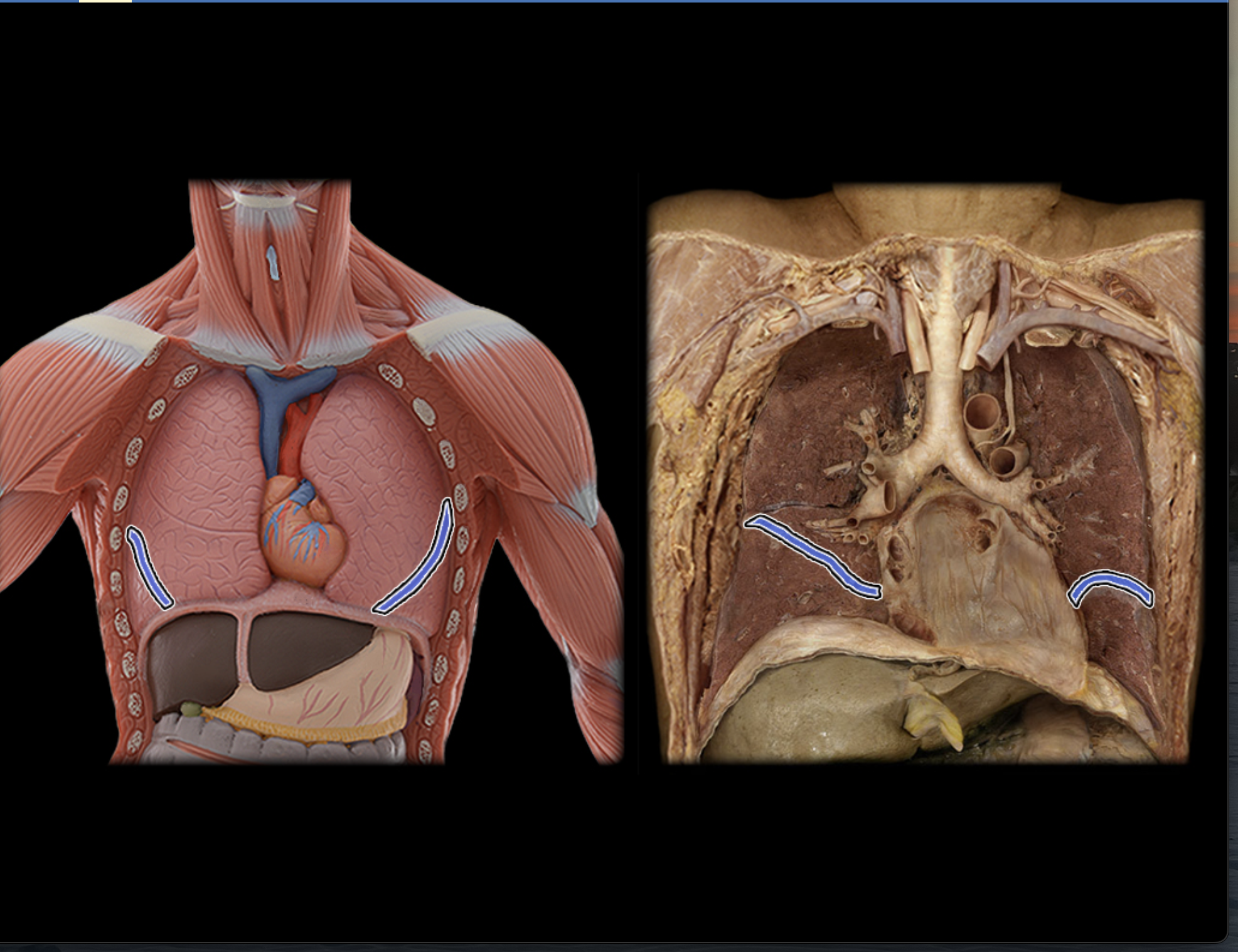
Cardiac notch
Parotid gland
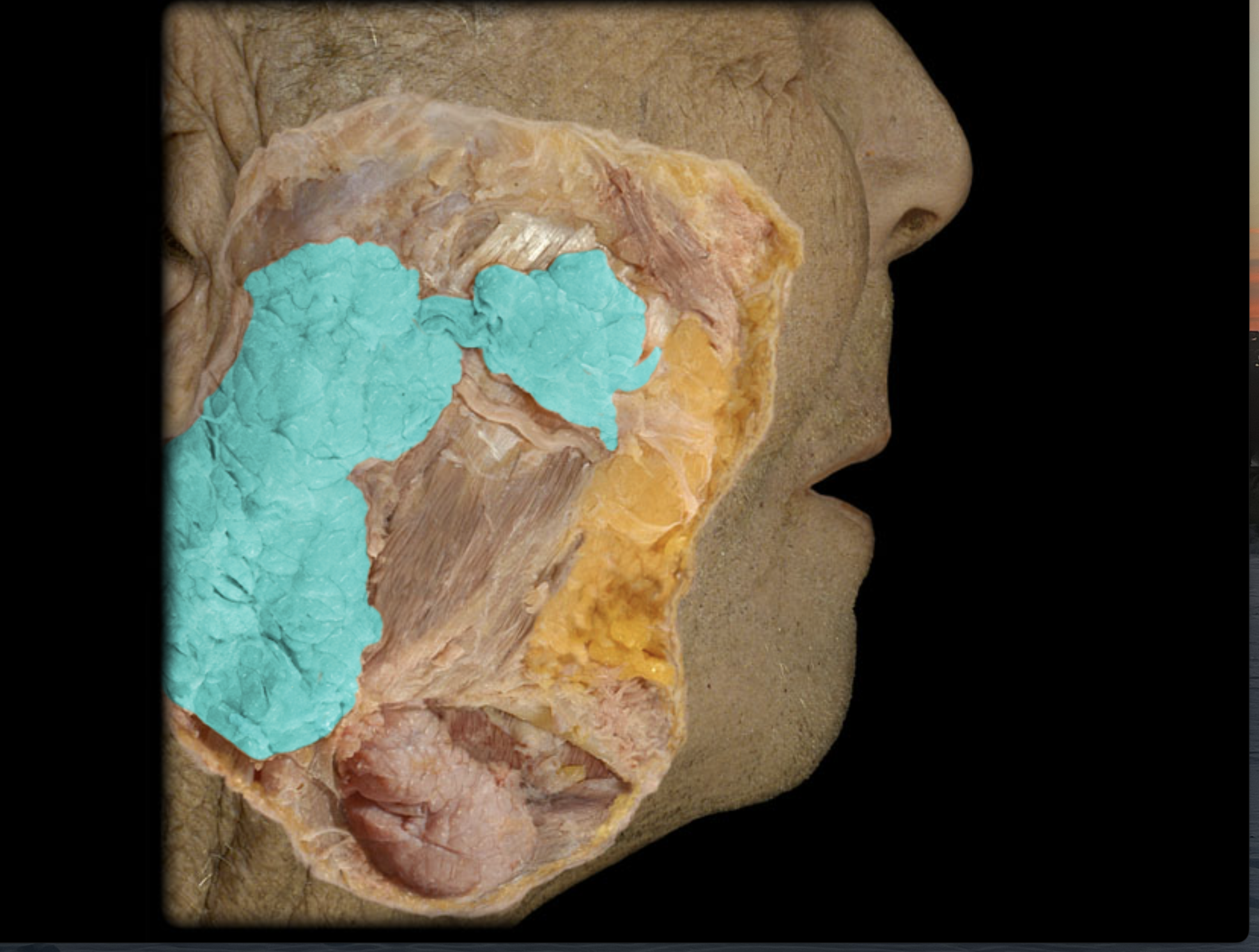
Parotid duct
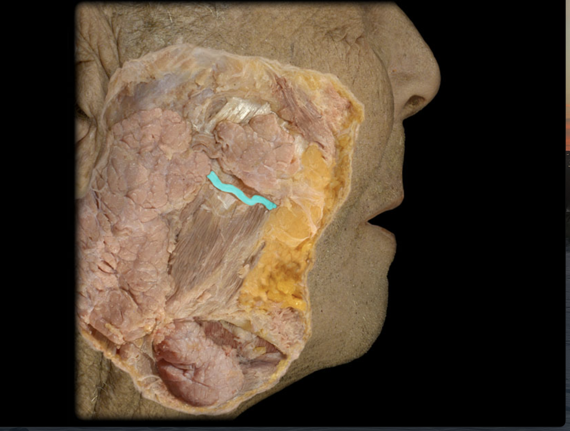
Submandibular gland
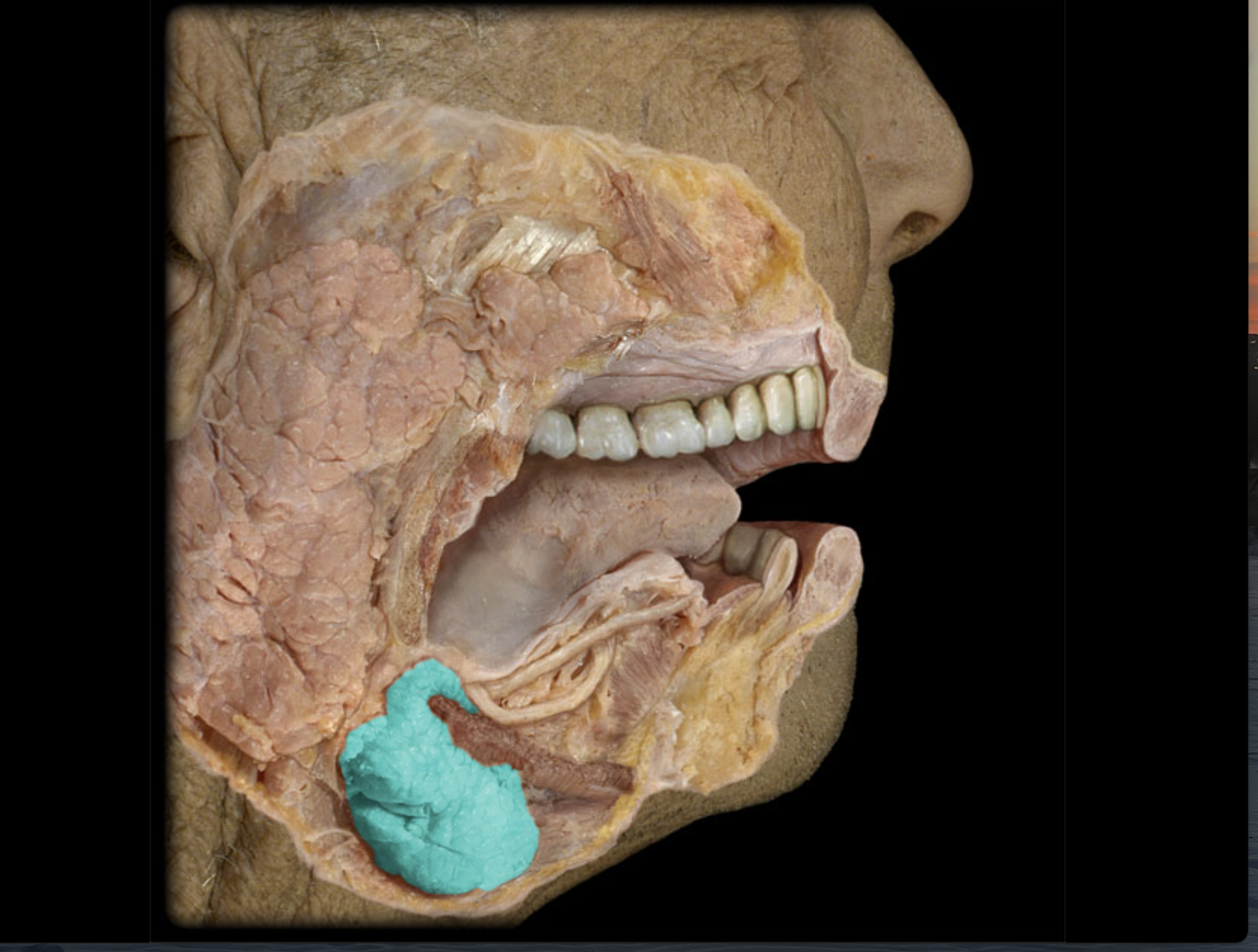
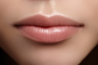
Labia (face)
Superior and inferior
Frena
Vertical strip that anchors tongue to mouth (middle)
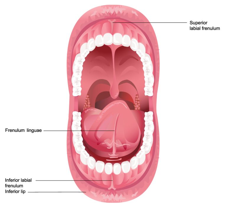
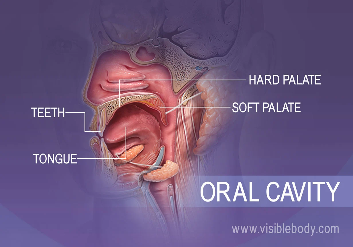
Oral cavity
The space within the mouth that contains the tongue, gums, and teeth, playing a vital role in digestion and speech.
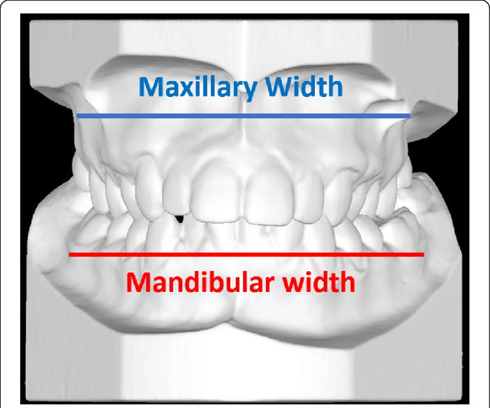
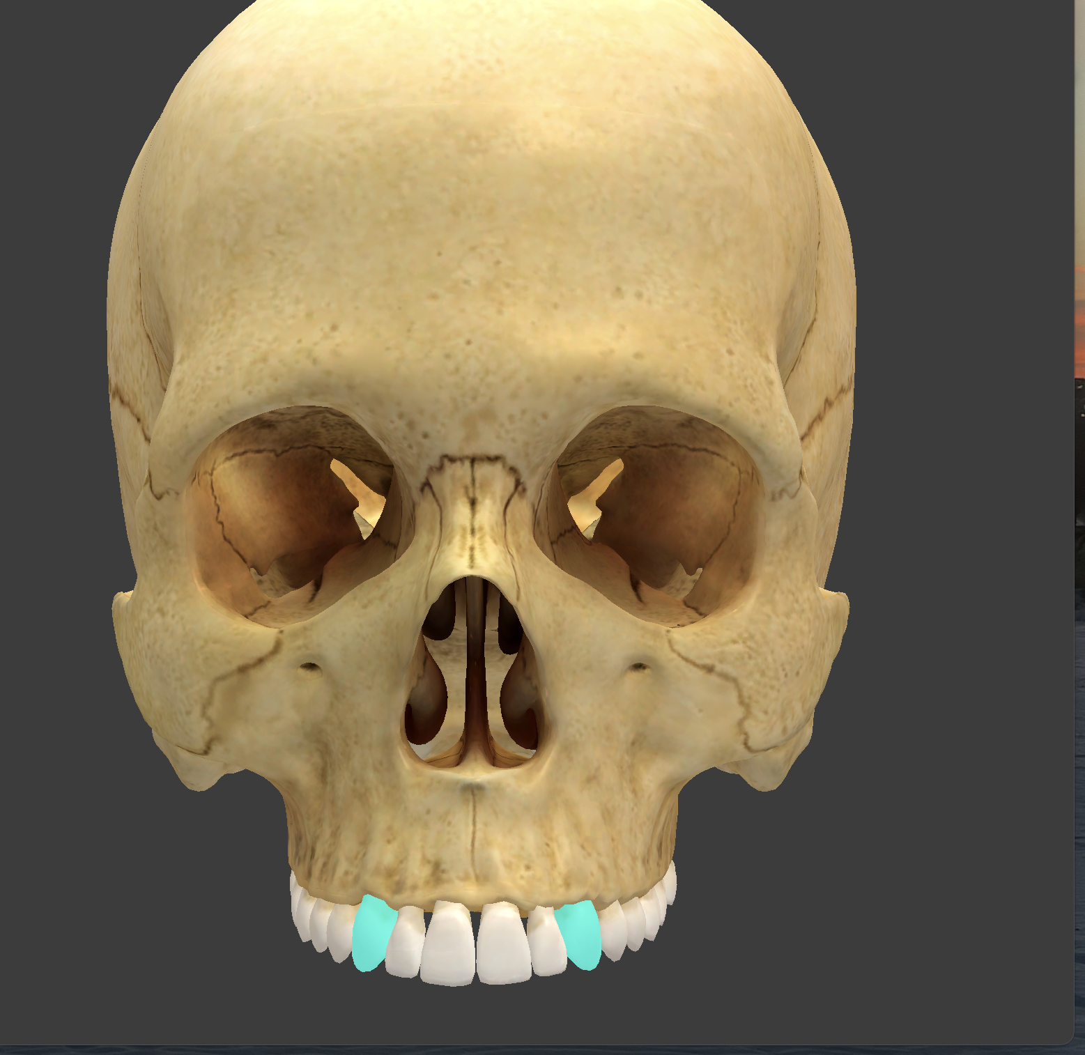
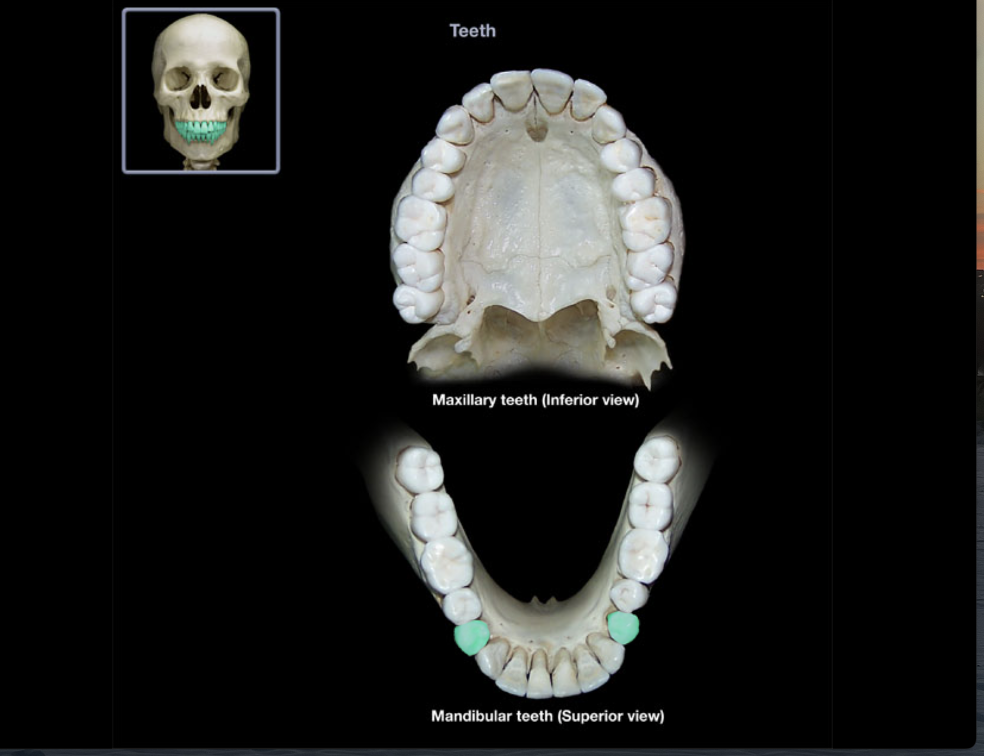
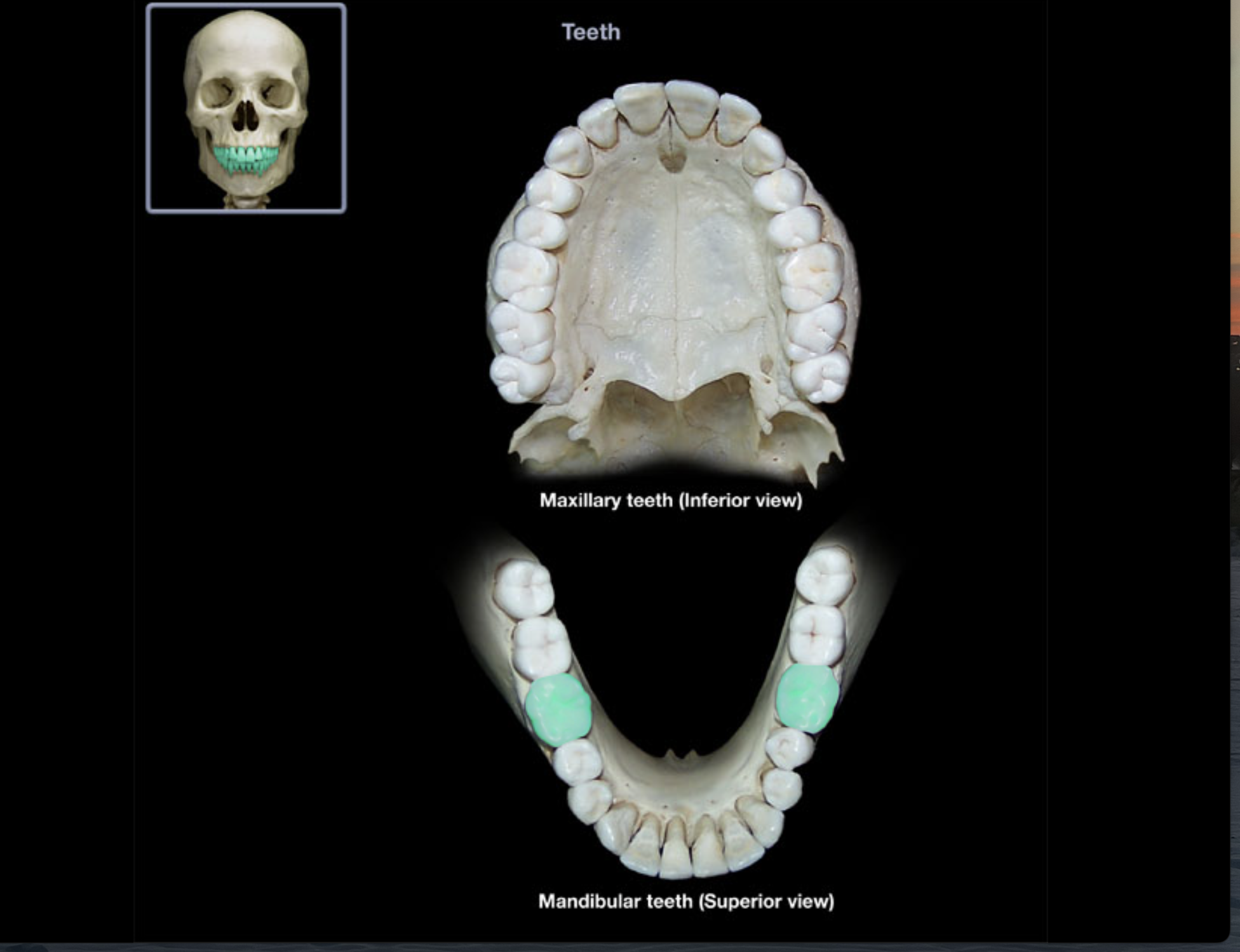
Maxillary and Mandibular teeth
Central incisors (2)

Lateral incisors (2)

Cuspids (canines)
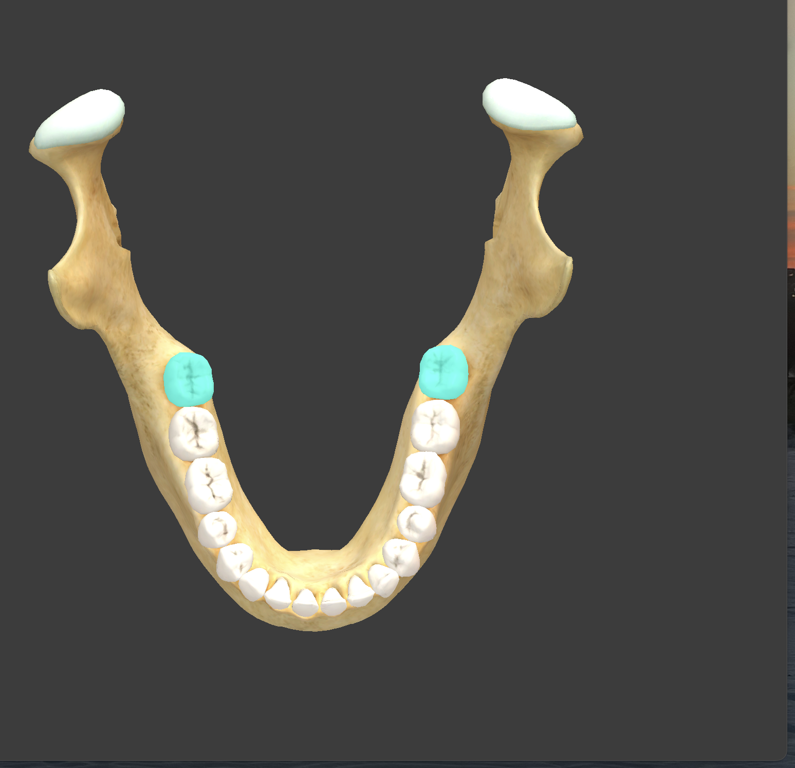
Bicuspids (premolars)
2 bumps
1st molars
3-4 bumps
2nd molars

3rd molars
-Note: not everyone has these, they are wisdom teeth and can be surgically removed
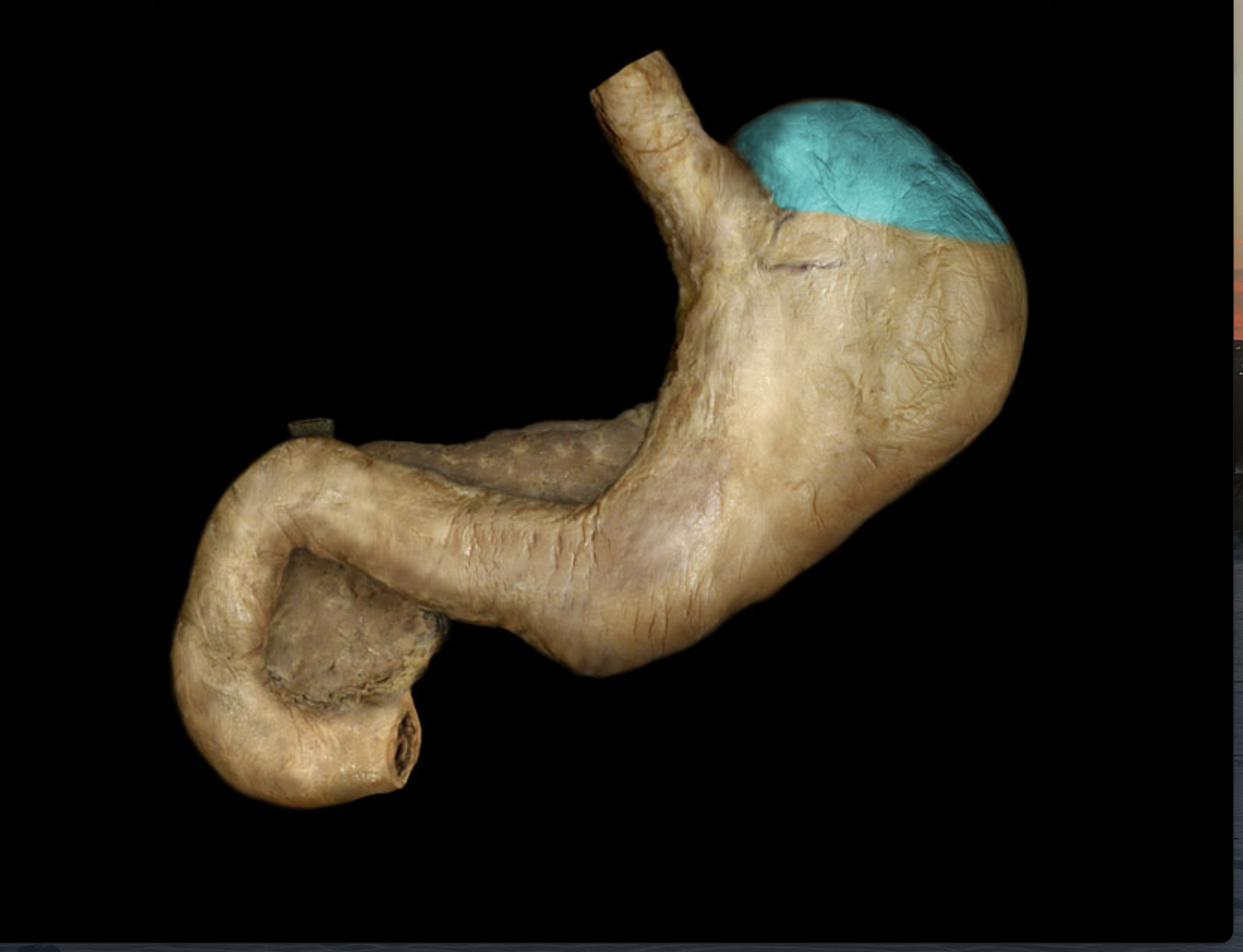
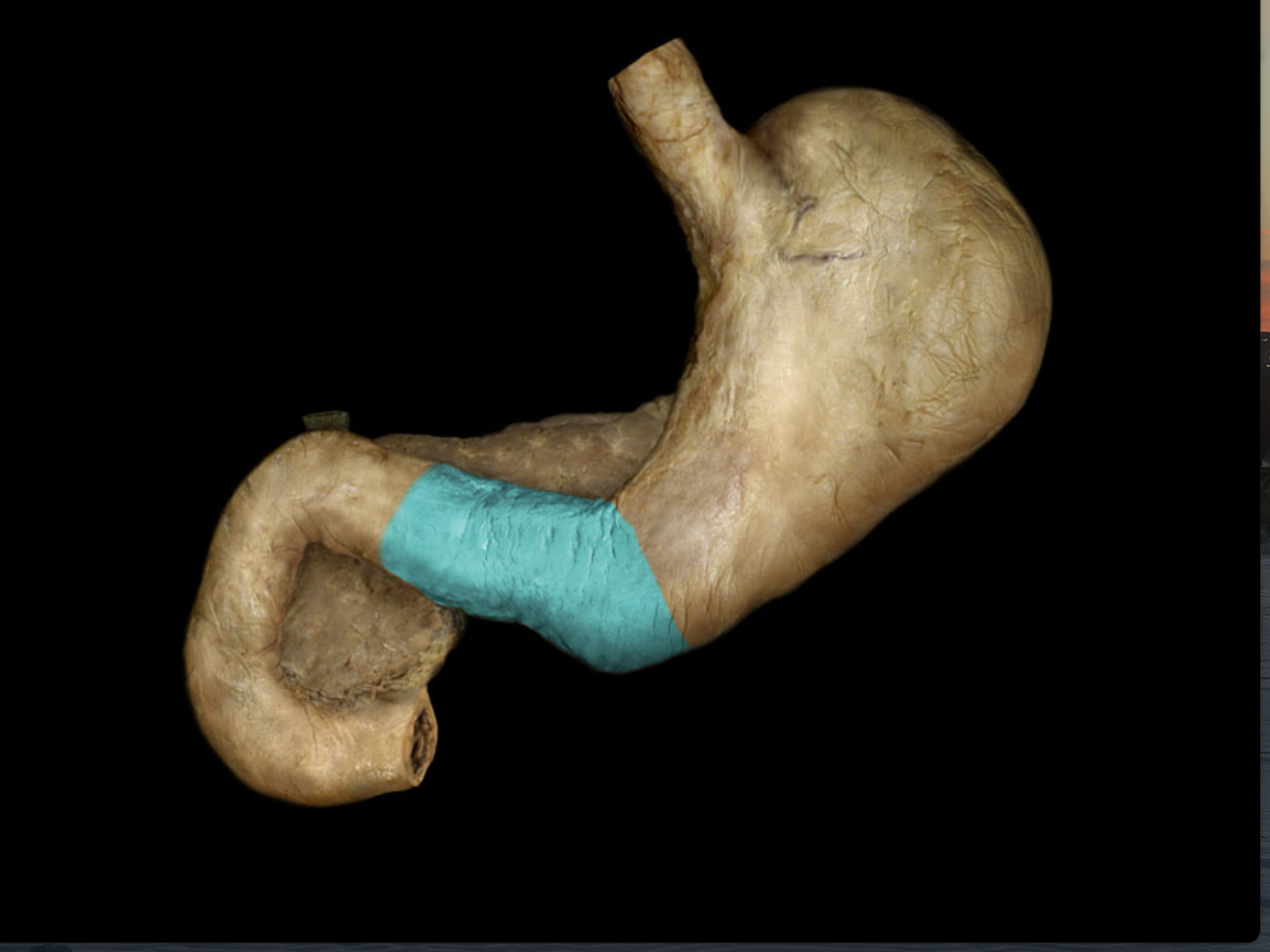
Esophagus
Slants slightly to left (of patient) which directly connects to stomach
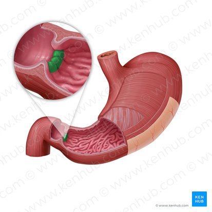
Stomach
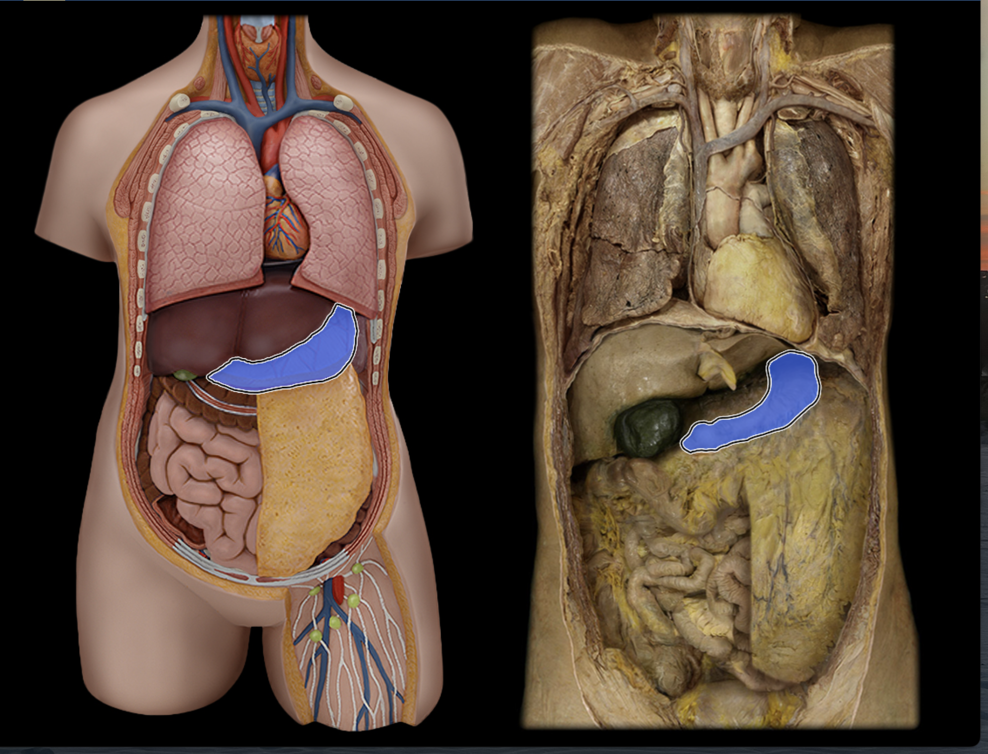
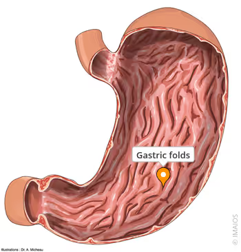
Rugae of stomach
1,16, 17, 32: 3rd molars
2, 15, 18, 31: 2nd molars
3, 14, 19, 30: 1st molars
4, 13, 20, 29: 1st bicuspids
5, 12, 21, 28: 2nd bicuspids
6, 11, 22, 27: Cuspids
7, 10, 23, 26: lateral incisors
8, 9, 24, 25: central incisors
Correctly identify the teeth for their number:

Cardia of stomach
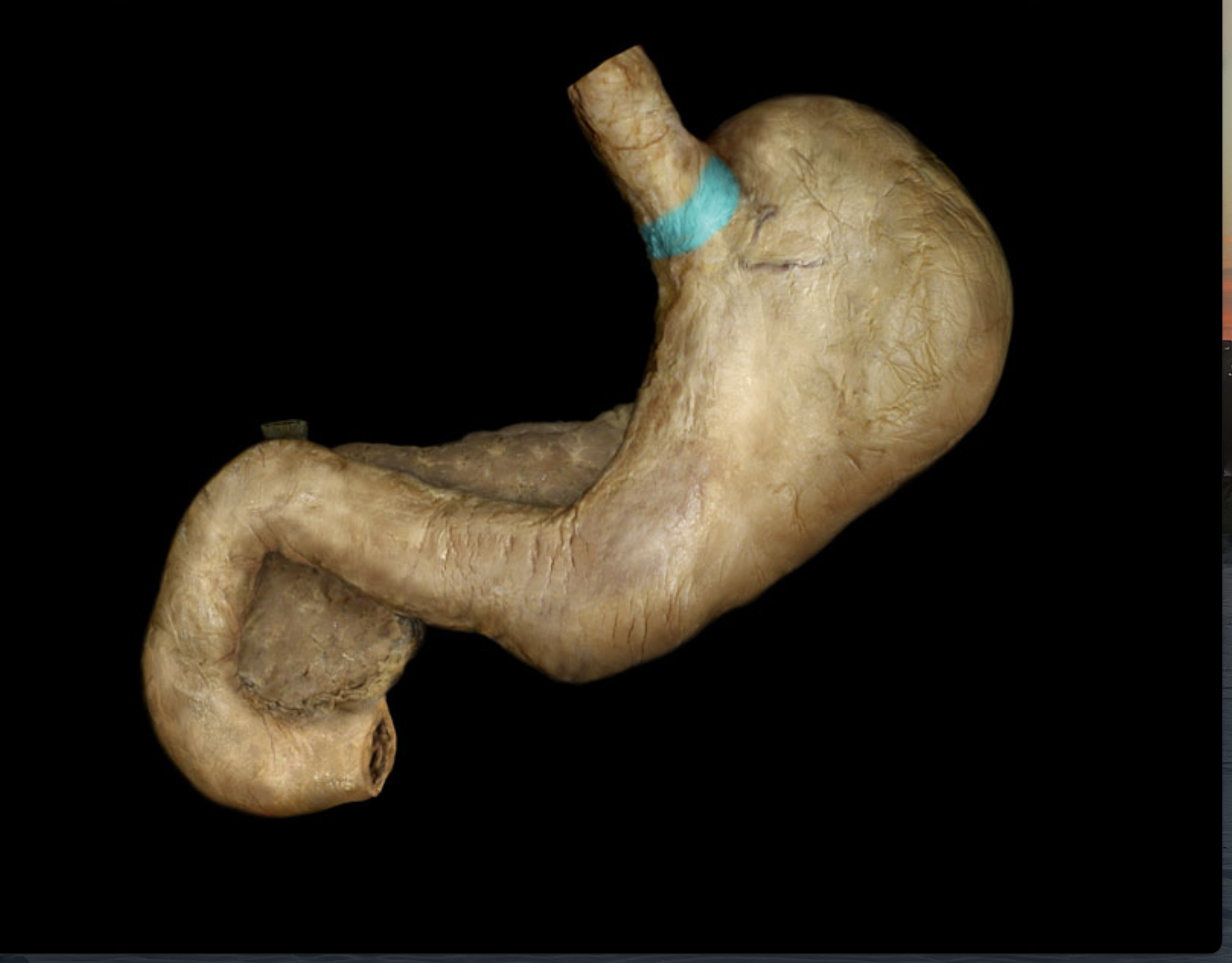
Fundus of stomach
Body of stomach
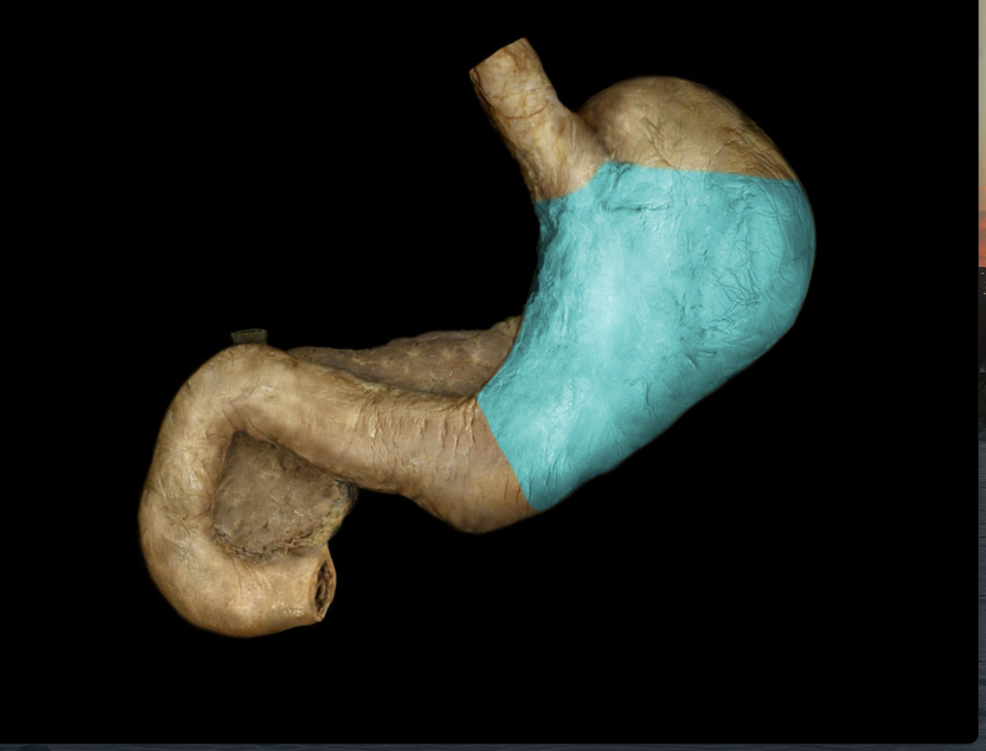
Pylorus of stomach
Pyloric valve
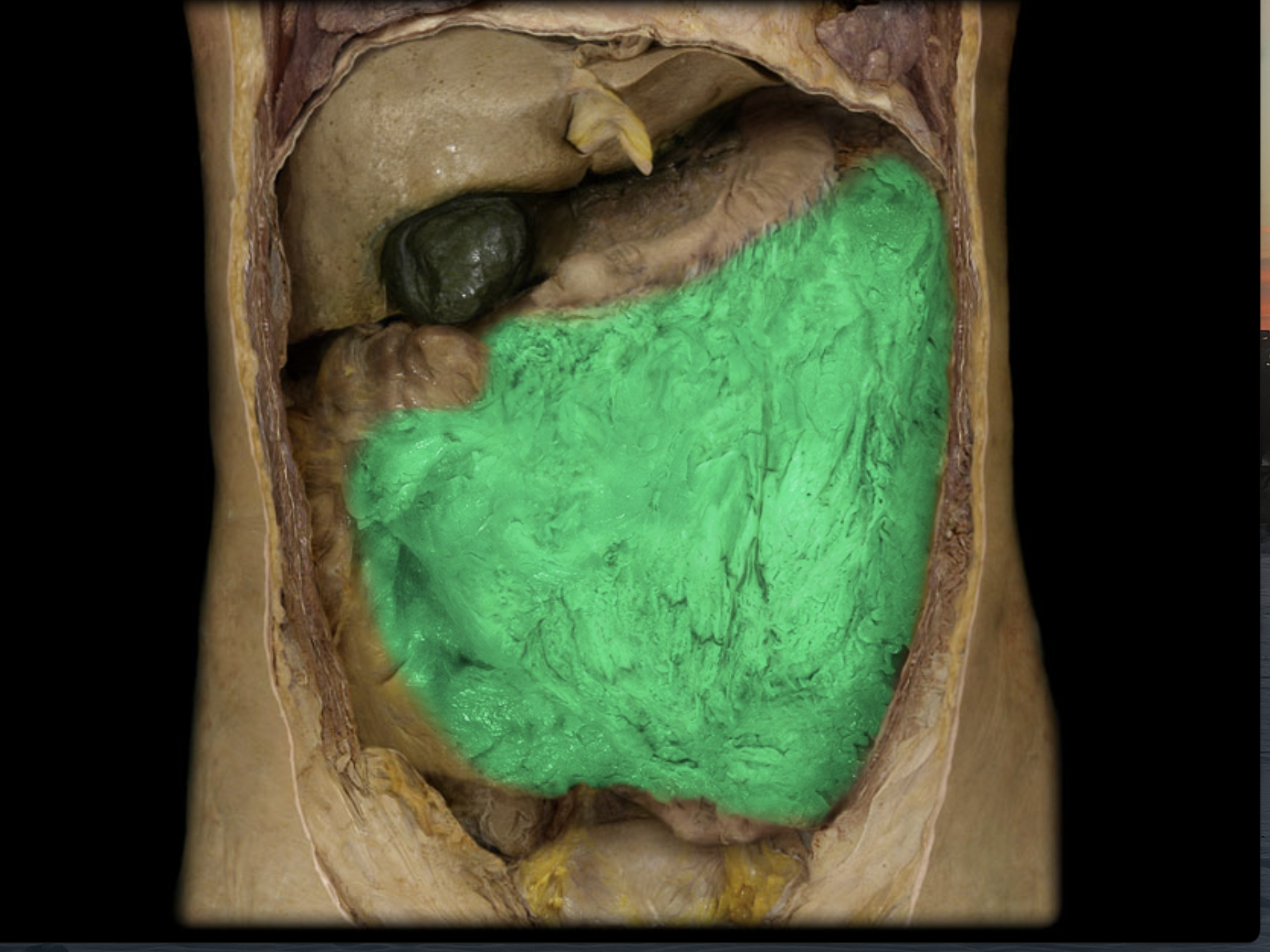
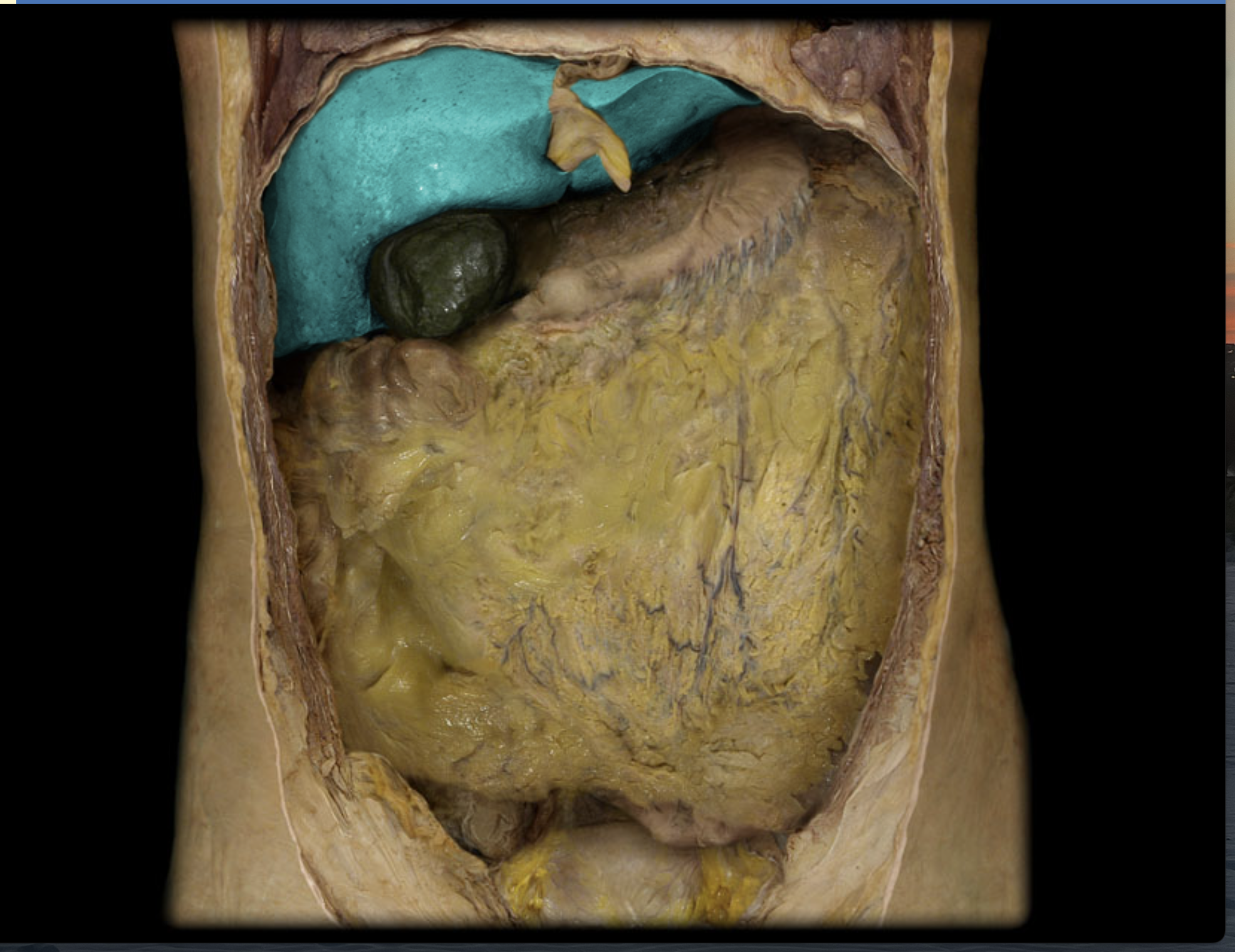
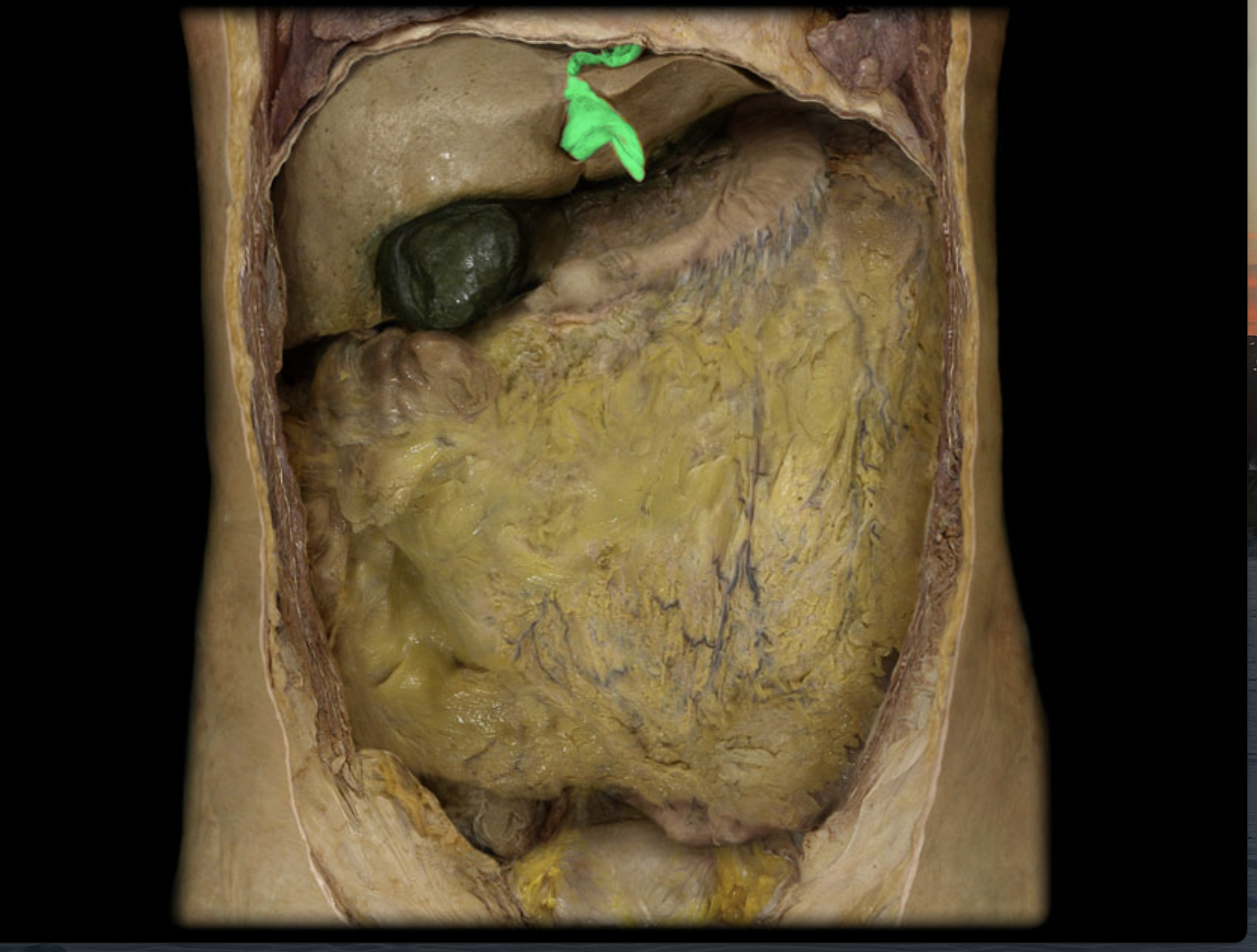
Greater omentum
Lesser omentum
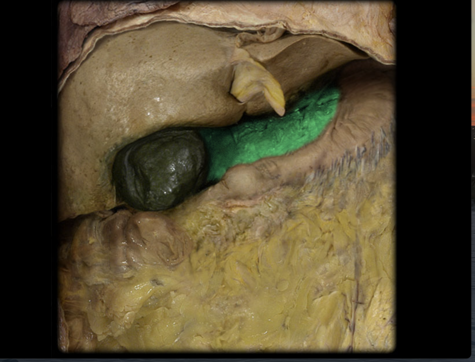
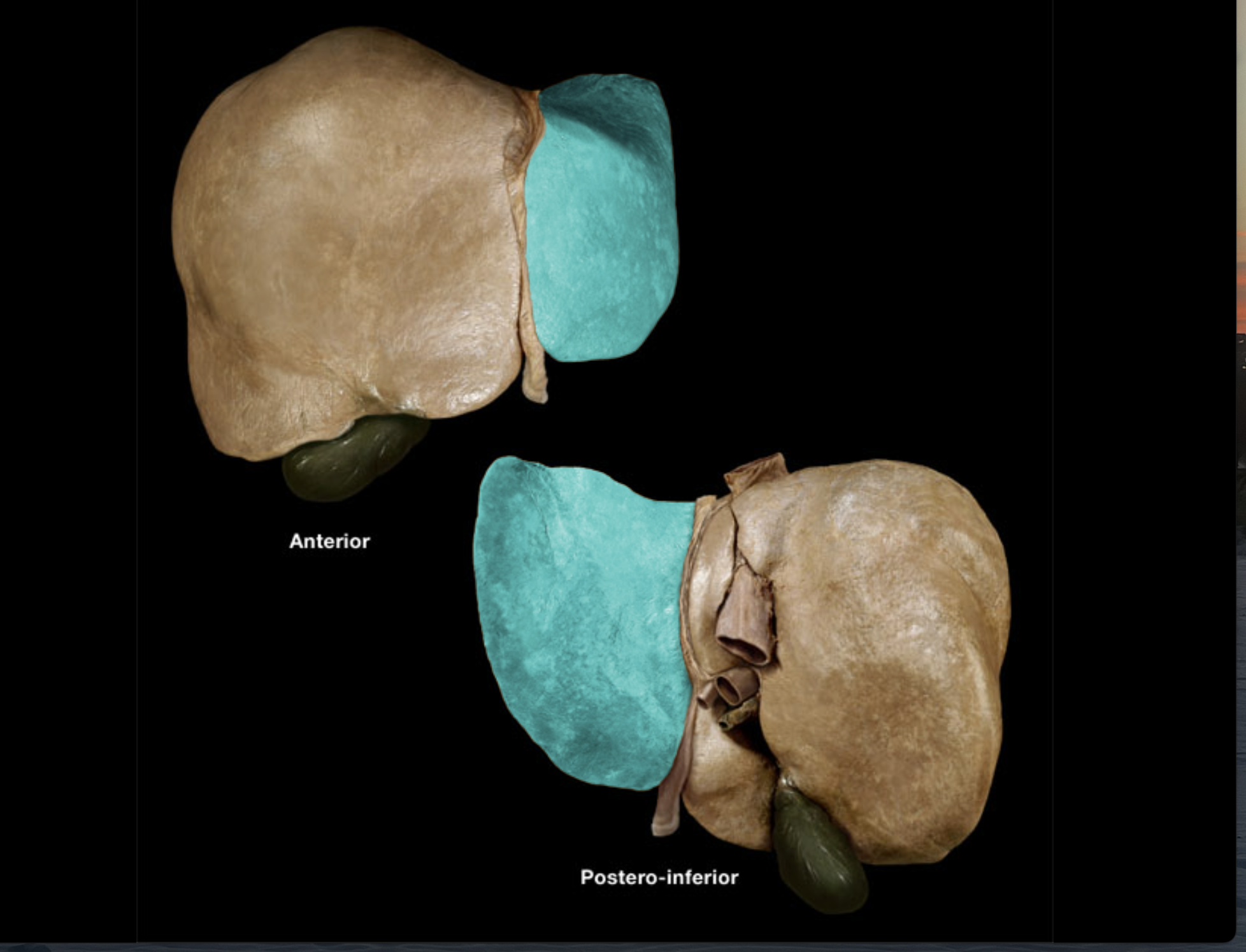
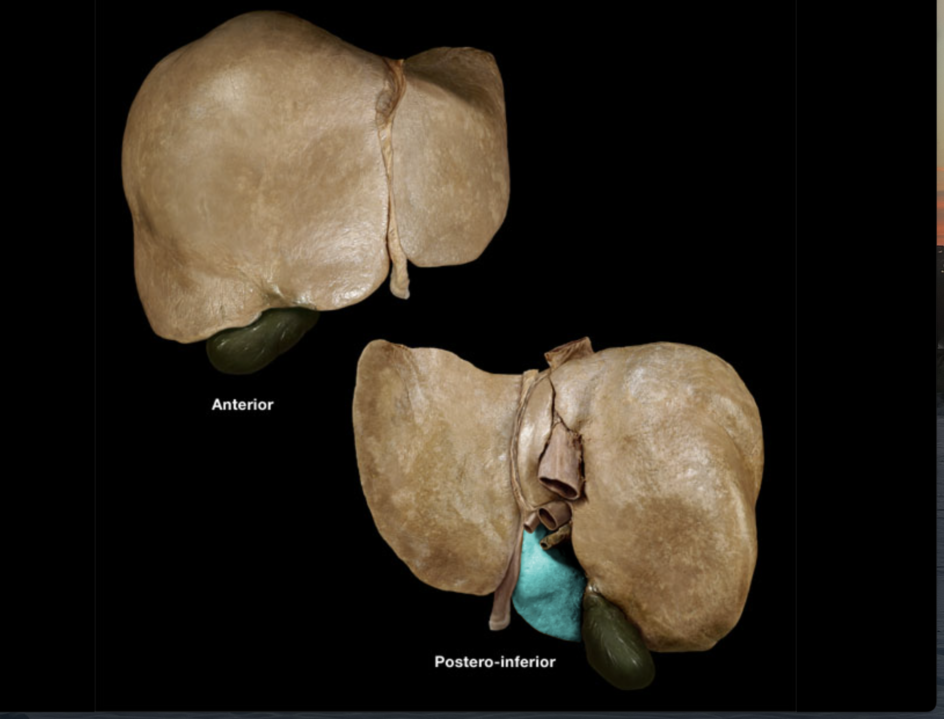
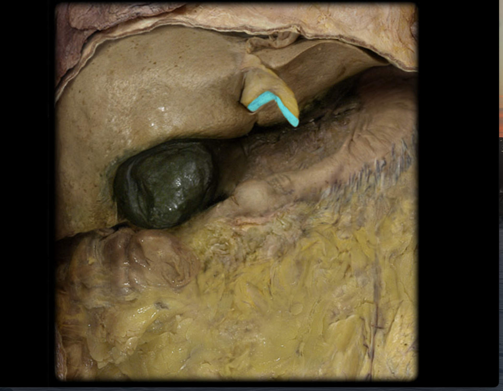
Liver
Right lobe of liver
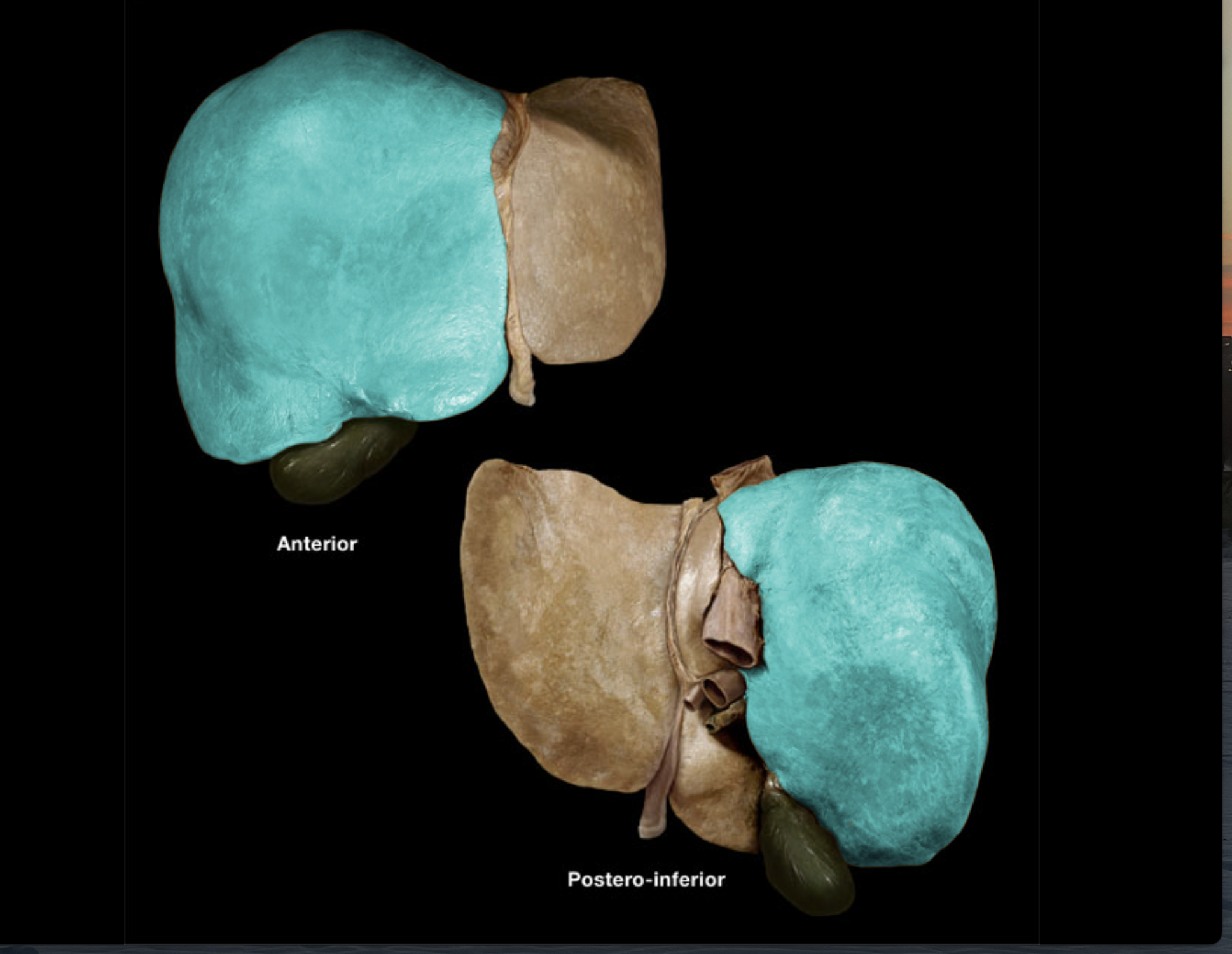
Left lobe of liver
Caudate lobe of liver
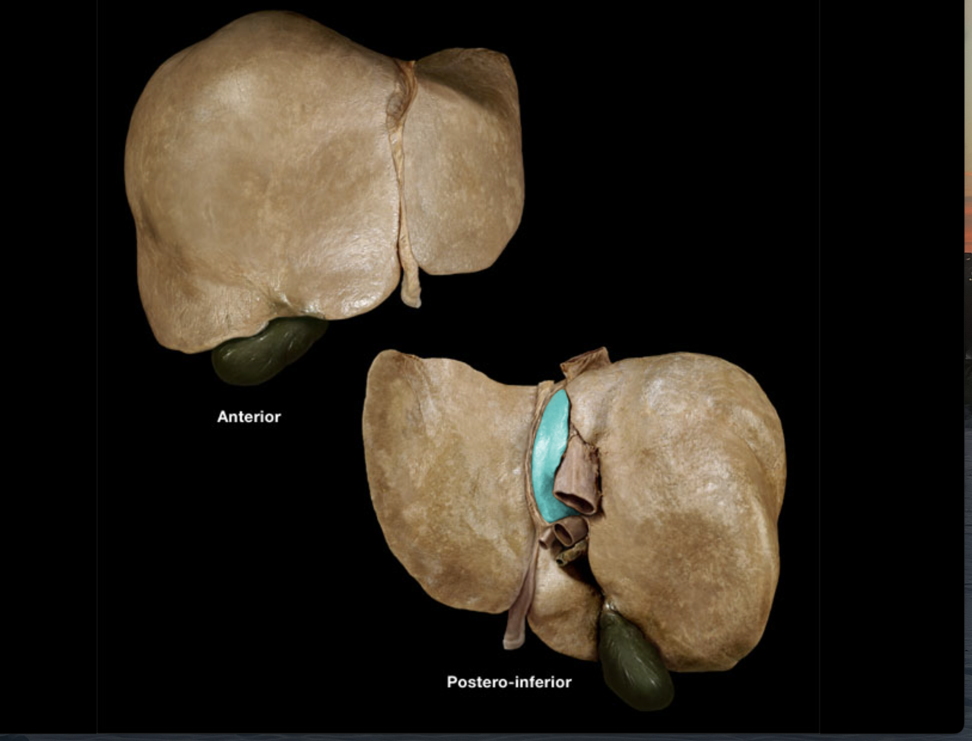
Quadrate lobe of liver
Falciform ligament
Round ligament
Hepatic portal vein
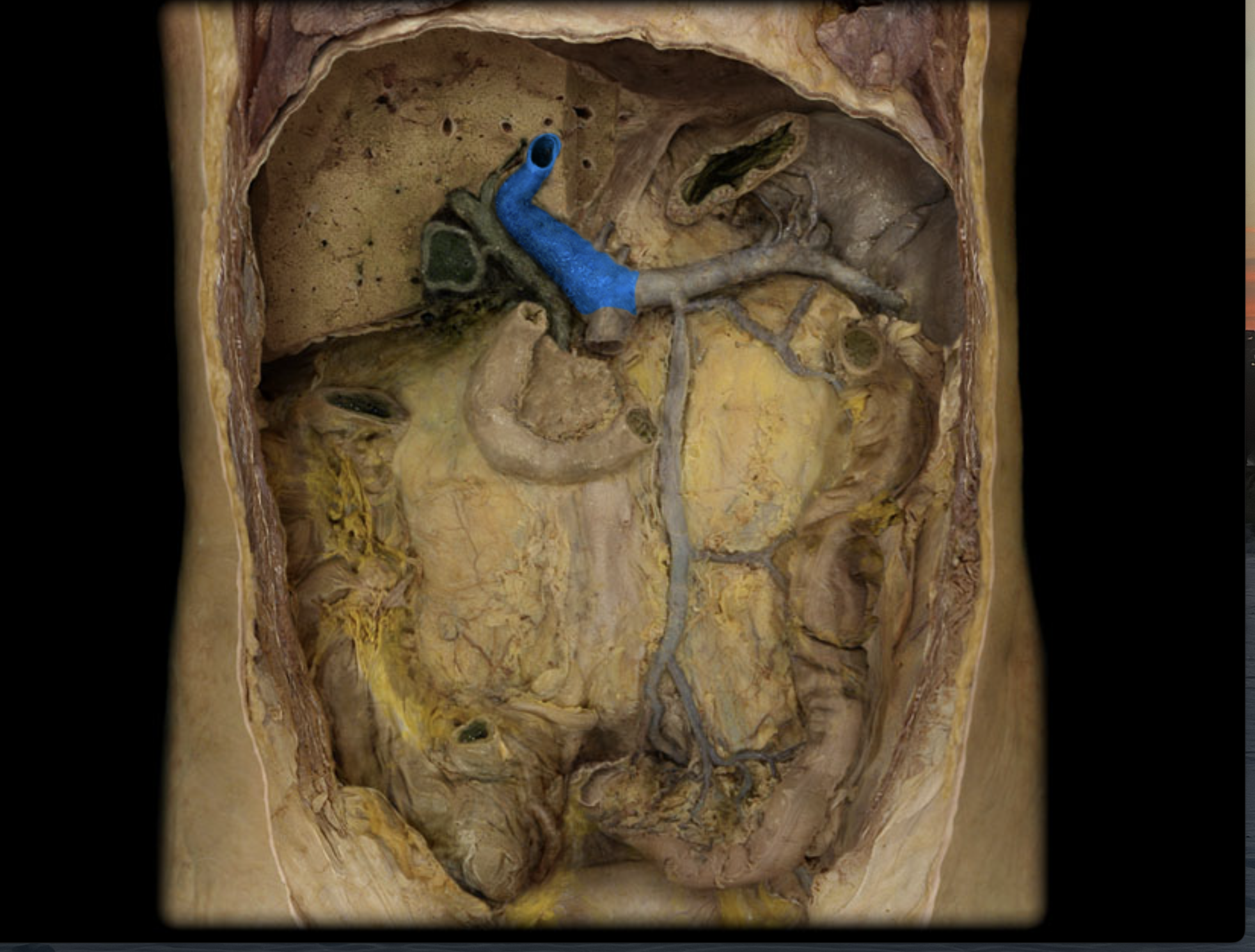
Hepatic artery proper
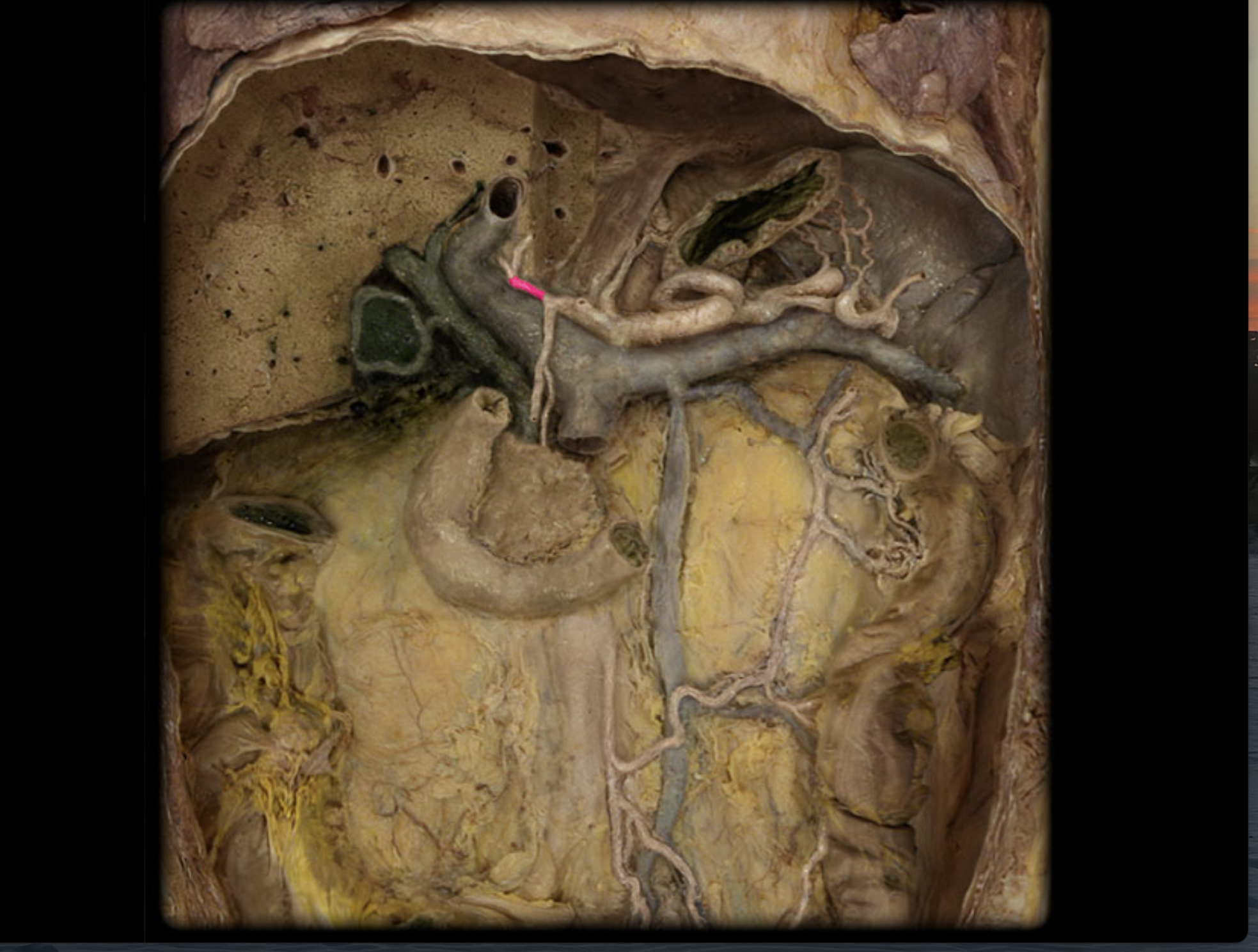
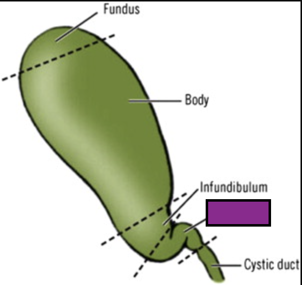
Gallbladder
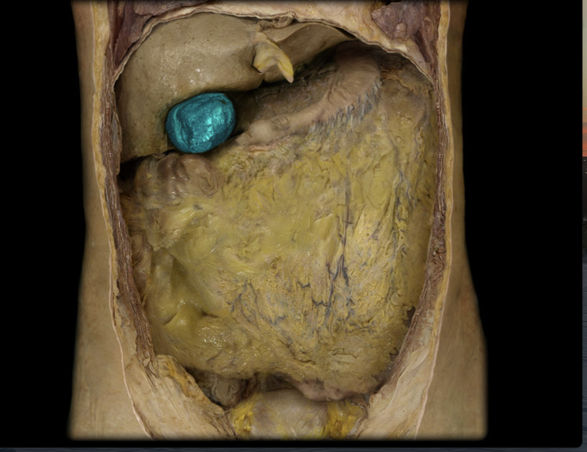
Fundus of gallbladder
(domed end)
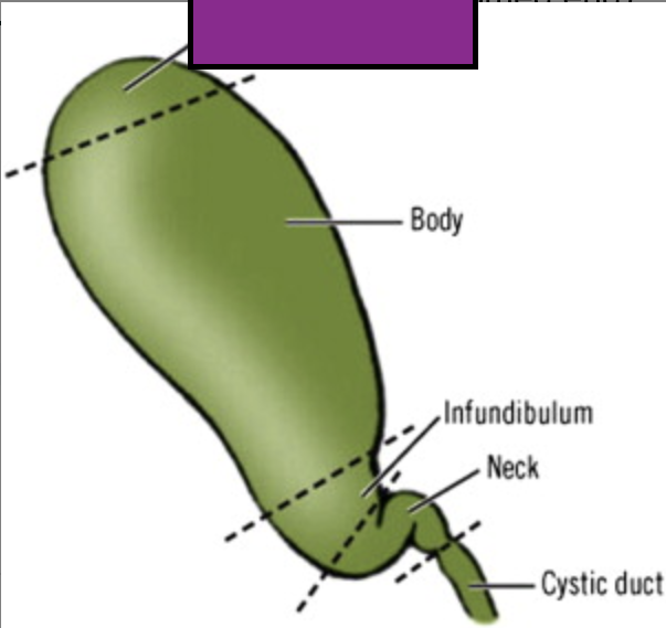
Body of gallbladder
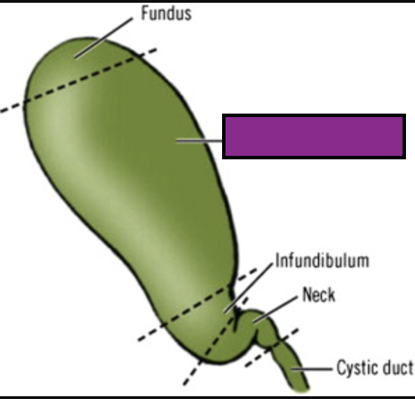
Neck of gallbladder