Genetics BCQ CH18
1/7
There's no tags or description
Looks like no tags are added yet.
Name | Mastery | Learn | Test | Matching | Spaced |
|---|
No study sessions yet.
8 Terms
1: What is the difference between a transition and a transversion? Which type of base substitution is more common?
Transition: A purine (A or G) is changed to the other purine, or a pyrimidine (T or C) is changed to the other pyrimidine.
Transversion: A purine is changed to a pyrimidine, or a pyrimidine is changed to a purine.
Which is more common?
Transitions are more common because it’s easier to chemically change a purine to another purine or a pyrimidine to another pyrimidine than to switch between purines and pyrimidines.
3: What is the difference between a missense mutation and a nonsense mutation? Between a silent mutation and a neutral mutation?
Missense mutation: A base substitution that changes a codon and results in a different amino acid being added to the protein.
Nonsense mutation: A base substitution that changes a codon to a stop codon, leading to early termination of the protein.
Silent mutation: A base substitution that changes the codon but does not change the amino acid (no effect on the protein).
Neutral mutation: A missense mutation where the amino acid change has little or no effect on protein function.
5: How do insertions and deletions arise?
Insertions and deletions arise from:
Strand slippage during DNA replication:
Template strand loop → causes deletion.
New strand loop → causes insertion.
Unequal crossover:
Misalignment at repetitive sequences during crossing over leads to one DNA molecule with an insertion and the other with a deletion.
15: List at least three different types of DNA repair and briefly explain how each is carried out.
Mismatch repair:
Repairs base-pair mismatches and slippage loops.
Mismatch-repair enzymes detect distortions in DNA.
They recognize the new strand by lack of methylation.
Exonucleases remove the error, DNA polymerase fills the gap, and DNA ligase seals the DNA.
Direct repair:
Repairs specific DNA damage like pyrimidine dimers and methylated bases.
Repair enzymes (e.g., photolyase or methyltransferase) directly restore the damaged nucleotide.
Nonhomologous end joining:
Repairs double-strand breaks in DNA without using a homologous template.
Proteins recognize and bind to the broken ends of DNA.
The ends are then joined together.
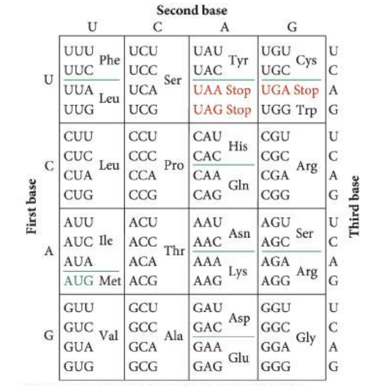
18: A codon that specifies the amino acid Gly undergoes a single-base substitution to become a nonsense mutation. In accord with the genetic code given in Figure 15.10, is this mutation a transition or a transversion? At which position of the codon does the mutation occur?
Gly codons: GGU, GGC, GGA, GGG.
A nonsense mutation occurs when a codon changes to a stop codon (UGA, UAA, UAG).
Only GGA can mutate to a stop codon (UGA) by changing the first position:
GGA → UGA.
The mutation involves a purine (G) changing to a pyrimidine (U), which is a transversion.

20: Hemoglobin is a complex protein that contains four polypeptide chains. The normal hemoglobin found in adults—called adult hemoglobin—consists of two alpha and two beta polypeptide chains, which are encoded by different loci. Sickle-cell hemoglobin, which causes sickle-cell anemia, arises from a mutation in the beta chain of adult hemoglobin. Adult hemoglobin and sickle-cell hemoglobin differ in a single amino acid: the sixth amino acid from one end in adult hemoglobin is glutamic acid, whereas sickle-cell hemoglobin has valine at this position. After consulting the genetic code provided in Figure 15.10, indicate the type and location of the mutation that gave rise to sickle-cell anemia.
Adult hemoglobin: Has glutamic acid at position 6 in the beta chain (codons: GAA or GAG).
Sickle-cell hemoglobin: Has valine at position 6 (codons: GUA or GUG).
The mutation changes GAG (glutamic acid) to GUG (valine), a single-base substitution.
This mutation is a transversion because it changes a purine (G) to a pyrimidine (U).
It is a missense mutation because it changes the amino acid at position 6.
43: A geneticist examines an ear of corn in which most kernels are yellow, but she finds a few kernels with purple spots, as shown here. Give a possible explanation for the appearance of the purple spots in these otherwise yellow kernels, accounting for their different sizes.
Yellow kernels: A Ds element is inserted in a pigment gene, inactivating it.
Ds can't move on its own, so the gene stays off—kernels stay yellow.
Purple spots appear if Ac element is present (from pollen).
Ac activates Ds to jump out of the pigment gene, restoring function.
Spot size depends on when Ds is excised:
Early excision → large purple spots.
Late excision → small purple spots.
48: Tay–Sachs disease (see p. 153 in Chapter 6) is a severe, autosomal recessive genetic disease that produces deafness, blindness, seizures, and, eventually, death at two to three years of age. The disease results from a defect in the HEXA gene, which encodes hexosaminidase A. This enzyme normally degrades GM2 gangliosides. In the absence of hexosaminidase A, GM2 gangliosides accumulate in the brain. The results of molecular studies showed that the most common mutation causing Tay–Sachs disease is a 4-bp insertion that produces a downstream premature stop codon. Results of further studies have revealed that the transcription of the HEXA gene is normal in people who have Tay–Sachs disease, but the HEXA mRNA is unstable. Propose a mechanism to account for how a premature stop codon could cause mRNA instability.
Tay–Sachs disease is caused by a 4-bp insertion in the HEXA gene, leading to a premature stop codon.
This early stop codon triggers nonsense-mediated mRNA decay (NMD).
Normally, ribosomes remove exon junction complexes (EJCs) during translation.
In Tay–Sachs, the stop codon is upstream of the EJC, so the ribosome stops too early and can’t remove the EJC.
Unremoved EJCs signal the cell to destroy the mRNA, making it unstable.