Balance Wk 1 bootcamp LG4
 Call Kai
 Learn
Learn Practice Test
 Spaced Repetition
 Match
Match Flashcards
 Knowt Play
1/175

There's no tags or description
Looks like no tags are added yet.
Name | Mastery | Learn | Test | Matching | Spaced | Call with Kai | Chat |
|---|
No analytics yet
Send a link to your students to track their progress
176 Terms
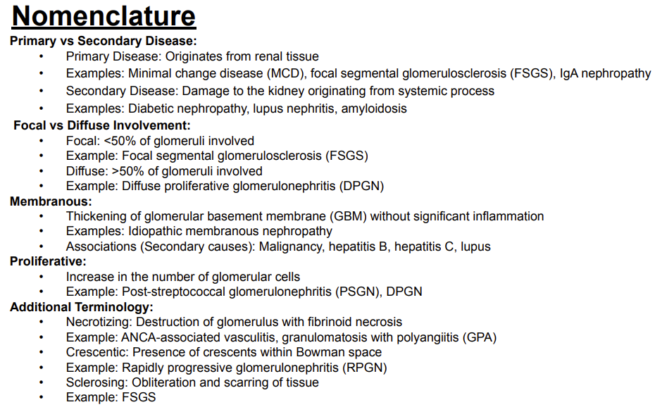
What is a primary renal disease?
Answer: A disease that originates from renal tissue.
Extra Information:
Primary means the disease process begins within the kidney itself.

What are examples of primary renal diseases?
Answer: Minimal change disease, focal segmental glomerulosclerosis, and IgA nephropathy.
Extra Information:
These diseases originate from renal tissue.

What is a secondary renal disease?
Answer: Kidney damage originating from a systemic process.
Extra Information:
The kidney is affected secondary to a disease process originating elsewhere in the body.
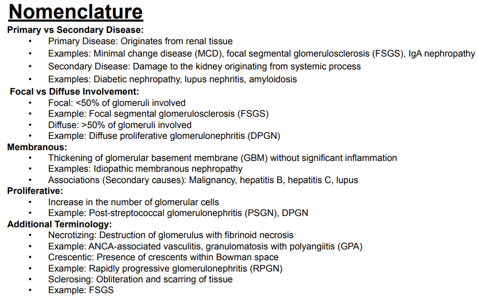
What are examples of secondary renal diseases?
Answer: Diabetic nephropathy, lupus nephritis, and amyloidosis.
Extra Information:
These represent kidney damage caused by systemic processes.

What does focal glomerular involvement mean?
Answer: Less than 50% of the glomeruli are involved.
Extra Information:
Focal segmental glomerulosclerosis is an example.

What disease is an example of focal glomerular involvement?
Answer: Focal segmental glomerulosclerosis.
Extra Information:
Focal means less than 50% of glomeruli are involved.
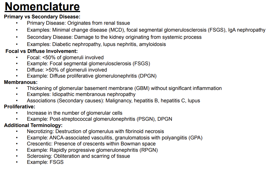
What does diffuse glomerular involvement mean?
Answer: Greater than 50% of the glomeruli are involved.
Extra Information:
Diffuse proliferative glomerulonephritis is an example.

What disease is an example of diffuse glomerular involvement?
Answer: Diffuse proliferative glomerulonephritis.
Extra Information:
Diffuse means greater than 50% of glomeruli are involved.
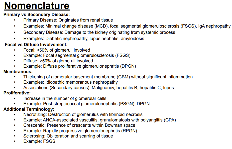
What does membranous mean when describing glomerular disease?
Answer: Thickening of the glomerular basement membrane without significant inflammation.
Extra Information:
Idiopathic membranous nephropathy is an example.

What secondary causes are associated with membranous disease?
Answer: Malignancy, hepatitis B, hepatitis C, and lupus.
Extra Information:
These are the secondary associations listed on the slide.

What does proliferative mean when describing glomerular disease?
Answer: An increase in the number of glomerular cells.
Extra Information:
This describes increased cellularity within the glomerulus.

What diseases are examples of a proliferative glomerular pattern?
Answer: Post-streptococcal glomerulonephritis and diffuse proliferative glomerulonephritis.
Extra Information:
Proliferative means there is an increased number of glomerular cells.

What does necrotizing mean when describing glomerular disease?
Answer: Destruction of the glomerulus with fibrinoid necrosis.
Extra Information:
This describes a destructive pattern of glomerular injury.

What diseases are examples of a necrotizing glomerular pattern?
Answer: ANCA-associated vasculitis and granulomatosis with polyangiitis.
Extra Information:
These are the examples listed for necrotizing glomerular disease.

What does crescentic mean when describing glomerular disease?
Answer: The presence of crescents within Bowman space.
Extra Information:
Rapidly progressive glomerulonephritis is an example.
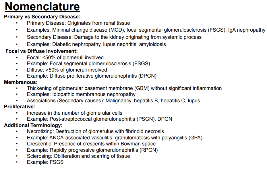
What disease is associated with a crescentic glomerular pattern?
Answer: Rapidly progressive glomerulonephritis.
Extra Information:
Crescentic refers to crescents within Bowman space.

What does sclerosing mean when describing renal pathology?
Answer: Obliteration and scarring of tissue.
Extra Information:
Sclerosis refers to a scarring pattern.
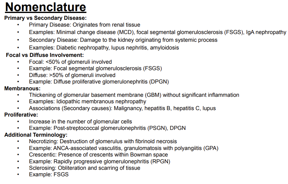
What disease is an example of a sclerosing pattern?
Answer: Focal segmental glomerulosclerosis.
Extra Information:
Sclerosing means obliteration and scarring of tissue.
What urine color is considered normal on gross examination?
Answer: Clear/yellow.
Extra Information:
Changes in urine color can suggest different underlying substances or conditions.

What can red, pink, or dark urine indicate?
Answer: Hematuria, myoglobinuria, or beets.
Extra Information:
These are the causes associated with red, pink, or dark urine on the slide.

What can dark brown urine indicate?
Answer: Liver disease or bilirubinuria.
Extra Information:
Dark brown urine is distinguished from red/pink urine and black urine on gross examination.

What condition is associated with black urine?
Answer: Alkaptonuria.
Extra Information:
This is the specific association for black urine on the slide.

What does cloudy urine suggest?
Answer: Possible infection or pyuria.
Extra Information:
Clear urine is considered normal on gross examination.
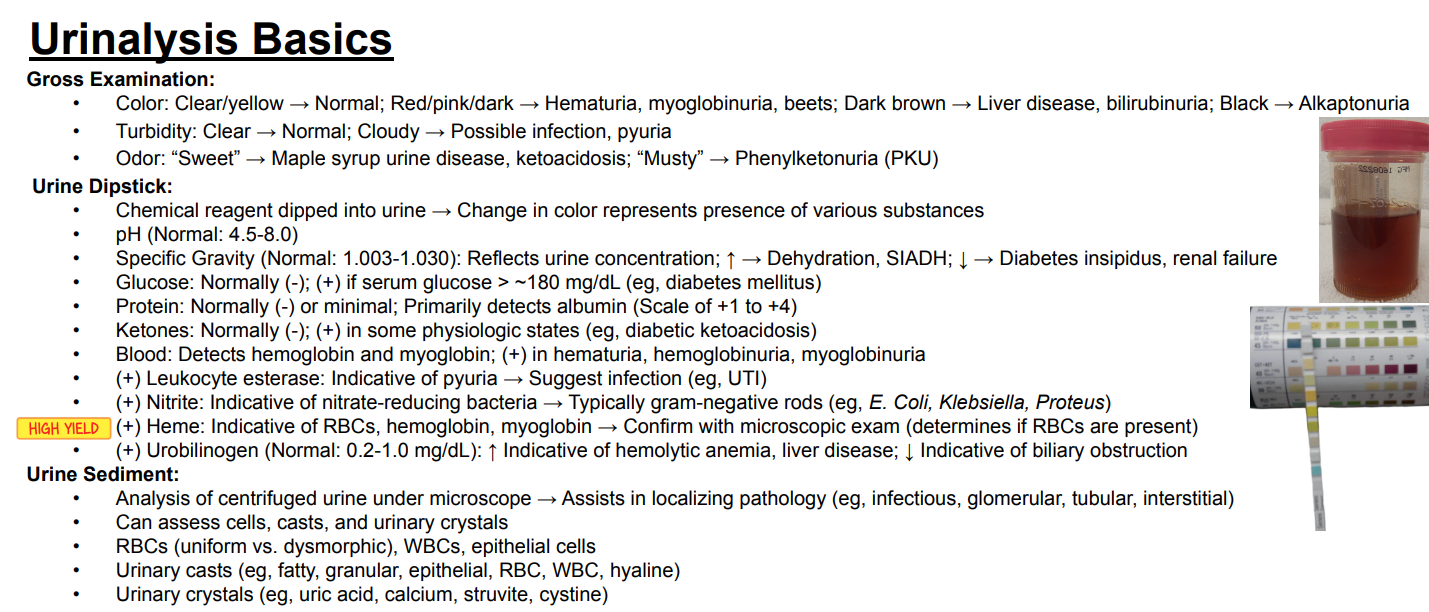
What conditions are associated with a “sweet” urine odor?
Answer: Maple syrup urine disease and ketoacidosis.
Extra Information:
Urine odor can provide clues to underlying metabolic abnormalities.

What condition is associated with a “musty” urine odor?
Answer: Phenylketonuria.
Extra Information:
Phenylketonuria is abbreviated PKU.
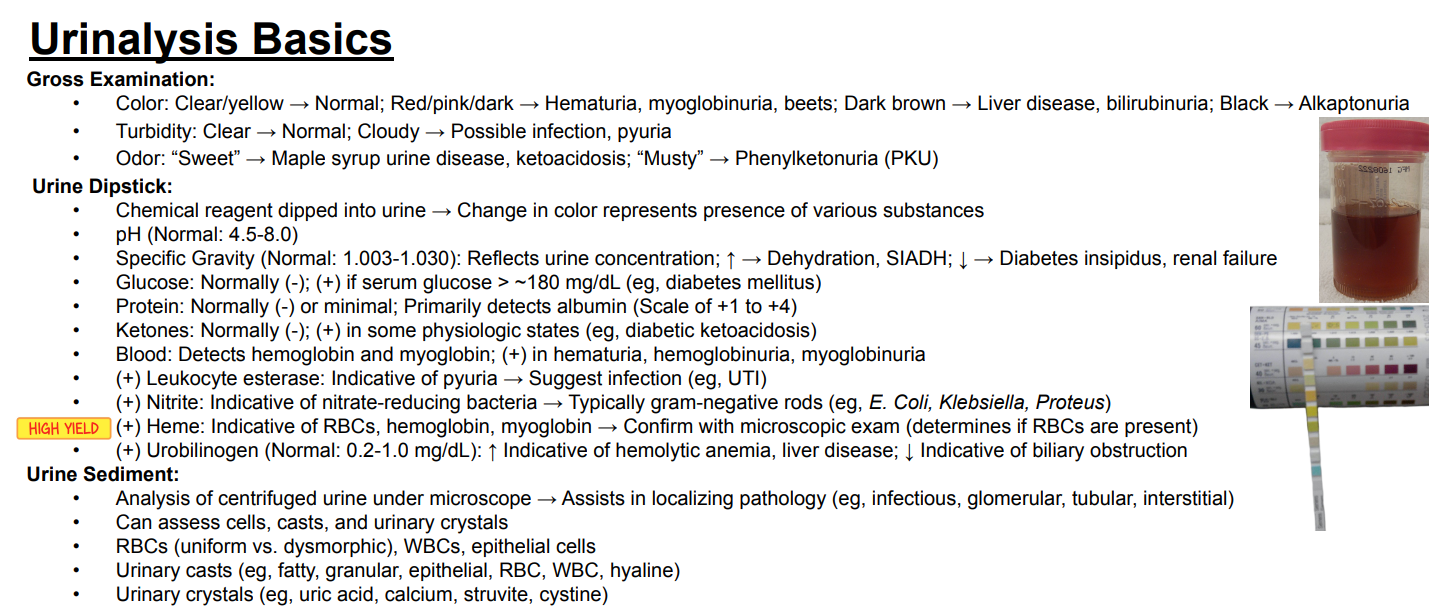
How does a urine dipstick detect substances in urine?
Answer: A chemical reagent is dipped into urine, and a color change represents the presence of various substances.
Extra Information:
Different dipstick components test different chemical properties of urine.

What is the normal urine pH range?
Answer: 4.5–8.0.
Extra Information:
Urine pH is one of the measurements obtained from a urine dipstick.

What is the normal urine specific gravity range?
Answer: 1.003–1.030.
Extra Information:
Specific gravity reflects urine concentration.

What does urine specific gravity reflect?
Answer: Urine concentration.
Extra Information:
The normal range on the slide is 1.003–1.030.
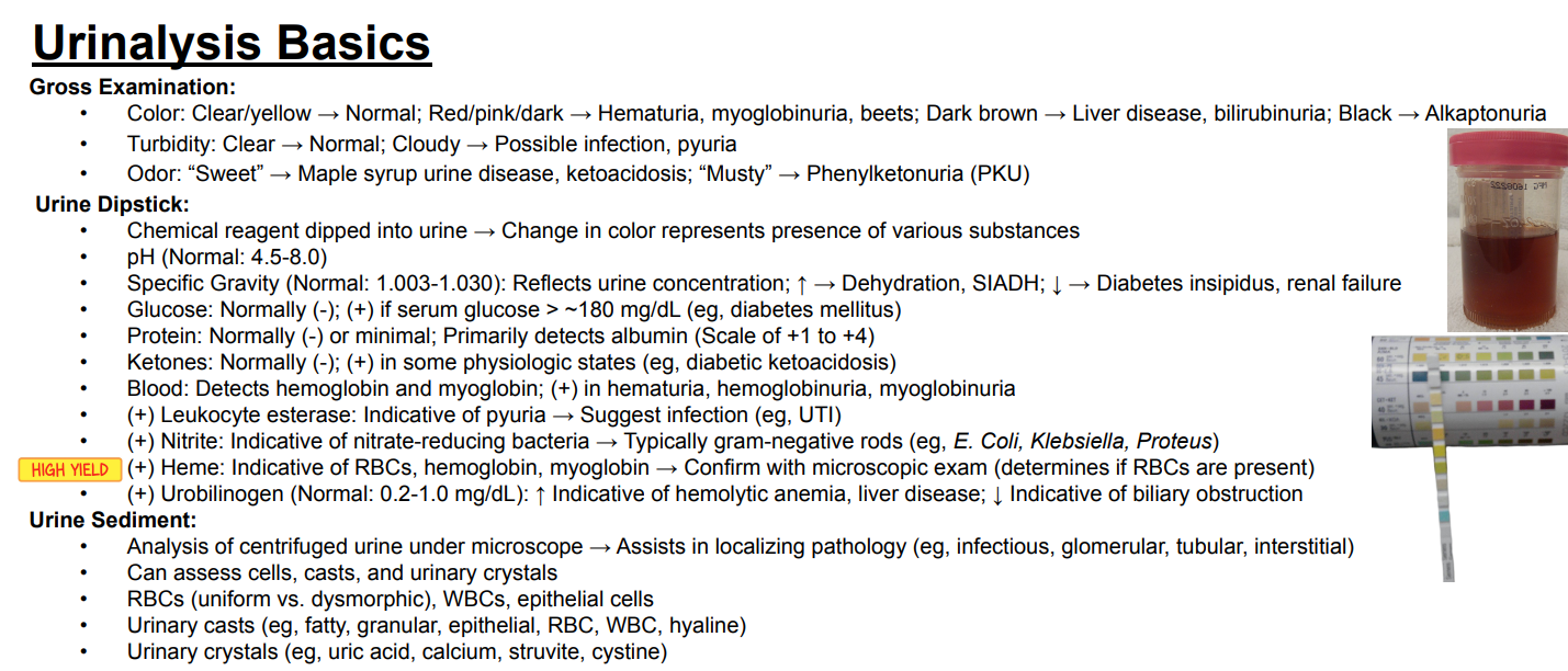
What conditions are associated with increased urine specific gravity?
Answer: Dehydration and syndrome of inappropriate antidiuretic hormone secretion.
Extra Information:
Increased specific gravity reflects more concentrated urine.
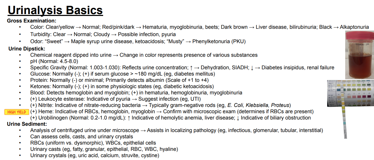
What conditions are associated with decreased urine specific gravity?
Answer: Diabetes insipidus and renal failure.
Extra Information:
Decreased specific gravity reflects less concentrated urine.
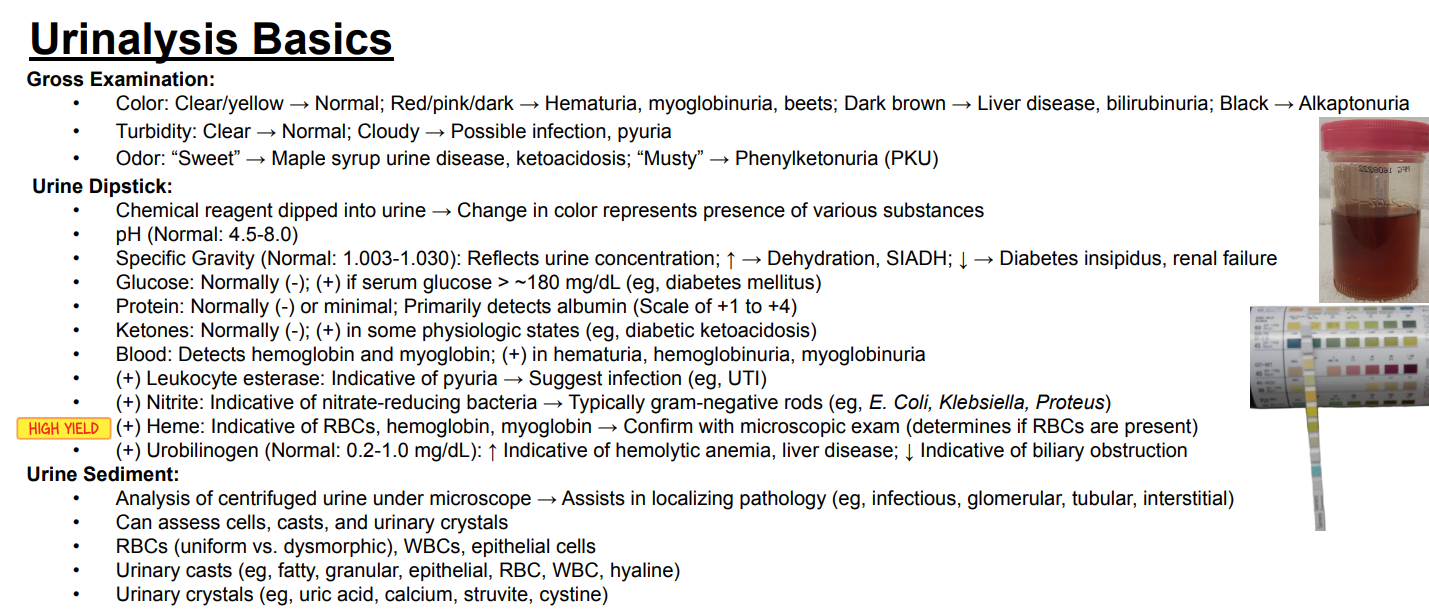
When does glucose become positive on urine dipstick according to the slide?
Answer: When serum glucose exceeds approximately 180 mg/dL.
Extra Information:
Urine glucose is normally negative.
Diabetes mellitus is an example associated with a positive result.
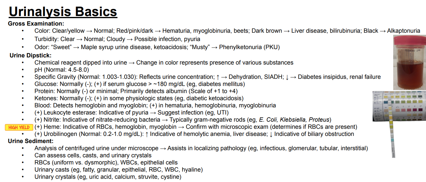
What does the protein portion of a urine dipstick primarily detect?
Answer: Albumin.
Extra Information:
Urine protein is normally negative or minimal.
Protein is graded on a scale from +1 to +4.

What is the normal urine ketone result, and when can ketones become positive?
Answer: Ketones are normally negative but can become positive in states such as diabetic ketoacidosis.
Extra Information:
Ketones are measured using the urine dipstick.

What does the blood portion of a urine dipstick detect?
Answer: Hemoglobin and myoglobin.
Extra Information:
It can be positive with hematuria, hemoglobinuria, or myoglobinuria.

What does a positive leukocyte esterase indicate?
Answer: Pyuria, which suggests infection.
Extra Information:
A urinary tract infection is an example given on the slide.
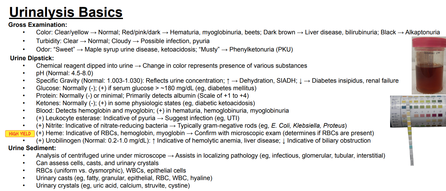
What does a positive urine nitrite test indicate?
Answer: Nitrate-reducing bacteria.
Extra Information:
These are typically gram-negative rods.
Examples on the slide include E. coli, Klebsiella, and Proteus.

What can cause a positive urine heme test?
Answer: Red blood cells, hemoglobin, or myoglobin.
Extra Information:
A positive heme result should be confirmed with microscopic examination.
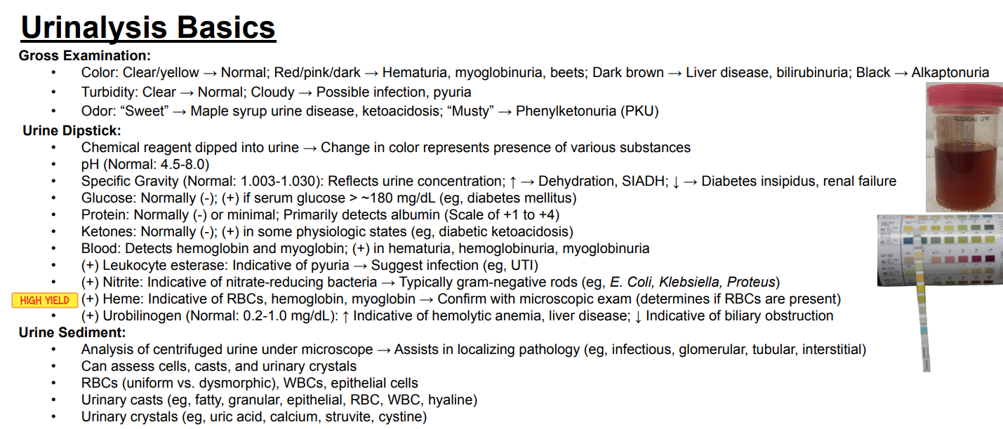
Why is microscopic examination performed after a positive urine heme result?
Answer: To determine whether red blood cells are actually present.
Extra Information:
Dipstick heme can be positive from red blood cells, free hemoglobin, or myoglobin.
Microscopy helps distinguish these possibilities by identifying red blood cells.
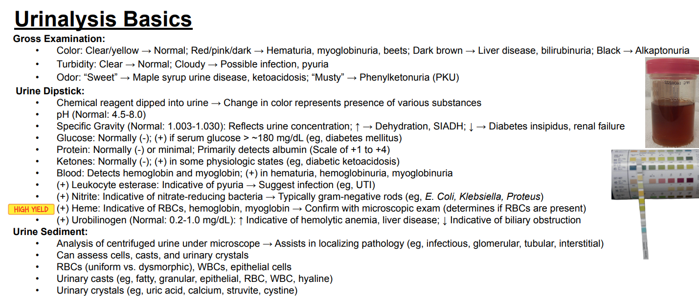
What is the normal urine urobilinogen range, and what do increased versus decreased levels suggest?
Answer: Normal is 0.2–1.0 mg/dL. Increased urobilinogen suggests hemolytic anemia or liver disease, while decreased urobilinogen suggests biliary obstruction.
Extra Information:
Urobilinogen is measured as part of urine dipstick testing.

What is urine sediment analysis, and what can it evaluate?
Answer: It is microscopic analysis of centrifuged urine that evaluates cells, casts, and urinary crystals.
Extra Information:
Cells include uniform or dysmorphic red blood cells, white blood cells, and epithelial cells.
Casts include fatty, granular, epithelial, red blood cell, white blood cell, and hyaline casts.
Crystals include uric acid, calcium, struvite, and cystine crystals.
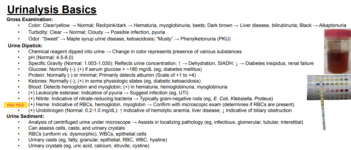
What is the clinical purpose of urine sediment analysis?
Answer: It helps localize urinary pathology.
Extra Information:
The slide lists infectious, glomerular, tubular, and interstitial pathology as examples.
Microscopy evaluates findings such as cells, casts, and crystals.

What are fatty casts composed of?
Answer: Lipid-laden renal tubular epithelial cells.
Extra Information:
Fatty casts are associated with heavy proteinuria and lipiduria.
What characteristic appearance is associated with fatty casts under polarized light?
Answer: A “Maltese cross” appearance.
Extra Information:
This finding is associated with lipid-containing fatty casts.

Which conditions are associated with fatty casts?
Answer: Nephrotic syndromes such as minimal change disease, focal segmental glomerulosclerosis, and membranous nephropathy.
Extra Information:
Fatty casts indicate heavy proteinuria and lipiduria.
What are granular casts?
Answer: Degenerated cellular casts, often derived from epithelial cells.
Extra Information:
They indicate tubular cell injury and necrosis.

What is the significance of “muddy brown” granular casts?
Answer: They are specific for acute tubular necrosis.
Extra Information:
Granular casts reflect tubular cell injury and necrosis.

What are hyaline casts composed of?
Answer: Tamm-Horsfall protein secreted by tubular epithelial cells.
Extra Information:
Hyaline casts are nonspecific.
When can hyaline casts be seen?
Answer: They can occur in healthy individuals, such as with dehydration, and may also occur in kidney disease.
Extra Information:
Their presence may indicate concentrated urine.
They are nonspecific.

Which urinary cast is nonspecific and may indicate concentrated urine?
Answer: Hyaline casts.
Extra Information:
They may be seen with dehydration.
They are composed of Tamm-Horsfall protein.

What are red blood cell casts composed of?
Answer: Red blood cells and mucoproteins.
Extra Information:
Their presence indicates bleeding originating from the glomerulus.

Which conditions are associated with red blood cell casts?
Answer: Glomerulonephritis and hypertensive emergency.
Extra Information:
Examples of glomerulonephritis on the slide include post-streptococcal glomerulonephritis and lupus nephritis.

What do red blood cell casts indicate about the location and mechanism of kidney injury?
Answer: Glomerular bleeding with disruption of the glomerular basement membrane.
Extra Information:
Red blood cell casts therefore point toward glomerular pathology.
What are white blood cell casts composed of?
Answer: White blood cells and Tamm-Horsfall protein.
Extra Information:
They indicate inflammation or infection within the renal interstitium or tubules.

Which conditions are associated with white blood cell casts?
Answer: Acute pyelonephritis, acute interstitial nephritis, and transplant rejection.
Extra Information:
These conditions involve inflammation or infection within the renal interstitium or tubules.

What do white blood cell casts indicate?
Answer: Inflammation or infection within the renal interstitium or tubules.
Extra Information:
Acute pyelonephritis and acute interstitial nephritis are important associations.

What are waxy casts?
Answer: Degenerative granular casts.
Extra Information:
They are associated with advanced or chronic kidney disease on the slide.
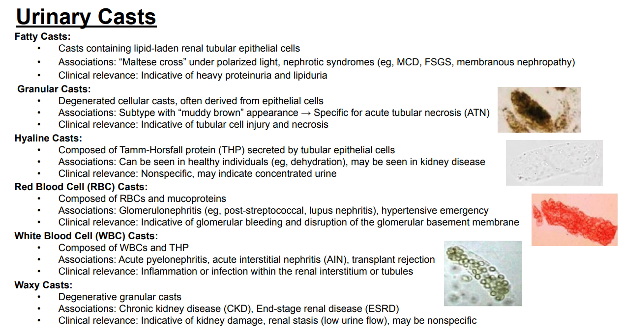
Which conditions are associated with waxy casts?
Answer: Chronic kidney disease and end-stage renal disease.
Extra Information:
Waxy casts can indicate kidney damage and renal stasis.

What is the clinical significance of waxy casts?
Answer: They can indicate kidney damage and renal stasis from low urine flow.
Extra Information:
Waxy casts may also be nonspecific.
They are associated with chronic kidney disease and end-stage renal disease.

Match the major urinary cast types with their key associations.
Answer: Fatty → nephrotic syndromes. Muddy brown granular → acute tubular necrosis. Hyaline → dehydration or concentrated urine. Red blood cell → glomerulonephritis. White blood cell → pyelonephritis or interstitial nephritis. Waxy → chronic kidney disease or end-stage renal disease.
Extra Information:
This card integrates the major cast-to-disease relationships from the slide.
Use the individual cards to recall each cast’s composition and clinical significance.

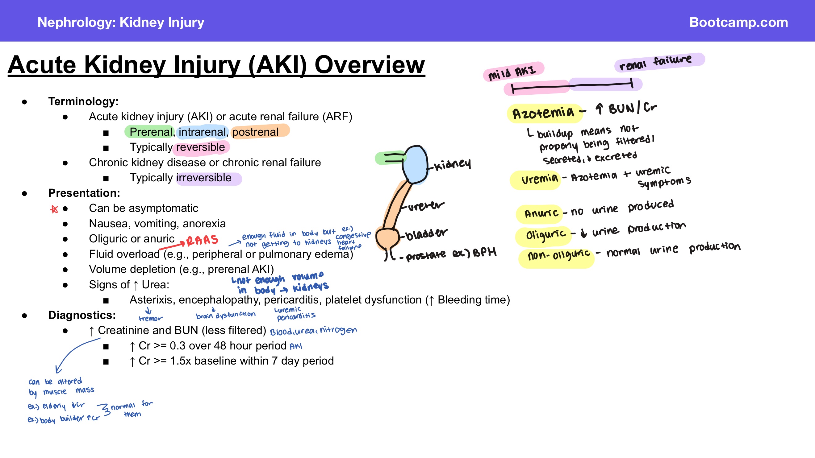
What are the two terms used on this slide for an acute decline in kidney function?
Answer: Acute kidney injury and acute renal failure.
Extra Information:
Acute kidney injury is abbreviated AKI.
Acute renal failure is abbreviated ARF.

What are the three major categories of acute kidney injury?
Answer: Prerenal, intrarenal, and postrenal.
Extra Information:
- These categories are used to classify acute kidney injury.

How does the reversibility of acute kidney injury generally compare with chronic kidney disease?
Answer: Acute kidney injury is typically reversible, while chronic kidney disease is typically irreversible.
Extra Information:
- The slide also refers to chronic kidney disease as chronic renal failure.
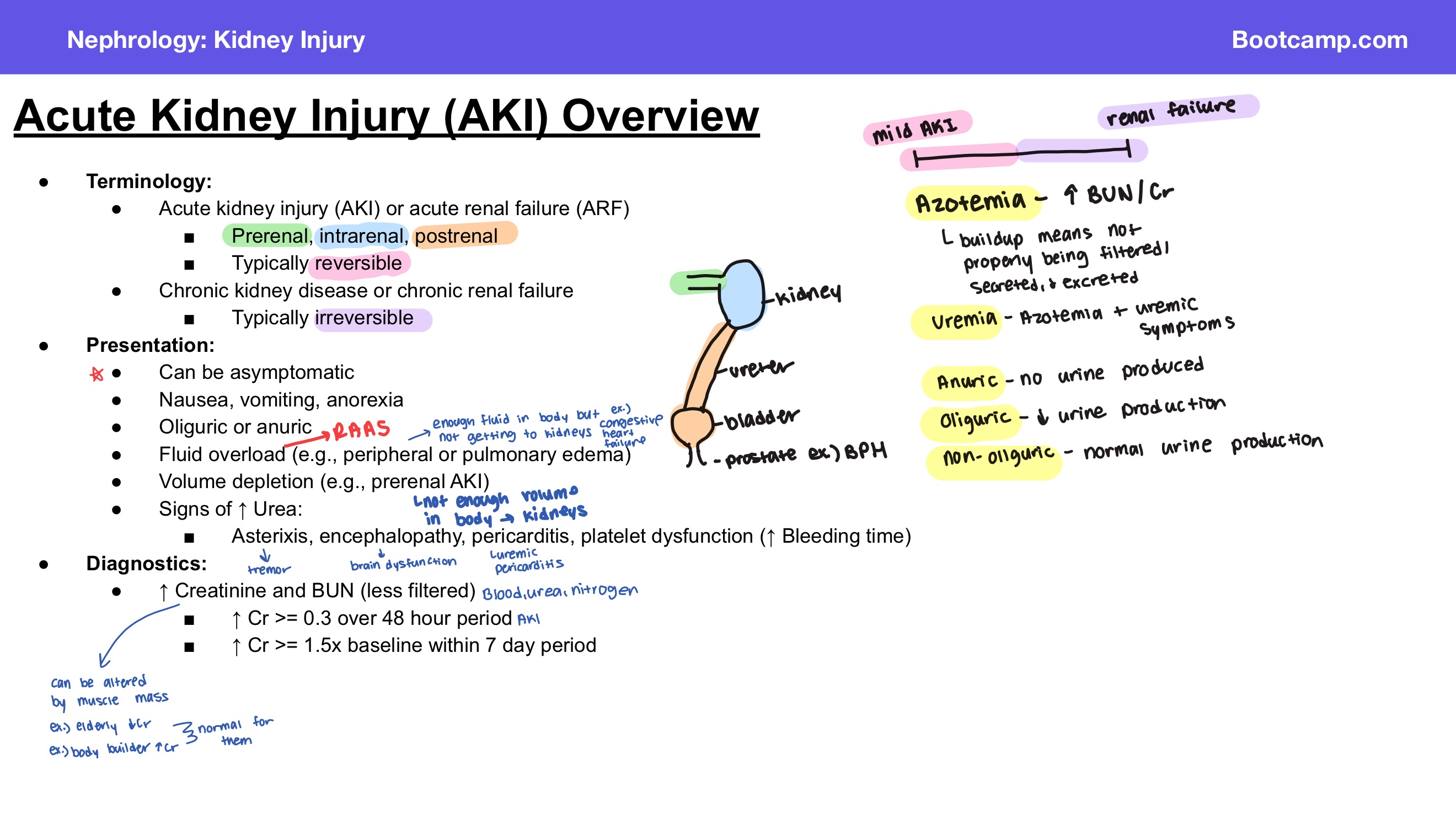
Can acute kidney injury be asymptomatic?
Answer: Yes, acute kidney injury can be asymptomatic.
Extra Information:
- When symptoms occur, the presentation can vary.

What gastrointestinal symptoms can occur with acute kidney injury?
Answer: Nausea, vomiting, and anorexia.
Extra Information:
- These are listed among the possible presentations of acute kidney injury.

What changes in urine output can occur with acute kidney injury?
Answer: Patients can be oliguric or anuric.
Extra Information:
- These terms describe reduced or absent urine output.

What findings can result from fluid overload in acute kidney injury?
Answer: Peripheral edema or pulmonary edema.
Extra Information:
- Fluid overload is one possible presentation of acute kidney injury.

Which type of acute kidney injury can present with volume depletion?
Answer: Prerenal acute kidney injury.
Extra Information:
- Volume depletion is given as an example associated with prerenal acute kidney injury.

What neurologic findings can occur with increased urea in acute kidney injury?
Answer: Asterixis and encephalopathy.
Extra Information:
- These are listed as signs associated with increased urea.
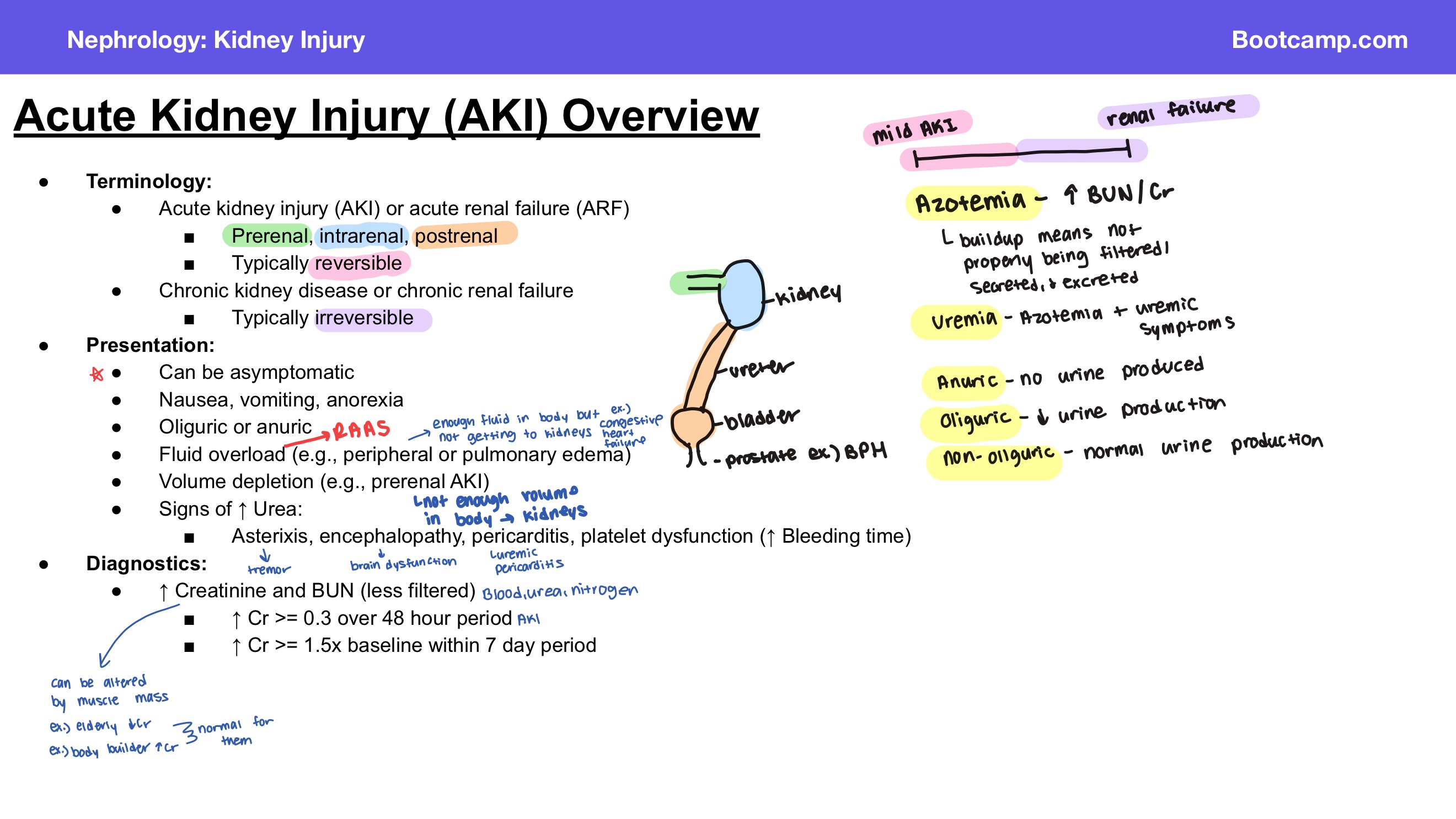
What cardiac finding can occur with increased urea in acute kidney injury?
Answer: Pericarditis.
Extra Information:
- Pericarditis is one of the signs associated with increased urea on the slide.

How can increased urea affect platelet function?
Answer: It can cause platelet dysfunction with increased bleeding time.
Extra Information:
- Platelet dysfunction is one of the signs associated with increased urea.

What laboratory values increase in acute kidney injury because less is filtered?
Answer: Creatinine and blood urea nitrogen.
Extra Information:
- Both increase as kidney filtration decreases.

What rise in creatinine over 48 hours meets the diagnostic criterion for acute kidney injury on this slide?
Answer: An increase in creatinine of at least 0.3 mg/dL over 48 hours.
Extra Information:
- This is one of the two creatinine-based diagnostic criteria listed on the slide.

What change in creatinine within 7 days meets the diagnostic criterion for acute kidney injury on this slide?
Answer: An increase in creatinine to at least 1.5 times baseline within 7 days.
Extra Information:
- This is the second creatinine-based diagnostic criterion listed on the slide.
What is the basic pathophysiology of prerenal acute kidney injury?
Answer: Decreased cardiac output and/or decreased effective circulating volume → decreased renal blood flow and glomerular filtration rate → renin-angiotensin-aldosterone system activation → decreased urinary sodium and water → increased urine osmolality.
Extra Information:
The kidney responds to decreased perfusion by conserving sodium and water.

What happens to renal blood flow and glomerular filtration rate in prerenal acute kidney injury?
Answer: Both renal blood flow and glomerular filtration rate decrease.
Extra Information:
- This occurs because of decreased cardiac output and/or decreased effective circulating volume.

Why is the renin-angiotensin-aldosterone system activated in prerenal acute kidney injury?
Answer: Decreased renal perfusion from decreased cardiac output and/or decreased effective circulating volume activates the renin-angiotensin-aldosterone system.
Extra Information:
- Activation leads to decreased urinary sodium and water.

How does prerenal acute kidney injury affect urinary sodium, urinary water, and urine osmolality?
Answer: Urinary sodium and water decrease, while urine osmolality increases.
Extra Information:
- These changes occur as the kidney attempts to conserve sodium and water.
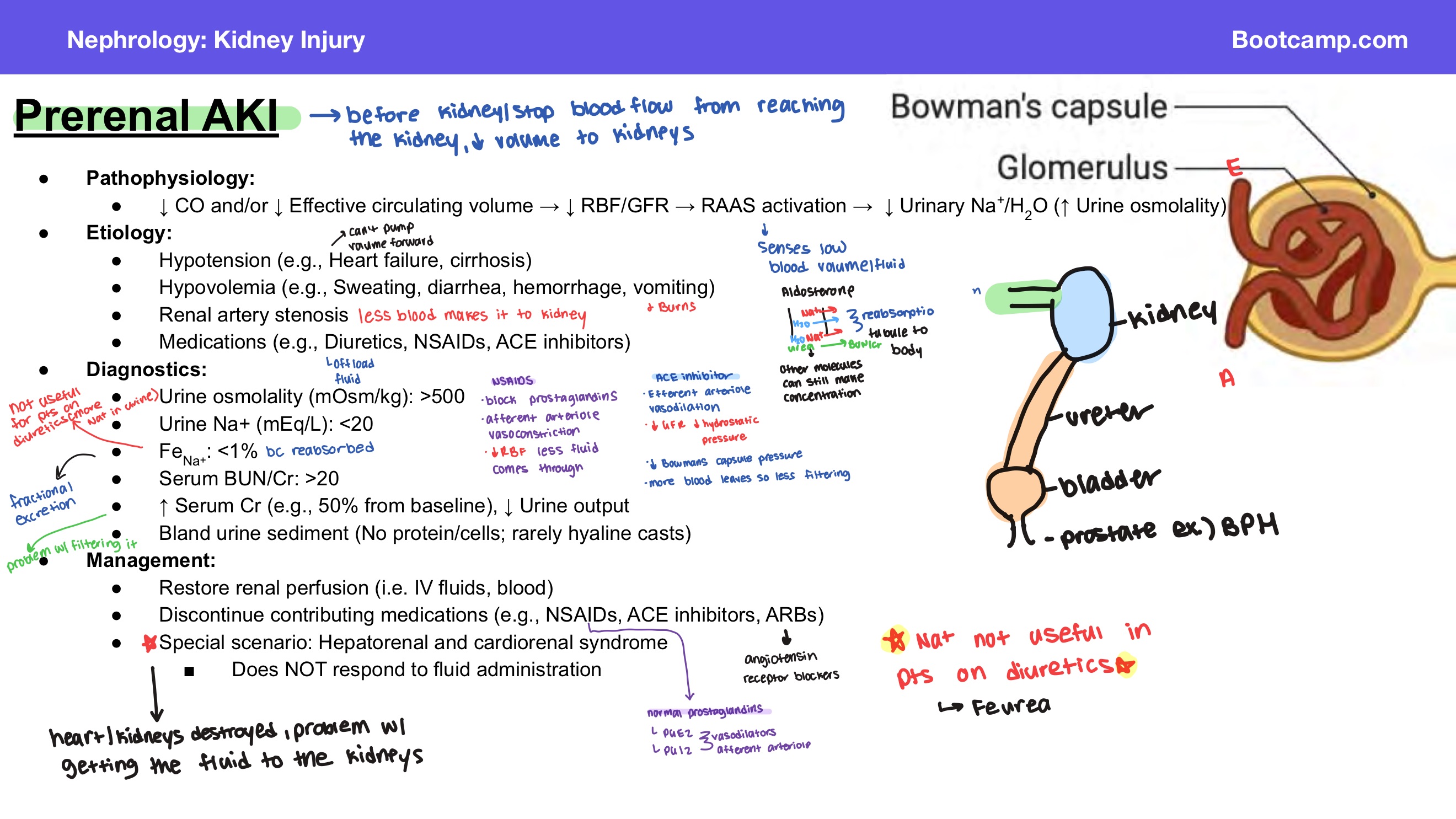
What hypotensive conditions can cause prerenal acute kidney injury according to the slide?
Answer: Heart failure and cirrhosis.
Extra Information:
- Hypotension is one cause of decreased renal perfusion.

What hypovolemic conditions can cause prerenal acute kidney injury?
Answer: Sweating, diarrhea, hemorrhage, and vomiting.
Extra Information:
- Hypovolemia decreases effective circulating volume and renal perfusion.

What vascular condition can cause prerenal acute kidney injury?
Answer: Renal artery stenosis.
Extra Information:
- Renal artery stenosis is listed as an etiology of prerenal acute kidney injury.

Which medications can contribute to prerenal acute kidney injury?
Answer: Diuretics, nonsteroidal anti-inflammatory drugs, and angiotensin-converting enzyme inhibitors.
Extra Information:
- Contributing medications may need to be discontinued during management.

What urine osmolality is expected in prerenal acute kidney injury?
Answer: Greater than 500 mOsm/kg.
Extra Information:
- The urine is relatively concentrated in prerenal acute kidney injury.

What urine sodium concentration is expected in prerenal acute kidney injury?
Answer: Less than 20 mEq/L.
Extra Information:
- The kidney conserves sodium in response to decreased renal perfusion.

What fractional excretion of sodium is expected in prerenal acute kidney injury?
Answer: Less than 1%.
Extra Information:
- Fractional excretion of sodium is abbreviated FeNa.

What serum blood urea nitrogen-to-creatinine ratio is expected in prerenal acute kidney injury?
Answer: Greater than 20.
Extra Information:
- This is one of the diagnostic findings listed on the slide.

What happens to serum creatinine and urine output in prerenal acute kidney injury?
Answer: Serum creatinine increases and urine output decreases.
Extra Information:
- The slide gives an approximately 50% increase in serum creatinine from baseline as an example.

What urine sediment is expected in prerenal acute kidney injury?
Answer: Bland urine sediment.
Extra Information:
- There is no protein or cells.
- Hyaline casts may rarely be present.

What is the main management goal in prerenal acute kidney injury?
Answer: Restore renal perfusion.
Extra Information:
- The slide gives intravenous fluids and blood as examples.
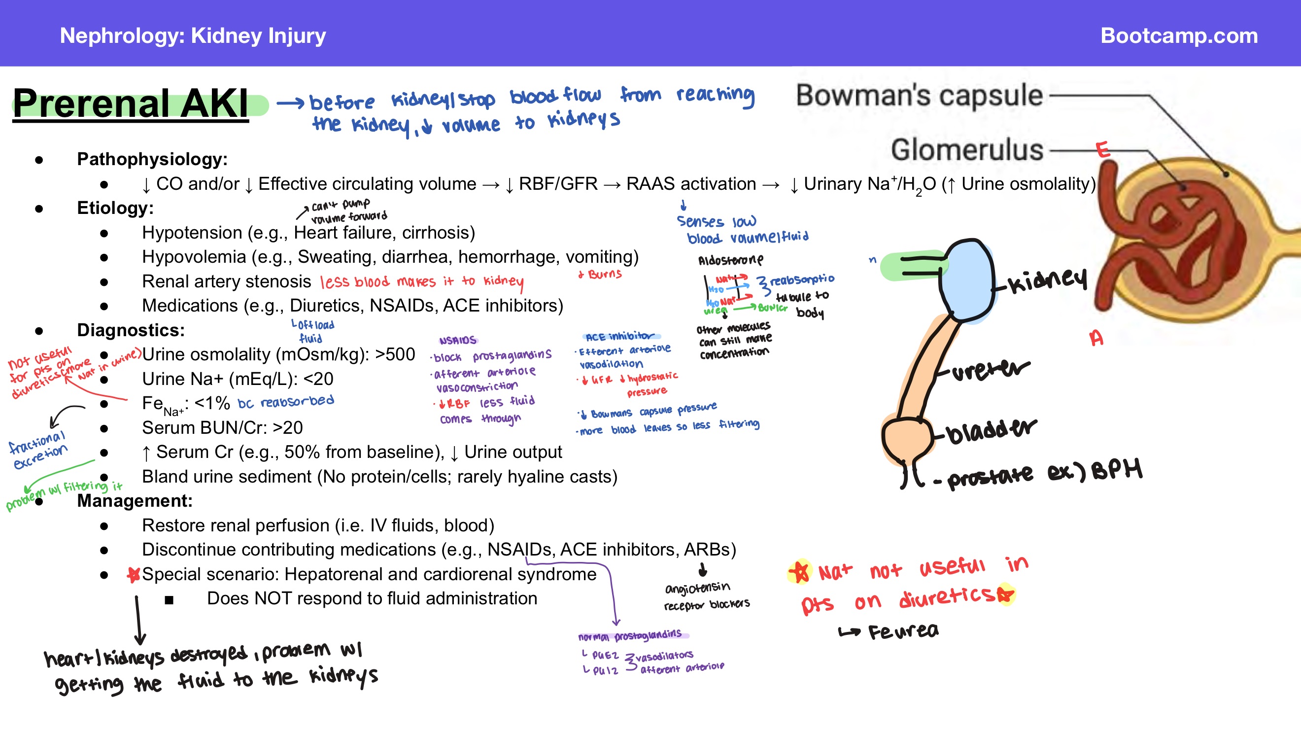
Which contributing medications should be discontinued in prerenal acute kidney injury according to the slide?
Answer: Nonsteroidal anti-inflammatory drugs, angiotensin-converting enzyme inhibitors, and angiotensin receptor blockers.
Extra Information:
- Discontinuing contributing medications is part of management.

Which special prerenal acute kidney injury syndromes do not respond to fluid administration?
Answer: Hepatorenal syndrome and cardiorenal syndrome.
Extra Information:
- These are identified as special scenarios on the slide.

What is the classic diagnostic pattern for prerenal acute kidney injury?
Answer: Urine osmolality greater than 500 mOsm/kg, urine sodium less than 20 mEq/L, fractional excretion of sodium less than 1%, serum blood urea nitrogen-to-creatinine ratio greater than 20, increased serum creatinine, decreased urine output, and bland urine sediment.
Extra Information:
- Think of the overall pattern as the kidney conserving sodium and water in response to decreased renal perfusion.

What is the basic pathophysiology of intrarenal acute kidney injury?
Answer: Renal tubular damage → decreased reabsorption capacity → increased urinary sodium and water → decreased urine osmolality.
Extra Information:
Damaged renal tubules cannot reabsorb sodium and water as effectively.

How does intrarenal acute kidney injury affect urinary sodium, urinary water, and urine osmolality?
Answer: Urinary sodium and water increase, while urine osmolality decreases.
Extra Information:
- This results from decreased tubular reabsorption capacity.
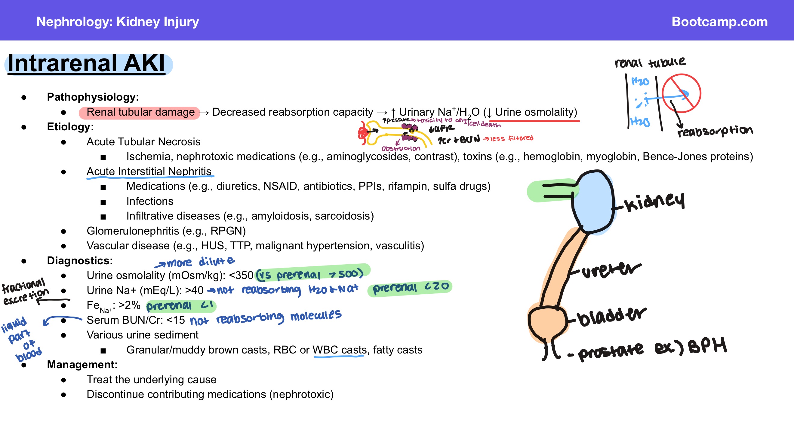
What are the four major categories of causes of intrarenal acute kidney injury on the slide?
Answer: Acute tubular necrosis, acute interstitial nephritis, glomerulonephritis, and vascular disease.
Extra Information:
- Each represents injury occurring within the kidney itself.

What can cause acute tubular necrosis?
Answer: Ischemia, nephrotoxic medications, and toxins.
Extra Information:
- Acute tubular necrosis is one cause of intrarenal acute kidney injury.

Which nephrotoxic medications are associated with acute tubular necrosis on the slide?
Answer: Aminoglycosides and contrast.
Extra Information:
These are examples of nephrotoxic causes of acute tubular necrosis.

Which toxins are associated with acute tubular necrosis on the slide?
Answer: Hemoglobin, myoglobin, and Bence-Jones proteins.
Extra Information:
- These are listed as toxin-related causes of acute tubular necrosis.

What are the major causes of acute interstitial nephritis on the slide?
Answer: Medications, infections, and infiltrative diseases.
Extra Information:
- Acute interstitial nephritis is one cause of intrarenal acute kidney injury.
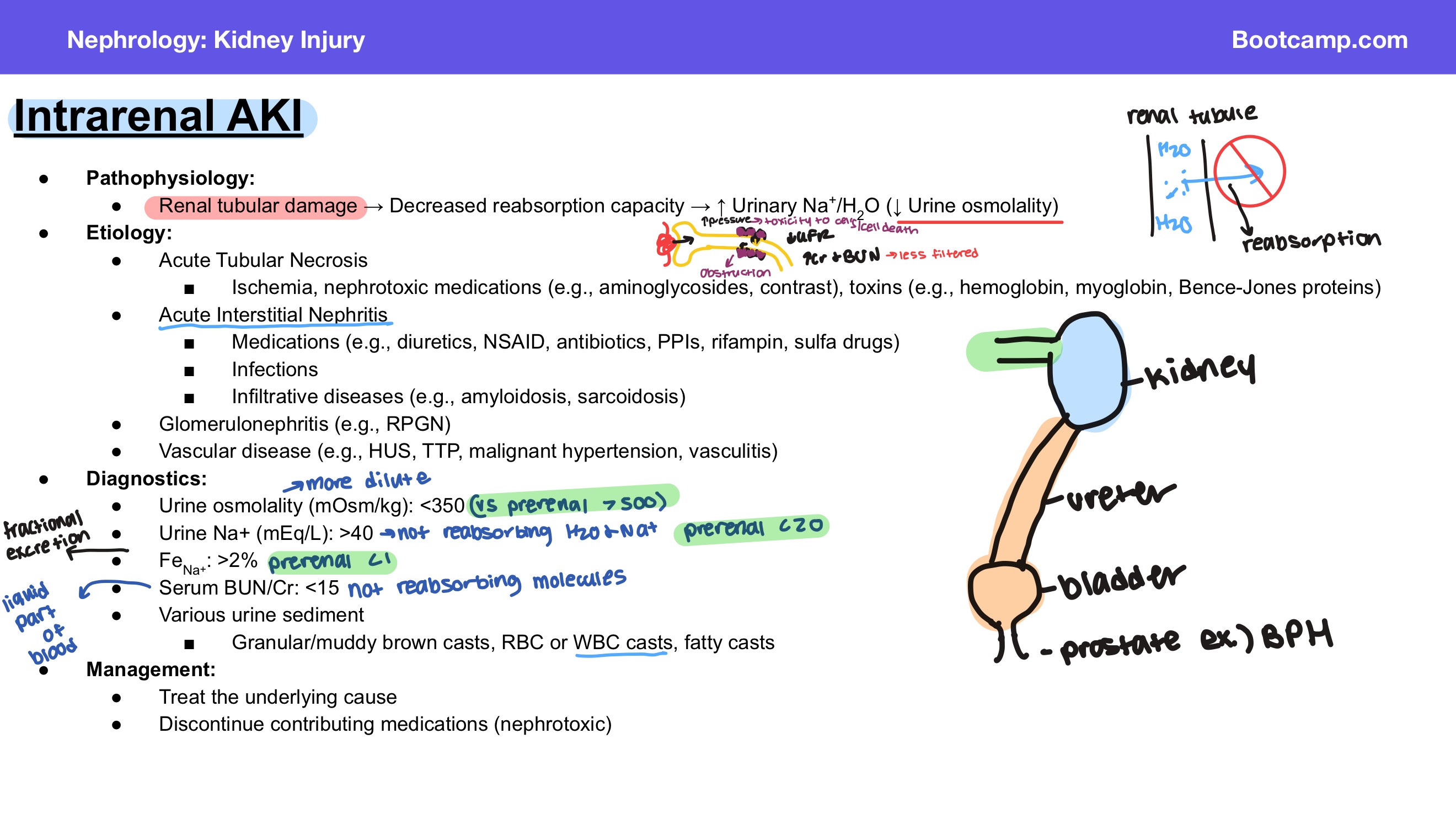
Which medications can cause acute interstitial nephritis according to the slide?
Answer: Diuretics, nonsteroidal anti-inflammatory drugs, antibiotics, proton pump inhibitors, rifampin, and sulfa drugs.
Extra Information:
- These are the medication examples specifically listed on the slide.
