LIFESCI 3K03 ALL
 Call Kai
 Learn
Learn Practice Test
 Spaced Repetition
 Match
Match Flashcards
 Knowt Play
1/265

There's no tags or description
Looks like no tags are added yet.
Name | Mastery | Learn | Test | Matching | Spaced | Call with Kai | Chat |
|---|
No analytics yet
Send a link to your students to track their progress
266 Terms
What are the three stages + areas of control of movement and what do they do?
Determines what needs to be done (Ready) - Prefrontal Cortex
Identifies the goal of the movement and best strategy to accomplish this goal
Plans the specific movement (Set) - Motor Cortex
Specific sequences of muscle activations and patterns required to do the movement
Execute the plan (Go) - Spinal Cord
Activation of the motor neurons to do the movement and make minor adjustments
What is the range of complexity in the 3 stages of control of movement?

What does the Prefrontal Cortex do?
Highest in the chain of command and greatest complexity
Identifies a goal and determines what needs to be done to accomplish this
“What you need to do”
Highly connected with sensory cortex
Executive Function (EF)
Higher cognitive processes for planning, organizing, and controlling thoughts, speech, and behaviors
Involves a wide-range of skills

What are the two components of Executive Function and Movement?
Goal-directed actions
Organizing
What is the goal and how does this relate to the current sensory state?
Planning
What will need to be done to accomplish the goal?
Directing
Sending this information to the next processing station (i.e., motor cortex)
Attention
Multitasking
Allocating attention among tasks performed simultaneously
Response inhibition
Respond effectively with distractions/irrelevant information
Similar to a second level of “sensory gating” (i.e., thalamus filtering)
How is EF and aging related?
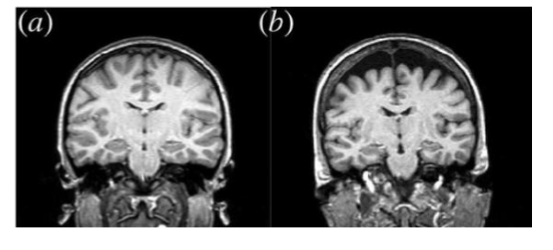
Executive function declines with age
Lesions in white matter
Loss of gray matter
Loss of dendritic branching
Changes are highly variable
Decline can be minimal in healthy aging
Influenced by things like lifestyle, education, genetics, etc.
What are the age related declines related to EF?
Aging related declines in:
Overall processing speed
Problem solving ability
Organizing, planning, directing (Goal-directed actions)
Controlling attentional resources
Multitasking and response inhibition (Attention)
Declines are not to a level of dysfunction in healthy aging
What are the changes in gait parameters with age?
In general, with aging we see:
Decrease in gait speed
Decrease in step length
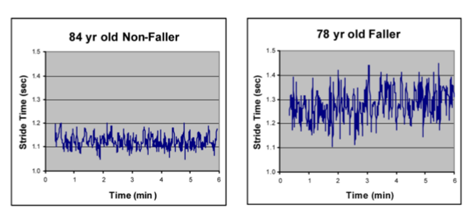
Increase in step time
Increase in variability in these parameters
However, healthy older adults may have little to no change
Reduced EF may be an important driver of these changes
What is Gait in relation to executive function?
Gait is a complex motor task that uses executive function
Not fully managed by CPGs (especially in humans)
EF needed to plan, organize, and direct of movements
Often must also divide attention to other tasks
EF allows effective division of attention between gait and other tasks
Dual-task or Multi-tasking (e.g., Walk and talk/text)
What are the important parts of the motor cortex in relation to control of movement?
Motor cortex is involved after you have decided what you need to do, in order to decide how you will do it
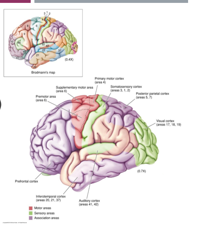
Supplementary Motor Area (SMA)
Premotor Area (PMA)
Primary Motor Cortex (M1)

How are the PMA & SMA involved in the planning of movement?
Created movement plans and holds them until ready to execute
Active just BEFORE movement occurs
Up to 1s before voluntary movement
Plans must be highly integrated with sensory information
Details of coding taking place remains unclear
Premotor Area (PMA)
Selection of best motor plans based on current sensory information (e.g. picking up an object)
Supplementary Motor Area (SMA)
More complex motor sequences often with bilateral connections
May be more internally driven (remembered sequences) (e.g. performing a dance)
How was the planning of movement tested?
Through experiment with monkeys
Measuring the planning of movement
Instruction stimulus
Red light where movement will need to be
PMA neuron begins firing
Trigger stimulus
Blue light tell it to act
PMA neuron stops firing soon after the action is made
Specific to the side required for movement

What is active when planning (or even watching) movement?
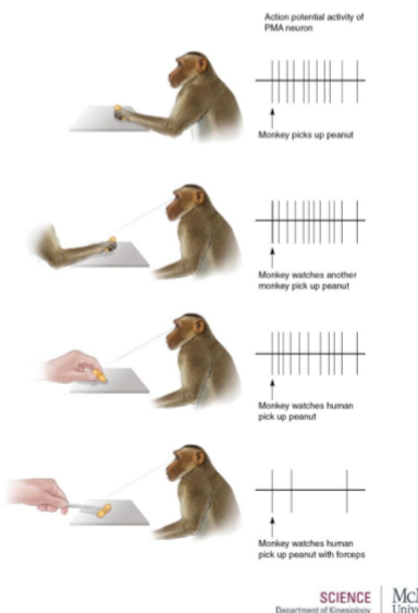
“Mirror neurons” exist in the PMA
Respond when movement is imagined or watched
May be part of an extensive brain system for understanding actions and intentions of others
Monkey’s motor neurons didn’t activate when seeing the peanut being picked up by a tool instead of the monkey hand or human hand
What does the primary motor cortex (M1) do in relation to movement?
Transforms the motor plan into specific movement patterns
Motor map masks the deeper complexity
Complex and overlapping neurons work together to control specific movements
Coding related to direction and force of movement

How does the coding of movement in the primary motor cortex work?
Movement direction is encoded by the collective activity of neurons
Many neurons are active for every movement
Activity of each cell represents a single “vote”
Direction of movement is based on a tally (and averaging) of votes
A single cell’s vote = Direction Vector
Tally of all cell votes = Population Vector
So…A population vector is based on MANY individual direction vectors
What is a direction vector?
Recordings from a single cell in M1 create a direction vector
Each cell has a “preferred” direction where firing rate is highest
Firing rate reduces as movement direction changes
Direction of the arrow stays the same for a given cell, but the length changes based on the direction of movement

How do you make a population vector from 2 direction vectors?
Based on testing we know cell 1 and cell 2’s preferred directions
Measure the firing rate in cell 1 & cell 2 in response to a particular motion
Create a population vector that represents the actual movement
Population vectors are created from many cells

What forces in relation to population vectors?
Population vectors generated in the direction in which force is needed
Population vectors based on normal movement

Population vectors based on moving against a resistive force (pushing in a specific direction)

Neural decoding in M1
We have a greater understanding of signals in M1
compared to prefrontal, SMA, and PMA
Still, decoding these overlapping signals remains extremely complex:
Requires interpreting inputs from potentially thousands of neurons
Relating to multiple muscle movements
What is the basal ganglia?
Group of subcortical nuclei which supports the selection and initiation of willed movements, while preventing unwanted movements
What are the 2 motor pathways of the basal ganglia?
Direct Pathway
Facilitates movement
Helps to select motor plans
Indirect Pathway
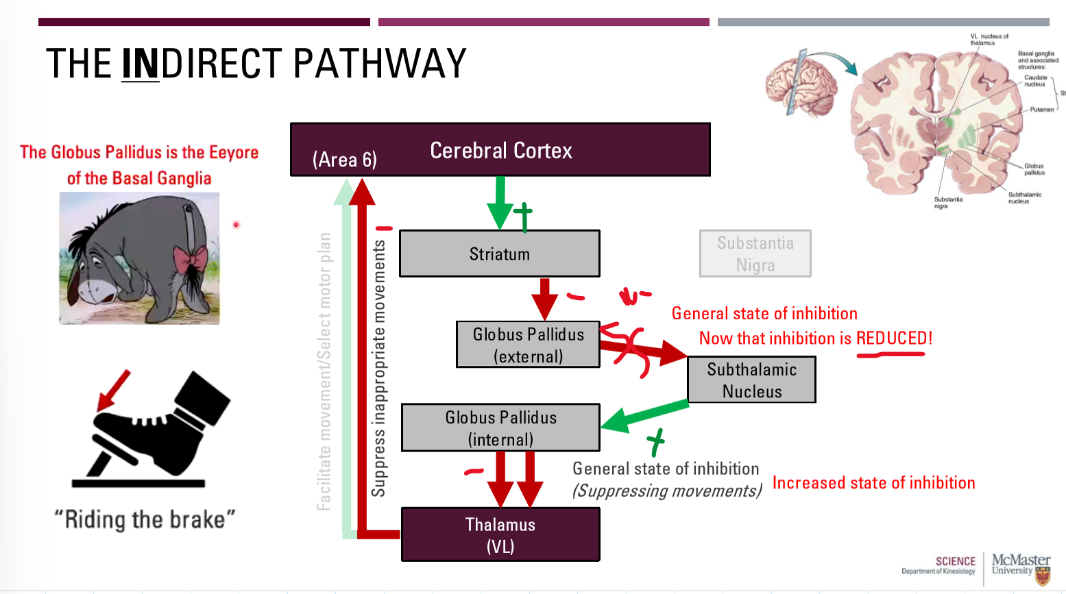
Inhibits movement
Help to suppress competing or inappropriate motor plans
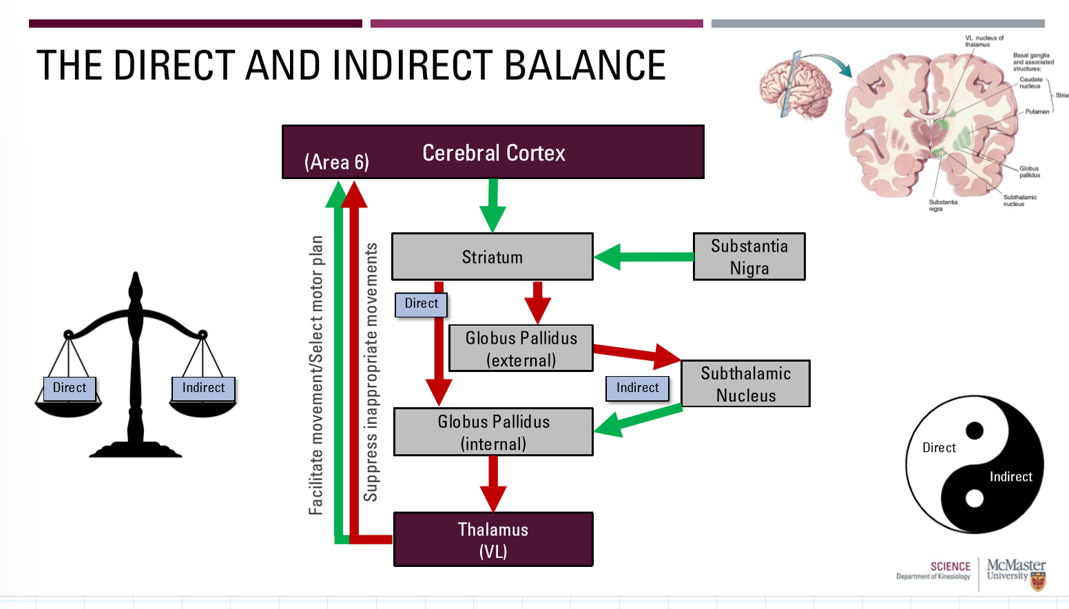
Need a healthy balance between the two

What is the direct pathway of the basal ganglia?

What is the indirect pathway of the basal ganglia?
What is the direct and indirect balance of the basal ganglia?
Direct = facilitates appropriate movements (Releasing the brake)
Indirect: Inhibits inappropriate movements (Riding the brake)
What is Parkinson’s Disease?
A disorder of the direct pathway
Difficulty stimulating wanted movement
Reduced direct pathway release of thalamus inhibition
Arises from a loss of dopaminergic neurons acting on the striatum in the direct pathway
What are hypokinetic disorders?
Reduced voluntary motor activity due to a disorder of the direct pathway
Bradykinesia - slowness of movement
Akinesia - lack of movement (e.g., freezing of gait)
How does a direct pathway disorder work?
Loss of dopaminergic neurons from Cerebral Cortex to Striatum (and from the substantia nigra to the striatum)
Levodopa supplements help maintain the pathway

What is Huntington’s disease?
A Disorder of the indirect pathway
Difficulty suppressing unwanted movements
Reduced indirect pathway = reduction in thalamus inhibition
Arises from a loss of striatum neurons acting on the globus pallidus external in the indirect pathway
Rare genetic disorder (<0.01% of people)
Symptoms arise in 30s or 40s
Neuron loss will also occur in other areas of the cerebral cortex, leading to:
Dementia
Personality changes
Death (~20 years after diagnosis)

What are hyperkinetic disorders?
Disorders of the indirect pathway that involve excessive involuntary motor activity
Chorea - spontaneous and uncontrollable movements
How does an indirect pathway disorder work?
Loss of neurons from striatum to GPe
Final overall inhibition cannot occur

What is the cortico-cerebellar loop and what are the 3 parts?
Proper execution of planned, voluntary, multi-joint movements
Necessary to fine tune the sequences (i.e., timing) of muscle contractions
3 parts:
Cortex to cerebellum
Within cerebellum
Cerebellum to cortex
What is the cortico-ponto-cerebellar pathway of the cortico-cerebellar loop?
Sensory and motor cortex axons form massive projection on pons
Pontine nuclei relay information to cerebellar cortex

What is ‘within the cerebellar cortex’ of the cortico-cerebellar loop?
Granule cells
Most numerous cells in the cerebellum
Excitatory/inhibitory output on Purkinje cells
Purkinje cells
Largest cells in cerebellum
Received thousands of synaptic inputs
Excitatory/Inhibitory output to deep cerebellar nuclei
Deep cerebellar nuclei
Excitatory or inhibitory output to thalamus

What is the ‘cerebello-thalamo-cortical pathway’ in the cortico-cerebellar loop?
Deep cerebellar nuclei relay to thalamus (VL)
VL relays information back to M1
Basal Ganglia was area 6 (premotor)
Here, M1 (area 4) signifies last chance tuning (timing, direction, force)

What results from cerebellar lesions?
Ataxia: uncoordinated and inaccurate movements
Summary of Brain Control
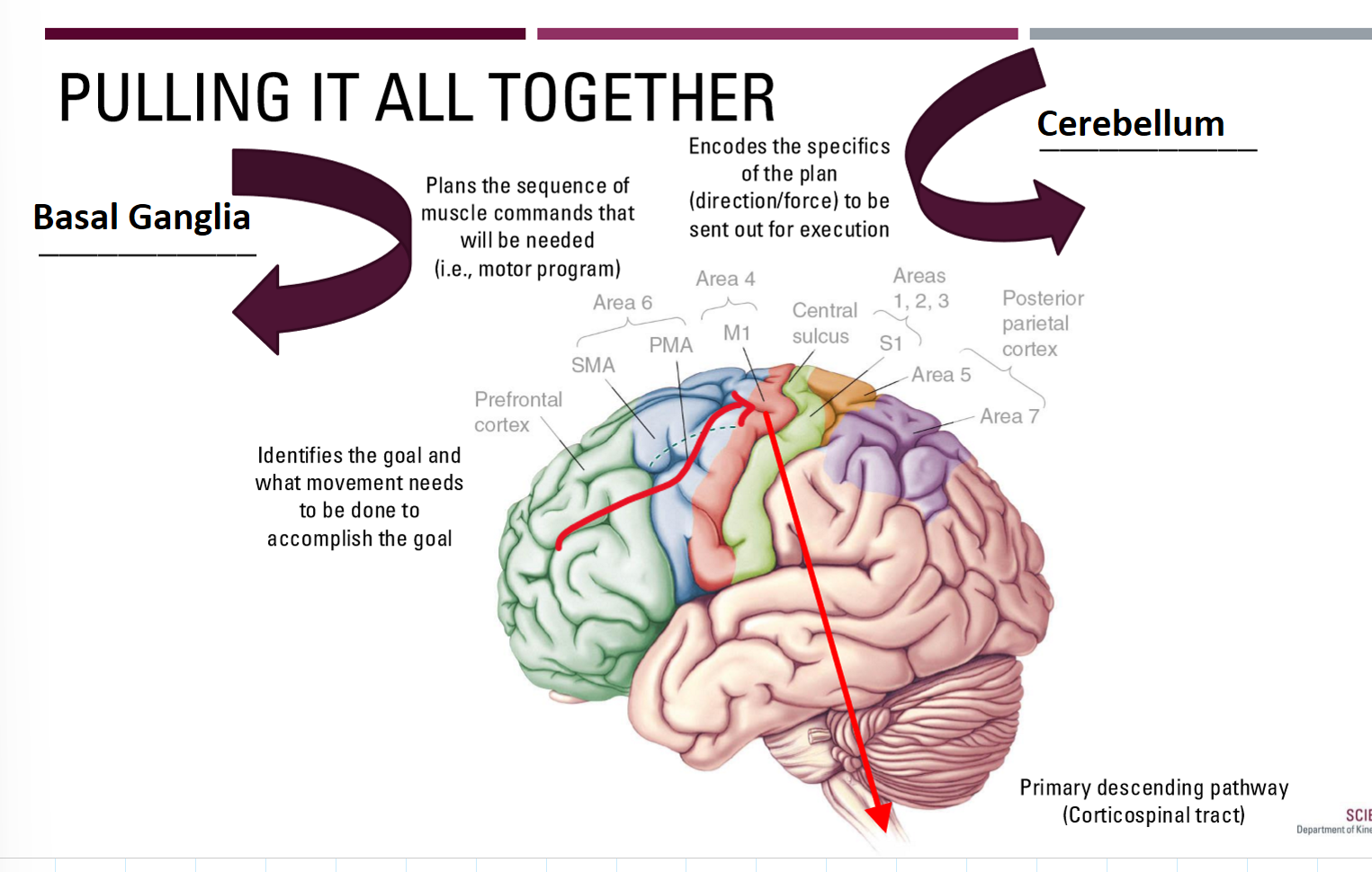
What are the two major groups of pathways in descending spinal tracts?
Lateral motor pathways
Commands for voluntary movements
Ventromedial motor pathways
Posture and reflex movements
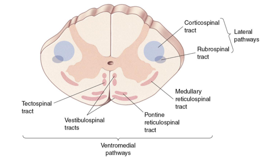
What is the corticospinal tract (CST)?
Primary pathway for voluntary motor control
Neck to feet
One of the largest and longest pathways
Pyramidal cells arising from the motor cortex
M1, but also premotor and other areas
Can be further divided into:
Lateral (limbs) vs. anterior (trunk, neck, shoulders)
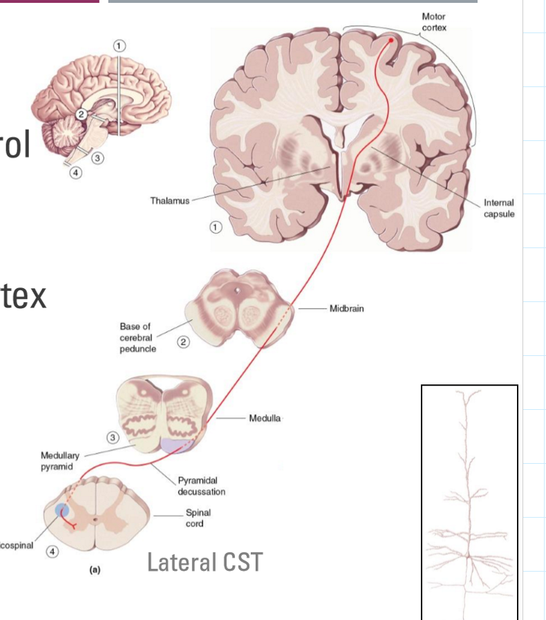
Lateral vs. Anterior CST
Lateral CST
90% of CST axons
Decussate in medullary pyramids
Control proximal/distal muscles (i.e., limbs)
Anterior CST
10% of CST axons
Decussate within spinal cord
Control axial muscles
Check slides for all diagrams

What is the lateral vs. anterior corticospinal tract pathway? (Draw it out)

How are the three different pathways impacted in Brown-Sequard Syndrome (Draw)?

What are the types of motor neurons?
Upper motor neurons
Cerebral cortex and brainstem
Can only connect to muscle through lower motor neurons
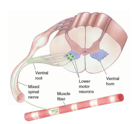
Lower motor neurons
Ventral horn of spinal cord
Direct connection to muscle
What do lower motor neurons involve?
Axons exit in ventral root
Can receive input from:
Upper motor neuron
Interneurons in spinal cord
Sensory input
Exist ventrally and join sensory fibers in spinal nerve
31 pairs classified in 4 segments (C-T-L-S-c)
*Mixed spinal nerve: both sensory and motor info coming in
What is the distribution of motor neurons in the spinal cord?
Not an even distribution
Cervical enlargement (C3-T1)
Lumbar enlargement (L1-S3)
These areas contain most of the motor neurons for distal and proximal muscles
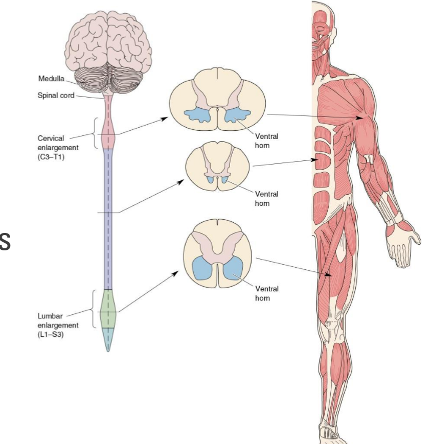
Distribution of motor neurons in spinal cord?
Organized at each level by area and function of the muscle they innervate
Axial muscles more medial than distal muscles
Flexors (decrease angle between bones) more posterior to extensors (increase angle between bones)
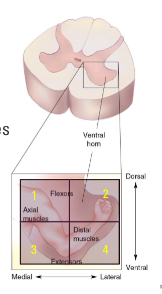
Flexors vs Extensors
Flexors: decrease the angle between bones
motor neuron will be more dorsal in the spinal cord
E.g. Hamstring (in-between distal and medial)
Extensors: muscles that increase the angle between our bons
motor neuron is most likely in the ventral area (3-4) of the spinal cord
E.g. Glutes (distal)
What are the two primary types of lower motor neurons?
Alpha motor neurons
Directly trigger the contraction of muscles for movement
Gamma motor neurons
Regulate muscle tone and control sensitivity of muscle spindles
Motor unit vs. Motor neuron pool?
Motor unit
Motor neuron and all the muscle fibers it innervates
Motor neuron pool
All the alpha motor neurons that innervate a single muscle

What are the steps of excitation-contraction coupling?
(electrical activity to mechanical contraction)
(A) Alpha motor neurons release ACh
(B) ACh produces large EPSP in muscle fiber
(C) EPSP evokes muscle action potential
(D) Action potential triggers Calcium ion release from Sarcoplasmic reticulum
(E) Fiber contracts (sliding filament model)
(F) Calcium ion reuptake
(G) Fiber relaxes

What is the sliding filament model of contraction?
Calcium ions binding to troponin allows myosin heads to bind to actin-myosin heads then pivot, causing filaments to slide

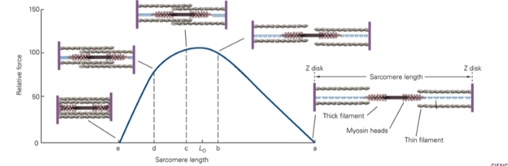
What is the force-length relationship?
A fundamental property of contractions
Describes the relationship of isometric muscle fiber forces with its length
Goldilocks zone for maximum number of cross-bridges (not too short, not too long)
Force output changes based on the speed it shortens (or lengthens)
What about power output in relation to force and velocity?
Power = force x velocity
If velocity = 0, then power = 0
In an isometric contraction, you have a lot of force, but the velocity on the x-axis 0, therefore there is no power
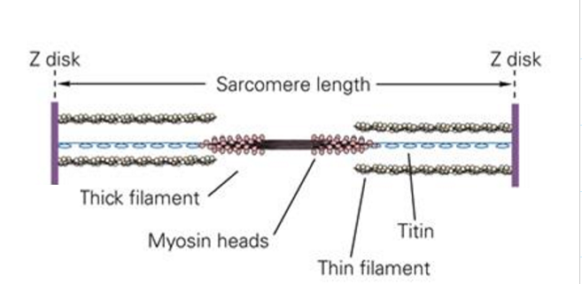
What is Titin?
“Spring” on the end of myosin filaments (connects the end of the thick filaments)
Provides elastic component in muscle fibers
Main source of passive force in a single fiber
Minimal source of passive force in complete muscle
What is residual force enhancement?
“When an active muscle is stretched, its isometric, steady-state force following the stretch is greater than the corresponding (same length, same activation) purely isometric contraction.”

What is residual force enhancement in relation to titin?
Titin appears to be an “adjustable” spring which plays an important role in residual force enhancement
Muscle activation can lead to a change in the stiffness and length of this spring
So… titin can help to increase force (+efficiency) in movements that involve activated/stretching muscles…

What is Electromyography (EMG)?
The quantification of a muscle(s) electrical activity (sum of action potentials)
Bipolar electrode configuration to record the difference in electrical activity
e.g., V1 - V2
…above the scope here
What are the applications of EMG?
Linear relationship to muscle force in isometric contractions
Provides amplitude of muscle activation (level of recruitment)
Provides timing of activation (activation patterns)
Fatigue and advance analyses
What are the differences in developmental changes with gait seen through EMG?
Considerable co-contraction of agonists and antagonists can be observed in in the infant
Brief periods of well-defined EMG activity can be seen in the adult
Muscle co-contraction returns in some older adults as a stabilizing feature

What are the limitations of EMG?
Not a direct relationship to muscle force (especially during motion)
Sensitive to differences in placement/processing
Surface EMG: Cross-talk between muscles, recording through skin/adipose
Fine-wire EMG - more accurate and gets intramuscular data
What can group 1 sensory neurons be divided into?
Ia axons & Ib axons
What are Ia axons?
Largest and fastest group I sensory neurons
Excitatory synapses with spinal interneurons and directly on alpha motor neurons
Muscle spindles (amount of stretch)
What are Ib axons?
Slightly smaller/slower group I sensory neurons
Inhibitory synapses with spinal interneurons
Golgi tendon organ (amount of force)
What are muscle spindles?
Sensory receptor
First thought to be a “muscle bud”
Sits within a muscle to measure the change in length
Small intrafusal muscle fibers
Parallel to primary muscle fibers (extrafusal)
Wrapped with a sensory neuron (Ia)

What is the stretch reflex (myotatic reflex)?
When a muscle is pulled (stretched), it pulls back (contracts)
Resists changes in muscle length
Important for maintaining limb position or posture
Monosynaptic stretch reflex
Primary sensory neuron (Ia - muscle spindle)
Primary motor neuron (alpha motor neuron)

What are the steps of the stretch reflex (myotatic reflex)?
Muscle is stretched (extrafusal & intrafusal)
Ia depolarizes from stretch
Action potential propagates along axon through dorsal root
Synapses with alpha motor neuron
Alpha motor neuron sends action potential to contract muscle
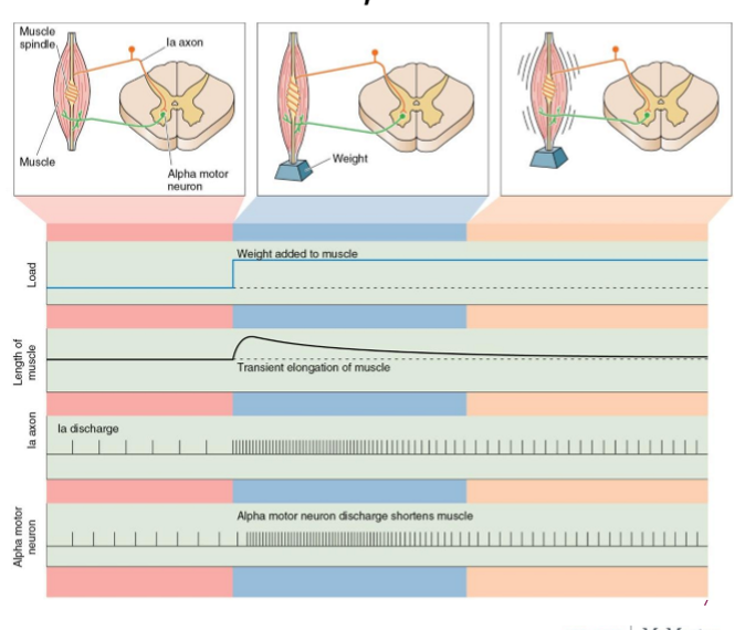
How do muscle spindles stay responsive to stretch?
Intrafusal fibers need the ability to contract, just like extrafusal fibers
Gamma motor neurons receive input from brain to keep intrafusal fiber taut

How does fine tuning muscle length with the gamma loop occur?
Descending command from brain sets first estimate
Coactivation of both alpha and gamma motor neurons (tuning for the initial guess)
Muscle spindle detects muscle is too long
Ia axons send signal to alpha motor neuron
Alpha motor neuron activate extrafusal fibers to shorten muscle

What is Gama bias?
Base level of firing for the intrafusal fibers to keep sensory “online”
Constant activity to keep the intrafusal fiber taut
Firing rate simply increases or decreases to compensate for changes in extrafusal fiber length
What is fusimotor gain?
Ability of the nervous system to adjust/fine tune the sensitivity to small changes
Ramping up/fine tuning the sensitivity of this loop to identify small changes
E.g. postural control
When a muscle stretches as you lean to one side, there is a stretch reflex to contract to get you back straight
How can we improve our fusimotor gain?
Balance training and plyometric training can help increase sensitivity (we can quickly/easily react to small changes)
What is the Gamma (motor) loop?
The loop between the muscle spindle (sensory fiber + gamma motor neuron) and muscle (alpha motor neuron)
What is the difference between static and dynamic response?
Static response:
Intrafusal fibers: Nuclear chain and static nuclear bag
Static gamma motor neurons
Type (group) II sensory fibers
Dynamic response:
Intrafusal fibers: Dynamic nuclear bag
Dynamic gamma motor neurons
Type (group) Ia sensory fibers

Why do we care about the static vs dynamic distinction?
Static response:
Primarily related to changes that are constant/predictable
Example: Static stretch (20-30s)
Inhibitory
Dynamic response:
Primarily related to changes that are quick/unpredictable
Example: Stretch reflex
Excitatory

Light green: static gamma motor neuron
Red: static (II sensory fiber), static, inhibitory
Dark green: Ia sensory fiber, dynamic, excitatory
Purple: Dynamic gamma motor neuron
Blue: Alpha motor neuron to extrafusal fibers
Static vs. Dynamic stretching?
Static stretch:
Likely to reduce muscle performance (if immediately prior to exercise)
Improves flexibility (may be required for specific taks)
activating type II sensory fibers (static response)
Dynamic stretching:
May increase muscle performance and sport specific performance
Activating type Ia sensory fibers (dynamic response)
Ramping up fusimotor gain → improving response to quick/unpredictable muscle length changes
However, if static stretching is integrated into a full warmup (aerobic, SS, DS, sport-specific activities), there is no evidence to say it is detrimental
What are Golgi tendon organs?
Sensory receptor
Sensory neuron (Ib) intertwined within the collagen fibers of tendons
Allows for the measure of force of contraction (strain of muscle)
Regulate muscle tension within optimal range
GTP fibers are in series rather than parallel
force must go directly through them
Synapse with inhibitory interneurons
Primarily helps to regulate muscle force
Allows for a protection of overload as last resort
What is the stretch-shortening cycle (SSC)?
Involves an eccentric stretching of the muscle, immediately followed by an enhanced concentric contraction
E.g. Vertical jump
Jump height:
Squat jump vs. countermovement jump
Mechanisms of the SSC?
Elastic energy
Stretch reflex
Neural potentiation
Active State
Mechanical potentiation
What is the elastic energy involved in the SSC?
Generated from all elastic components, but tendons dominate the storage and release of elastic energy
>0.5s may negate all effects of elastic energy
More elastic energy = fast eccentric immediately followed by fast concentric w/ minimal pause
What is the stretch reflex of the SSC?
Fast lengthening of muscle (eccentric contraction)
Dynamic nuclear bag convey sensory information of the stretch of the muscle through type Ia sensory neurons
Firing rate is dependent on the speed/amount of stretch
Improved fusimotor gain can help to maximize sensitivity of the reflex
What is neural potentiation in relation to the SSC?
Electromechanical decay
Picking up the “slack” in the unit
30-100ms
What is the active state of the SSC?
Time for cross-bridges to build up force
100ms - 1s
What is mechanical potentiation of the SSC?
Residual force enhancement
What is involved in a fast SCC?
Fast eccentric phase, with an explosive concentric phase
Stiff joint, high activation in eccentric, very little loss of force during amortization
Contract time <250 ms
Primary mechanisms:
Elastic energy and stretch reflex, as well as mechanical potentiation (i.e., RFE)
What is involved in a slow SSC?
Slower eccentric phase with less explosive concentric phase
Less stiff joint, longer contact times, greater neuromuscular activity in the concentric phase
Contact time >250 ms
Primary mechanisms:
Active state, neural potentiation, and mechanics of contractile component
Slow SSC allows for:
More work to be done by contractile unit over a greater distance (work = force x distance)
too fast it may “fall down” the power curve
Spinal interneurons can design simple to highly complex patterns of movement based on input from:
Primary sensory axons
Descending axons from brain
Collaterals of lower motor neuron axons
Other interneurons
What is reciprocal inhibition?
Reflex arc using spinal interneurons to support simple monosynaptic reflexes (e.g., stretch reflex)
Contraction of one muscle accompanied by relaxation of its antagonist muscle
What is the withdrawal reflex?
Reflex arc used to withdraw limb from aversive stimulus
Excitatory input for multiple ipsilateral motor units
What about staying upright?
Opposite leg has to take on load
What is the crossed-extensor reflex?
Reflex arc for extensors and flexors on opposite side
Excitatory and inhibitory for ipsilateral and contralateral motor units
What are central pattern generators?
Circuitry for many rhythmic movements resides in Spinal cord
Complex network of sensory, motor and interneurons
Controlled by two “half-centers” of spinal neurons
Mutually inhibiting halves that produce alternating bursts of flexor and extensor activity
Rhythmic activity will continue as long as input exists
Circuitry for walking resides in the lumbar/sacral spinal cord
Descending control (input from brain) initiates and adapts CPGs
can influence both rhythm and pattern generation neural pools
Rhythm vs. pattern
Patterns are continually adjusted from higher and lower inputs
Where do we have evidence of Central pattern generators?
Evidence from animal spinal transections
Measured bursts of rhythmic and coordinated activity arising for input that was constant
What are the pathways of central pattern generators?
Rhythm Generators
Setting the timing of patterns to be generated
Pattern Generators
Sending the required pattern of muscles activations
What is Gait retraining?
Changing the mechanics of how you walk or run
Easy to override motor patterns or CPGs
Very difficult to “rewrite” them
CPG evidence in cats
Sensory information may also influence BOTH rhythm and pattern
Stepping in cats
Proception
Amount of stretching can regulate the stance phase
Cats can match speed of treadmill, through CPG alone (after training / stimulation)
Sensory information from the skin
Stimulus to dorsal side of paw causes pattern with increased flexion to avoid a tripping hazard

What is fictive movement?
Primary source of evidence for CPGs
“Resemble” locomotion, but are not true locomotion…
Reflex theory in CPGs
Reflexes are still present
We said the reflexed can “influence” the rhythm and pattern of the CPG, but…
What if the reflexes themselves are doing all the work
Simulations suggest this is possible (in theory)
Evidence vs Evidence against for CPGs
Lots of evidence for:
Animal models
Stepping reflexes in infants as foundation for adult locomotion
Spontaneous or evoked activation patterns SCI patients
Effective circuitry for controlling robotic locomotion
Evidence against…
No direction locomotion example in humans (vs. cats)
Most is “fictive”
Could simply be reflex-based
So…do humans really use CPGs in walking?
“Our interpretation is that, taken together, these facts underpin the view that CPGs do exist in the human spinal cord.”
Perhaps a better question for future research is to what extent do CPGs control our locomotion?
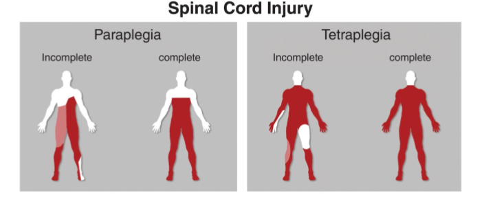
What are Spinal Cord Injuries (SCI)?
Produce sensory, motor, and/or autonomic disfunction
Complete vs. incomplete
Paraplegia vs. tetraplegia (quadriplegia)
Can be from direct trauma or vascular insufficiency