Chapter 11: Functional Organization of Nervous Tissue
1/103
There's no tags or description
Looks like no tags are added yet.
Name | Mastery | Learn | Test | Matching | Spaced |
|---|
No study sessions yet.
104 Terms
functions of the nervous system
Maintaining homeostasis: stimulate or inhibit multiple activities to help maintain homeostasis
Receiving sensory input: sensory receptors monitor internal and external stimuli
Integrating information: brain and spinal cord process sensory input and initiate responses
controlling muscles and glands: skeletal muscles contract only when stimulated by the nervous system. it also controls the secretions from many glands as well as causes the contractions of cardiac and some smooth muscles to occur more rapidly or slowly
Establishing and maintaining mental activity: center of mental activities such as consciousness, thinking, memory, and emotion
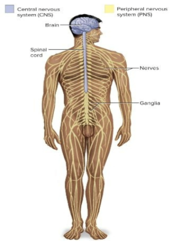
two major subdivisions of nervous system
Central and peripheral nervous systems
Central nervous system (CNS)
Brain and spinal cord which connect with each other at the foramen magnum of the skull
Enclosed by cranium and vertebral column
Tract —> bundle of axons in CNS
Nucleus —> bundle of cell bodies in CNS

Peripheral nervous system (PNS)
Carries information to CNS from different tissues of the body and carries commands from the CNS that alter body activities
Entire nervous system except the brain and spinal cord; composed of nerves and ganglia
Nerve —> bundle of axons in PNS
Ganglion—> bundle of cell bodies in PNS
Tract
bundle of axons in central nervous system
nucleus
bundle of cell bodies in central nervous system
nerve
bundle of axons in the peripheral nervous system
ganglion
bundle of cell bodies in the peripheral nervous system
Sensory (afferent)
carries signals from receptors to CNS
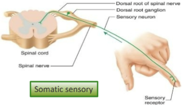
Somatic sensory: carries signals from receptors in the skin, muscles, bones, and joints
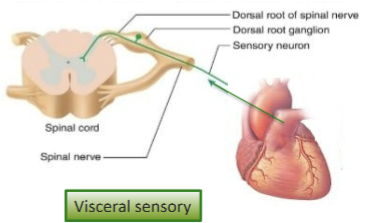
Visceral sensory: carries signals from the viscera (heart, lungs, stomach, and urinary bladder)
Motor (efferent)
Carries signals from CNS to effectors (glands and muscles that carry out the body’s response)
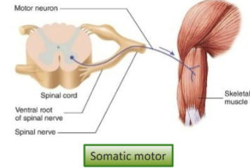
Somatic motor: carries signals to skeletal muscles; output produces voluntary muscular contraction and involuntary somatic reflexes
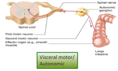
Visceral motor (autonomic nervous system): carries signals to glands, cardiac and smooth muscle and produces involuntary visceral reflexes
somatic sensory
carries signals from receptors in the skin, muscles, bones, and joints
visceral sensory
carries signals from the viscera to the CNS, including organs like the heart, lungs, and stomach.
somatic motor
Carries signals from the CNS to skeletal muscles, resulting in voluntary movements and involuntary reflexes.
visceral motor (autonomic nervous system)
carries signals to glands, cardiac and smooth muscle and produces involuntary visceral reflexes
Consists of sympathetic and parasympathetic divisions
Sympathetic
tends to arouse body for action, accelerates heartbeat and respiration, inhibits digestive and urinary systems
Parasympathetic
Tends to have calming effect, slows heart rate and breathing, stimulates digestive and urinary systems
Cells of the nervous system
Neurons: electrically excitable cells which consists of cell body and axons; conduct APs and transmit signals to other neurons or effector organs and can secrete a neurotransmitter to stimulate the next cell
Glial cells: support and protect the neurons
Neurons
electrically excitable cells which consists of cell body and axons; conduct APs and transmit signals to other neurons or effector organs and can secrete a neurotransmitter to stimulate the next cell
Glial cells
support and protect the neurons
Neuron structure
Electrically excitable cells which consist of nucleus/ cell body (soma), dendrites and axons
cell body: typical cell functions such as protein synthesis and housekeeping; contain Nissl bodies (rough ER)
dendrites: extensions of cell body and receives signals from other neurons and conduct small electric currents to the cell body
axons: arise from axon hillock and APs generated in trigger zone, contain cytoplasm (axoplasm) and its plasma membrane (axolemma). Terminate at presynaptic terminal which store vesicles with neurotransmitters
axons
arise from axon hillock and APs generated in trigger zone, contain cytoplasm (axoplasm) and its plasma membrane (axolemma). Terminate at presynaptic terminal which store vesicles with neurotransmitters
dendrites
extensions of cell body and receives signals from other neurons and conduct small electric currents to the cell body
cell body
typical cell functions such as protein synthesis and housekeeping; contain Nissl bodies (rough ER)
Types of neurons
functional and structural
Functional classification
Sensory or afferent: detect stimuli and transmit information (conduct APs) towards the CNS
Interneurons or association neurons: lie entirely within CNS connecting motor and sensory pathways, conduct APs within CNS from one neuron to another
Motor or efferent: conduct APs signals from CNS and send signals out to muscles and gland cells (the effectors)
motor or efferent
conduct APs signals from CNS and send signals out to muscles and gland cells (the effectors)
interneurons or association neurons
lie entirely within CNS connecting motor and sensory pathways, conduct APs within CNS from one neuron to another
sensory or afferent
detect stimuli and transmit information (conduct APs) towards the CNS
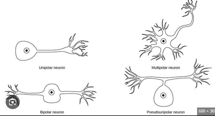
Structural classification
multipolar: many dendrites and an axon; most neurons in CNS; motor neurons
Bipolar: a dendrite and an axon; sensory in retina of the eye, nose, inner ear
Pseudo- unipolar: appears to have an axon and no dendrite; sensory cells from skin and organs to spinal cord
Anaxonic: many dendrites, no axons; found in brain and retina
Anaxonic
many dendrites, no axons; found in brain and retina
Pseudo- unipolar
appears to have an axon and no dendrite; sensory cells from skin and organs to spinal cord. Involved for sensory pathways
Bipolar
a dendrite and an axon; sensory in retina of the eye, nose, inner ear
Multipolar
many dendrites and an axon; most neurons in CNS; motor neurons
Glial cells of the CNS
Astrocytes, Ependymal cells, Microglia, and Oligodendrocytes
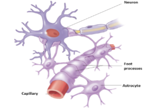
Astrocytes
Most abundant
Create supportive framework
Promote formation of right junctions between capillaries which helps form the blood- brain barrier
Communicate electrically with neurons
Monitor activity; regulate blood flow to match metabolic need
Star- shaped with many cytoplasmic extensions from the cell body
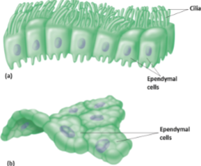
Ependymal cells
Secrete and circulate cerebrospinal fluid
Line the internal cavities (ventricles) of brain and central canal of spinal cord
Cuboidal epithelium with cilia
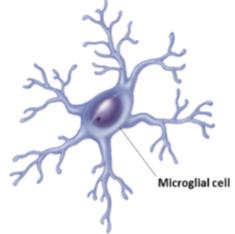
Microglia
Small glial cells with many extensions
Macrophages
Phagocytize neurotic tissue, micro-organisms, foreign substances (almost like the “immune cells”)
maintains health and function
CNS
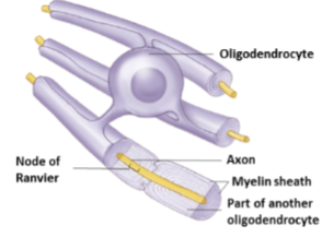
Oligodendrocytes
Have broad cytoplasmic extensions that wrap many times around axons
Extensions form insulating materials called myelin sheath
Glial cells of the PNS
Schwann cells and Satellite cells
Schwann cells
Broad cell the wraps many times around a portion of an axon
Forms the myelin sheath for the portion of the axon it wraps
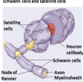
Satellite cells
Surround neuron cell bodies in ganglia
Provide support and nutrition

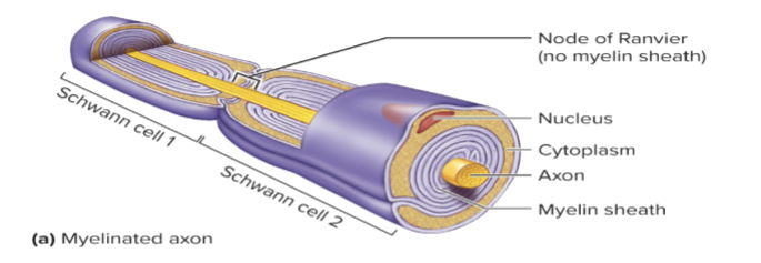
Myelinated axons
Extensions wrap around the axon repeatedly to form many phospholipid rich layers. Periodic gaps where the axons are uncovered are called nodes of Ranvier
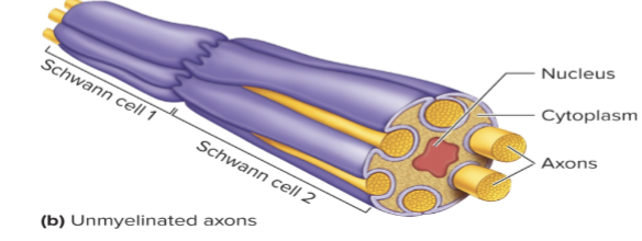
Unmyelinated axons
Axons rest in invaginations of associated Schwann cells or oligodendrocytes but are not wrapped by them
Gray matter
Unmyelinated axons, cell bodies, dendrites. Integrative function
White matter
Myelinated axons. Propagate APs
CNS
clusters of neuron cell bodies are nuclei; bundles of myelinated axons are nerve tracts
PNS
clusters of cell bodies are ganglia; bundles of axons with their connective tissue sheaths are nerves
Establishing RMP, where is K+ and Na+
K+ is higher inside the cell than outside, and the concentration of Na+ is higher outside the cell than inside. The plasma membrane is more permeable to K+ than other positively charged ions. The negative charge inside the cell attracts positively charged K+.
changing the RMP
Change in ion concentration gradients or ion permeability of the plasma membrane can either cause depolarization which is the movement of EMP toward zero or hyperpolarization which is the movement of RMP away from zero
Example: Decrease Extracellular (K+) Depolarization or Hyperpolarization?
Hyperpolarization
Example: Increase Extracellular (K+) Depolarization or Hyperpolarization?
Depolarization
Example: Open K gated channels: Depolarization or Hyperpolarization?
Hyperpolarization
Example: Open Na gated channels: Depolarization or Hyperpolarization?
Depolarization
Example: Open Ca gated channels: Depolarization or Hyperpolarization?
Depolarization
Example: Open Cl gated channels: Depolarization or Hyperpolarization?
Hyperpolatization
Graded (local) potentials
a small change in membrane potential localized to one area of plasma membrane. They result from:
ligands binding to receptors, changes in charge across membrane, mechanical stimulation, temperature changes, spontaneous change in permeability
A stimulus causes changes in permeability of the membrane to Na+, K+, or Cl- which results in either depolarization or in hyperpolarization
Depolarization
Excitatory (makes a neuron more likely to fire an action potential)
Hyperpolarization
inhibitory (makes a neuron less likely to fire an action potential)
Decremental
Graded potentials are conducted in a decremental fashion, meaning that their magnitude decreases as they spread over the plasma membrane
Summate
Depolarizing graded potentials can combine (summate) to cause an action potential
Action potentials
series of permeability changes when a graded potential causes depolarization of membrane
All or none principle
no matter how strong the stimulus, if it is greater than threshold, then AP will occur
Non-decremental
Do not get weaker with distance
Magnitude is constant along the axon (or muscle fiber)
Operation of Gates: Action Potential
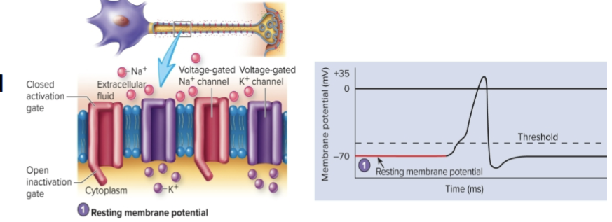
Resting membrane potential
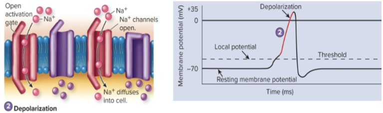
Depolarization
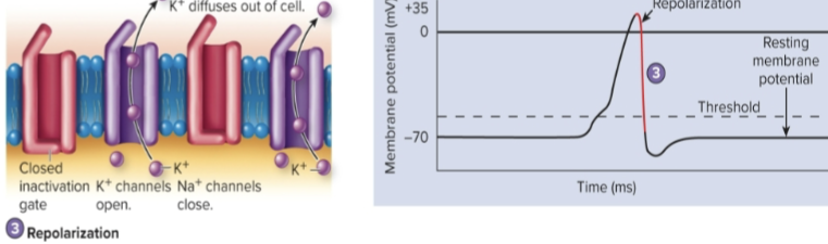
Repolarization
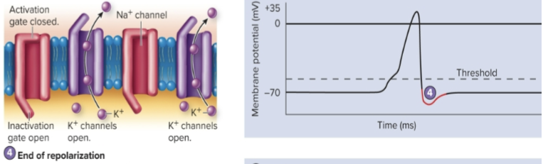
End of repolarization and afterpotential
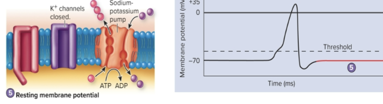
Resting membrane potential
Resting membrane potential (first step)
Na+ gated channels and most K+ gated channels are closed. The outside of the plasma membrane is positively charged compared to the inside
Depolarization
Na+ channels open. K+ channels begin to open more slowly. Depolarization results because the inward movement of Na+ makes the inside of the membrane more positive
Repolarization
Na+ channels close and additional K+ channels continue to open. Na+ movement into the cell stops, and K+ movement out of the cell increases, causing repolarization
End of repolarization and afterpotential
Voltage- gated Na+ channels are closed. Closure of the activation gates and opening of the inactivation gates reestablish the resting condition for Na+ channels. Diffusion of extra K+ through late closing voltage- gated channels produces a slight hyperpolarization (afterpotential)
Resting membrane potential (last step)
The resting membrane potential is reestablished by Na/K pump after the voltage-gated K+ channels close
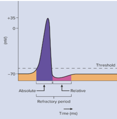
Refractory Period
Once an AP is produced at a given point on the membrane, that area becomes less sensitive to further stimulation
Two parts: Absolute refractory period and Relative refractory period
Absolute refractory period
the time during an actions potential when a second stimulus, no matter how strong, cannoy initiate another action potential. It lasts almost from the beginning of AP until near the end of repolarization. It ensures that once an AP has begun both the depolarization and repolarization phases are completed or nearly completed before another AP can begin

Relative refractory period
follows the absolute refractory period and is the time during which a strong stimulus or stronger- than- threshold stimulus can evoke another action potential. It ends when the membrane potential has returned to the resting level
Action Potential Frequency
Number of potentials produced per unit of time to a stimulus
Subthreshold stimulus, Threshold stimulus, Maximal stimulus, Submaximal stimulus, and Supramaximal stimulus
Subthreshold stimulus
does not cause a graded potential that is great enough to initiate an AP

Threshold Stimulus
causes a graded potential that is great enough to initiate an AP
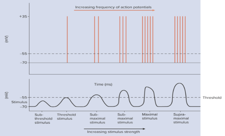
Maximal stimulus
just strong enough to produce a maximum frequency of APs

Submaximal stimulus
all stimuli between threshold and the maximal stimulus strength

Supramaximal stimulus
any stimulus stronger than a maximal stimulus. These stimuli cannot produce a greater frequency of APs than a maximal stimulus

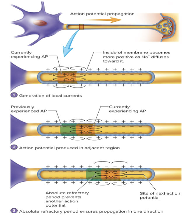
Continuous Conduction
Propagation of APs- Unmyelinated axon
1. when an action potential is produced, the inside of the membrane becomes more positive than outside. This diffusion of Na+ is called a local current
2. as a result of the local current, the part of the membrane immediately adjacent to the action potential depolarizes
3. if an action potential propagation is initiated at one end of the axon, it is propagated down the axon. The absolute refractory period ensures one-way propagation of an action potential because it precents the local current from stimulating the production of the action potential in the reverse direction
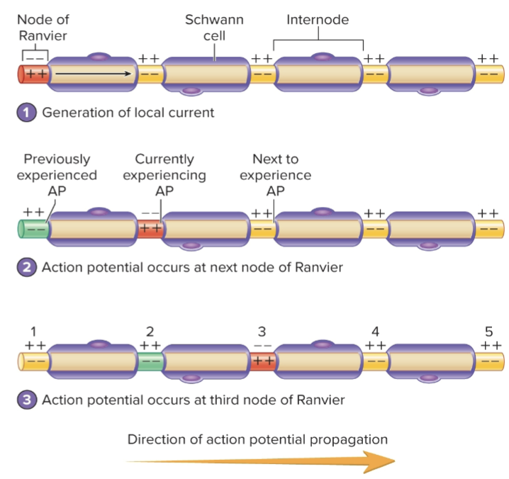
Saltatory Conduction
Propagation of APs- myelinated axon
1. an action potential (orange) at a node of Ranvier generates local currents (black arrows). The local currents flow to the next node of Ranvier because the myelin sheath of the Schwann cell insulates the axon of the internode
2. When the depolarization caused by the local currents reaches threshold at the next node of Ranvier, a new action potential is produced (orange)
3. Action potential propagation is rapid in myelinated axons because the action potentials are produced at successive nodes of Ranvier (1-5) instead of at every part of the membrane along the axon
The speed of AP conduction along an axon depends on three factors:
Myelination of axon: APs are conducted more rapidly in myelinated than unmyelinated axons
Thickness of myelin sheath: Heavily myelinated axons conduct APs more rapidly than lightly myelinated ones
Diameter of axon: Large diameter axons have more surface area with more voltage gated Na channels and conduct signals more rapidly than small- diameter axons
The synapse
Junction between two cells and is the site where APs in one cell cause action potentials in another cell
Types of cells in synapse
Presynaptic: cell that transmits signal toward the synapse
Postsynaptic: target cell receiving the signal
Presynaptic
cell that transmits signal toward the synapse
Postsynaptic
Target cell receiving the signal
Electrical synapse
Gap junctions allows graded current to flow between adjacent cells where the membranes are connected by protein tubes called connexons
AP in one cell causes AP in the next cell, almost as if the tissue were one cell allowing APs to be conducted rapidly between cells allowing the cells’ activity to be synchronized
Found in cardiac muscle and many types of smooth muscle
Chemical synapse
It consists of a presynaptic terminal, synaptic cleft and postsynaptic membrane
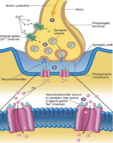
Neurotransmitter release
Action potentials arriving at the presynaptic terminal cause voltage-gated Ca2+ channels to open
Neurotransmitter removal
Method depends on neurotransmitter/ synapse
Acetylcholine (ACh) or Norepinephrine (NE)
Acetylcholine (ACh)
Acetylcholinesterase splits ACh into acetic acid and choline. Choline recycled within presynaptic neuron
Norepinephrine (NE)
Recycled within presynaptic neuron or diffuses away from synapse. Enzyme monoamine oxidase (MAO). Absorbed into circulation, broken down in liver
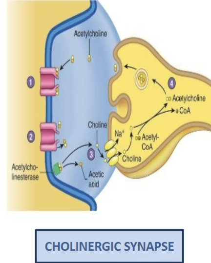
Removal of Neurotransmitter (ACh) from Synaptic Cleft (Cholinergic Synapse)
ACh molecules bind to their receptors
ACh molecules unbind from their receptors
Acetylcholinesterase splits ACh into choline and acetic acid, which prevents acetylcholine from again binding to its receptors. Choline is taken up by the presynaptic terminal
Choline is used to make new acetylcholine molecules that are packaged into synaptic vesicles
Acetic acid is used to make AcetylCoA
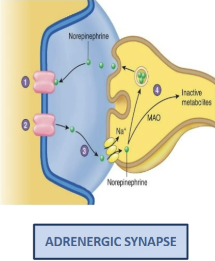
Removal of Neurotransmitter (NE) from Synaptic Cleft
Norepinephrine binds to tits receptor
Norepinephrine unbinds from its receptor
Norepinephrine is taken up by the presynaptic terminal, which prevents norepinephrine from again binding to its receptor
Norepinephrine is repackaged into synaptic vesicles or broken down by monoamine oxidase (MAO)
Classification of Neurotransmitters
Based on:
Chemical structure
Their effect on the postsynaptic membrane (the target cell; excitatory or inhibitory)
The mechanism of action at their target (inotropic or metabotropic)
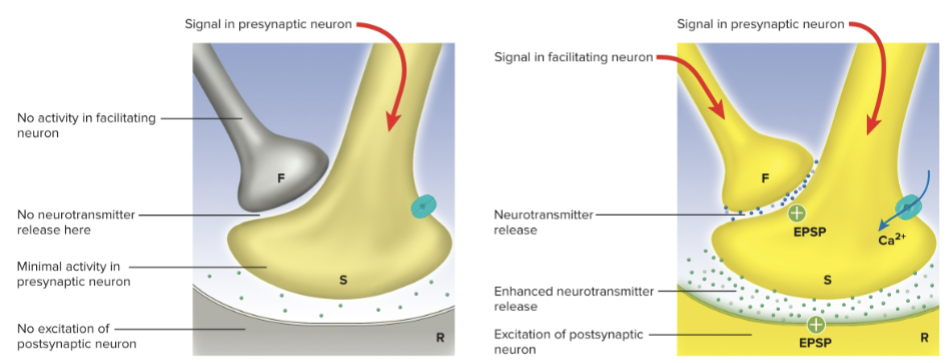
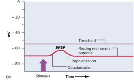
Excitatory Postsynaptic Potentials (EPSPs)
Depolarization of postsynaptic membrane occurs (due to increased permeability to Na+) and depolarization might reach threshold producing an AP and cell response
Neurons releasing neurotransmitter substances that cause EPSPs are excitatory neurons
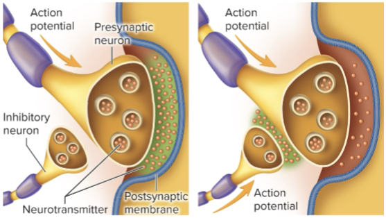
Inhibitory Postsynaptic Potential (IPSP)
Hyperpolarization of postsynaptic membrane occurs (due to increased permeability of K+ and Cl-) and no AP generated as moving membrane potential farther from threshold
Neurons releasing neurotransmitter substances that cause IPSPs are inhibitory neurons
Neuromodulators
Chemicals produced by neurons that influence the likelihood of APs in postsynaptic cell. Act by increasing or decreasing the amount of neurotransmitter released by the presynaptic neuron
A neuromodulator that decreases the release of an excitatory neurotransmitter will decrease the likelihood of an AP in postsynaptic cell
Axoaxonic synapses
axon of one neuron synapses with the presynaptic terminal (axon) of another
Presynaptic inhibition
decrease in amount of neurotransmitter released from presynaptic terminal
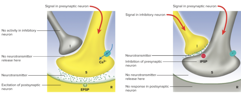
Presynaptic facilitation
increase in amount of neurotransmitter released from presynaptic terminal