AN141 Lab Exam
1/23
There's no tags or description
Looks like no tags are added yet.
Name | Mastery | Learn | Test | Matching | Spaced | Call with Kai | Chat |
|---|
No analytics yet
Send a link to your students to track their progress
24 Terms
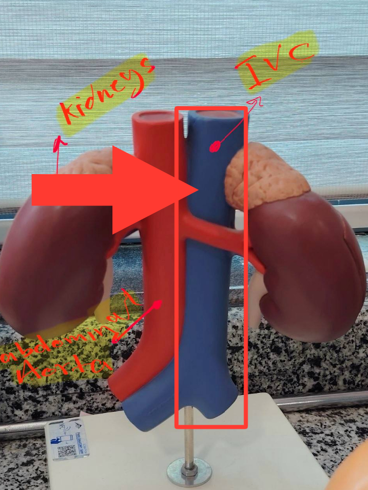
Inferior Vena Cava
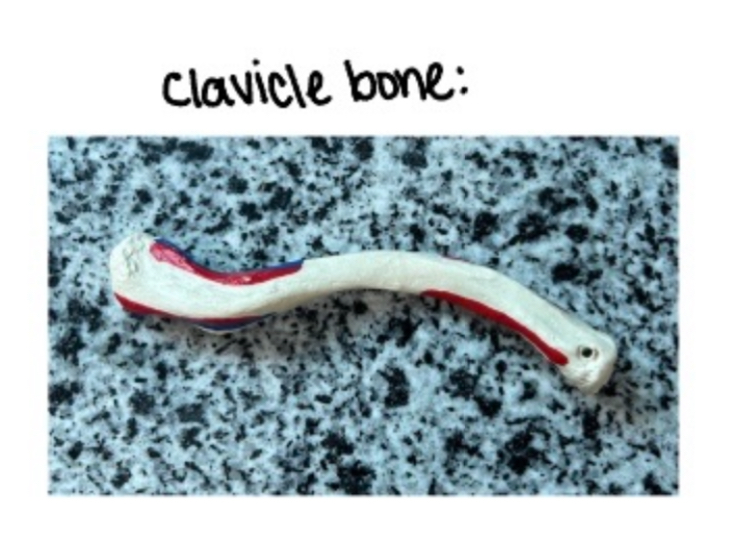
Clavicle
Medial end thick; Lateral end flat. Conoid tubercle inferior. Medial 2/3 convex anteriorly.
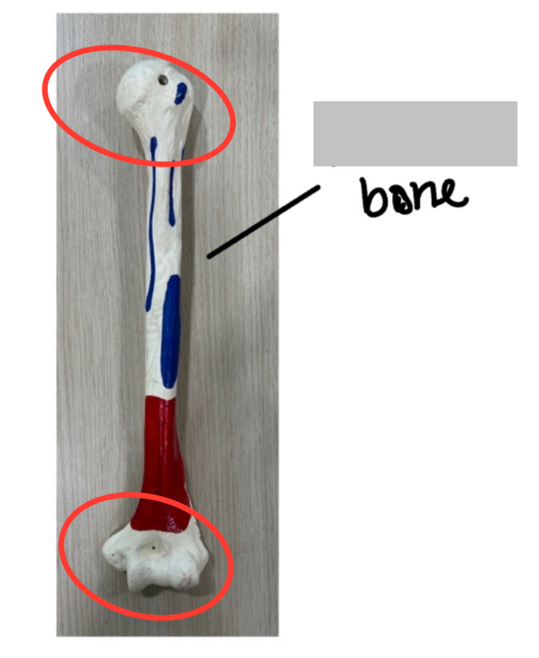
Humerus
Head faces medial and posterior. Olecranon fossa posterior. Bicipital groove anterior.
1. Head points medially, upward, and backward.
2. Bicipital groove is anterior.
3. Olecranon fossa is posterior (big deep hole at the distal end). If the head faces your right and the bicipital groove faces you, it's a left humerus. The deep hole (olecranon fossa) is always at the back.
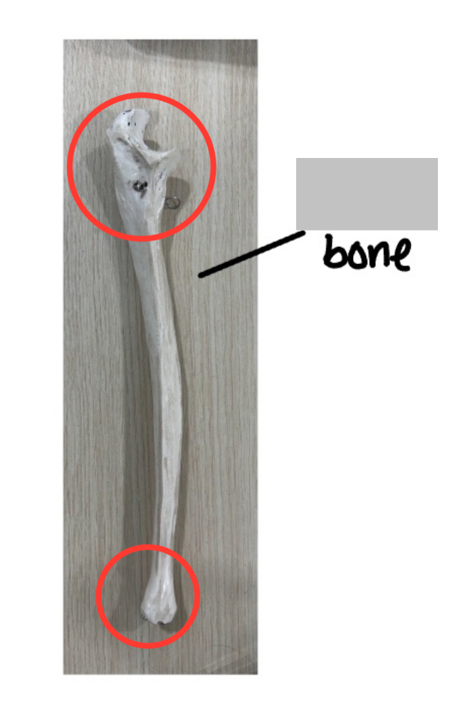
Ulna
Olecranon posterior. Trochlear notch anterior. Radial notch lateral. Head distal.
1. Big hook (olecranon) is at the proximal/posterior end.
2. Radial notch is on the lateral side of the coronoid process.
3. Trochlear notch faces anteriorly. The hook is back. The trochlear notch opens forward. The radial notch is a small dip facing outward; if it's on your right → left ulna. Remember: the radial notch faces the radius.
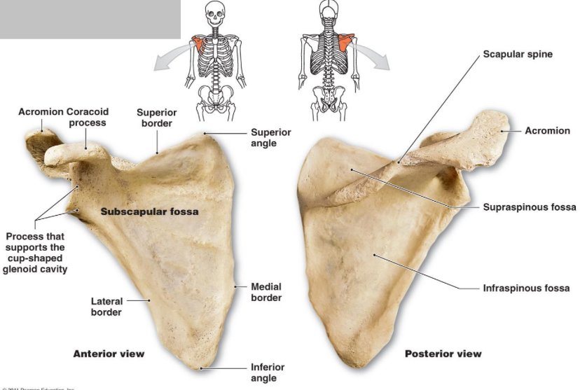
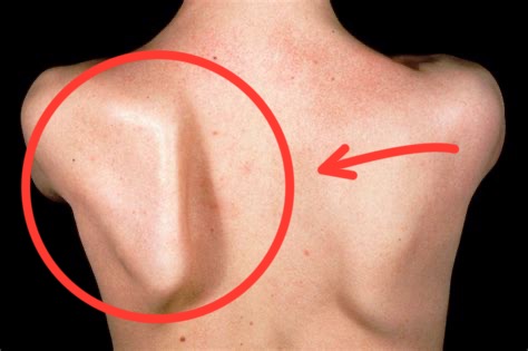
Scapula
1. Glenoid cavity faces outward (lateral) and slightly forward.
2. Spine is posterior.
3. Coracoid process points anteriorly. Hold it with the spine toward you and the glenoid pointing to the side you're naming. Right glenoid = right scapula. The big flat subscapular fossa is anterior
Radius
1. Disc-shaped head is at the proximal end (top).
2. Styloid process is at the distal end, on the lateral side.
3. Radial tuberosity faces anteriorly and medially. Hold it like a dagger: head up, the pointy styloid points to the thumb side. If styloid is on your right → right radius. The tuberosity points toward the other bone (medial).
Fibula
1. Head is at the proximal end (small knob).
2. Lateral malleolus is at the distal end, pointed laterally (outer ankle).
3. The malleolar fossa (pit) is posterior on the lateral malleolus. The little knob is the top. The long pointy end is the lateral ankle bone. If it's on your left, it's a left fibula. The pit behind the lateral malleolus confirms it's posterior.
Tibia
1. Tibial tuberosity is a big bump on the anterior side, just below the knee.
2. Medial malleolus is the thick bump on the medial side of the distal end (inner ankle).
3. Soleal line is a ridge on the posterior upper shaft. The bump in front (shin) is anterior. The inner ankle bone (bigger) is medial. If the medial malleolus is on your right → right tibia.
Scapula
Femur
1. Head faces medially, up, and slightly forward.
2. Linea aspera is a rough ridge on the posterior shaft.
3. Patellar surface is a smooth groove on the anterior distal end. Head points medially. The linea aspera is the back. The smooth groove (for kneecap) is the front. If head points to your right and groove faces you → left femur.
Fibula
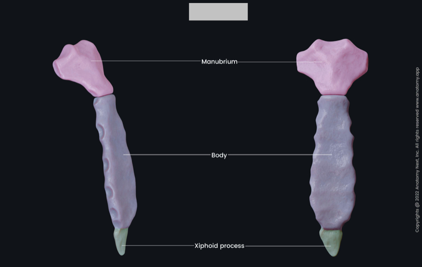
Sternum
Manubrium (top), Body (middle), Xiphoid (bottom). Sternal angle (ridge) is where 2nd rib attaches.
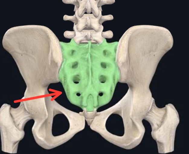
Sacrum
Smooth concave surface = anterior (pelvic). The sacral hiatus (U-shaped gap) is posterior, at the bottom. The auricular surfaces are lateral (ear-shaped for SI joint).
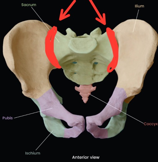
Sacroiliac (SI) Joint
Synovial (anterior) + Syndesmosis (posterior) type. Sacrum + Ilium (hip bone)
Very limited movement; transfers weight from spine to legs.
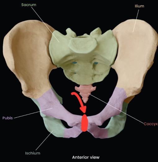
Pubic symphysis
Secondary Cartilaginous (Symphysis) type. Involves Right + Left Pubic Bodies.
Minimal movement; slight widening during pregnancy.
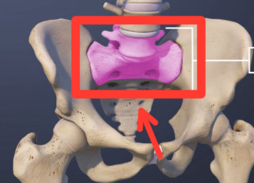
Lumbosacral joint
Secondary Cartilaginous (Symphysis) type. Involves L5 vertebra + Sacrum (base). Limited movement; disc and facet joints.
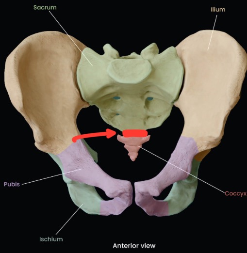
Sacrococcygeal Joint
Secondary Cartilaginous (Symphysis) type. Involves Sacral apex + Coccyx. Slight movement; can move in childbirth.
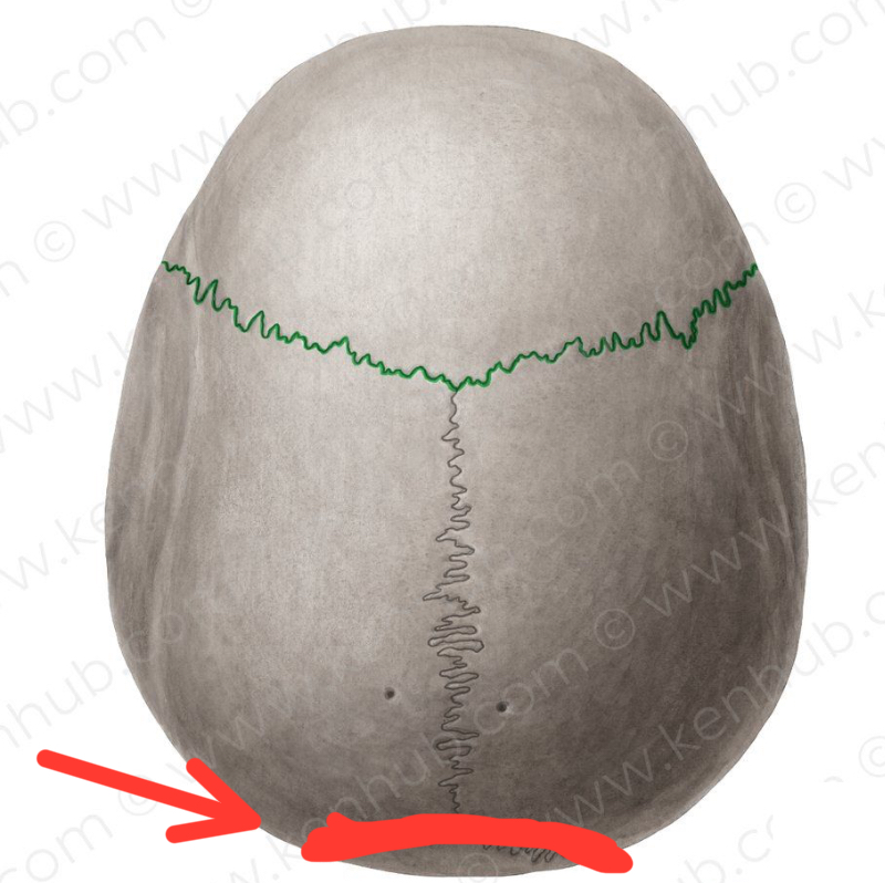
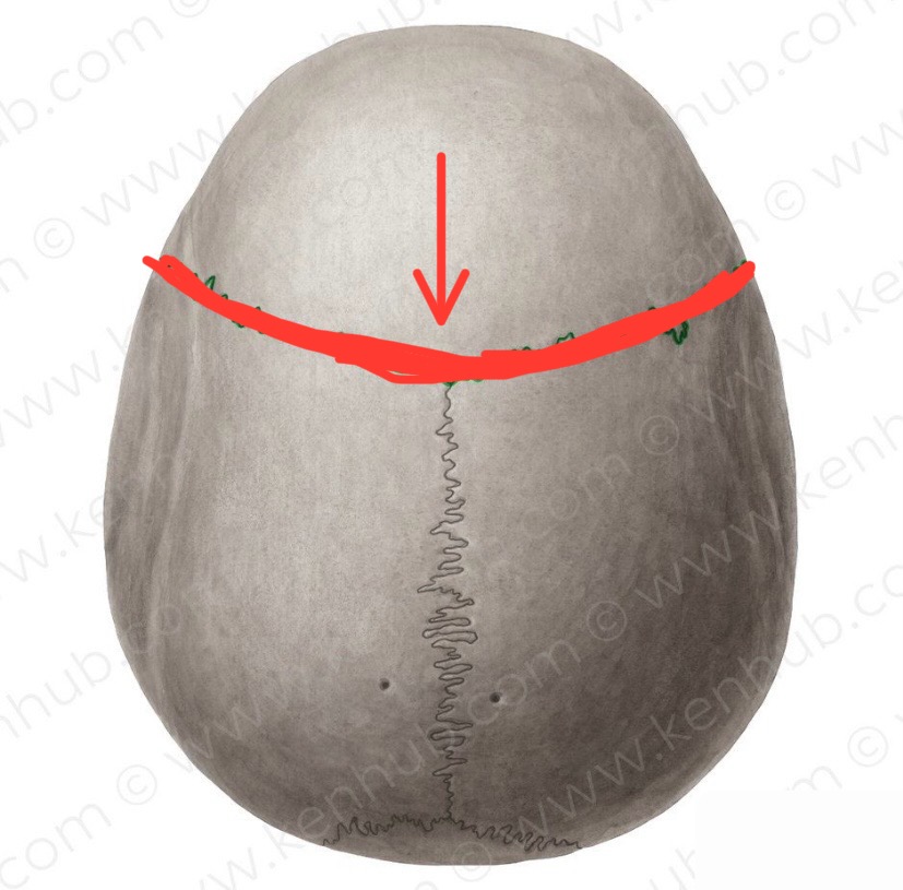
Lambdoid
Parietal (both) + Occipital Back of skull (curved Λ shape)
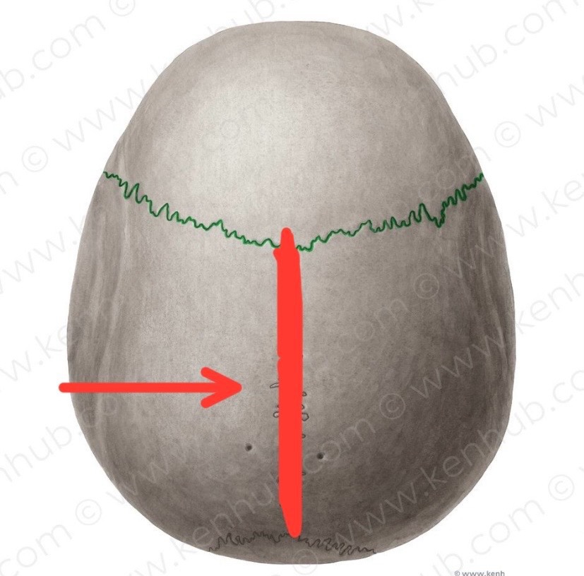
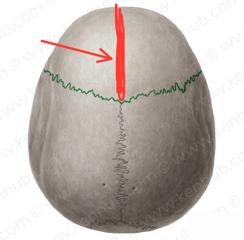
Sagittal
Right Parietal + Left Parietal Midline, top of skull
Coronal
Frontal + Parietal (both)
Crosses skull transversely
Metopic (Frontal)
Two halves of frontal bone Midline of forehead (closes in childhood; may persist)
Squamous
Parietal + Temporal Side of skull (flat overlapping)
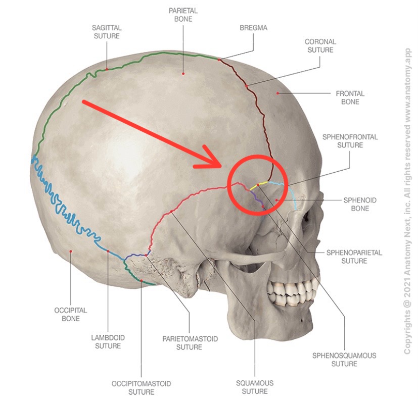
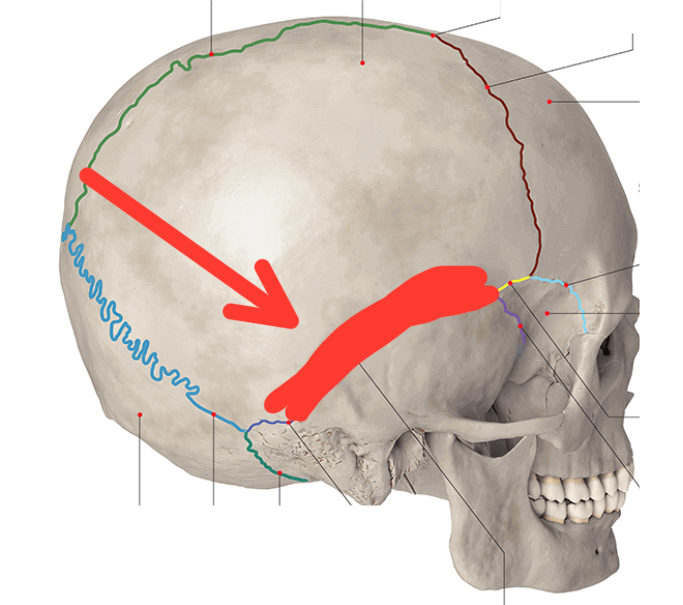
Pterion (junction)
The Pterion is the H-shaped meeting of Frontal, Parietal, Temporal, Sphenoid bones. It's a suture junction, not a suture itself, but it’s a critical landmark because the middle meningeal artery runs beneath it. Fracture → epidural haematoma.