5 Interpretation of Dental Radiographs
 Call Kai
 Learn
Learn Practice Test
 Spaced Repetition
 Match
Match Flashcards
 Knowt Play
1/69

There's no tags or description
Looks like no tags are added yet.
Name | Mastery | Learn | Test | Matching | Spaced | Call with Kai | Chat |
|---|
No analytics yet
Send a link to your students to track their progress
70 Terms
Interradicular bone
What is 1?
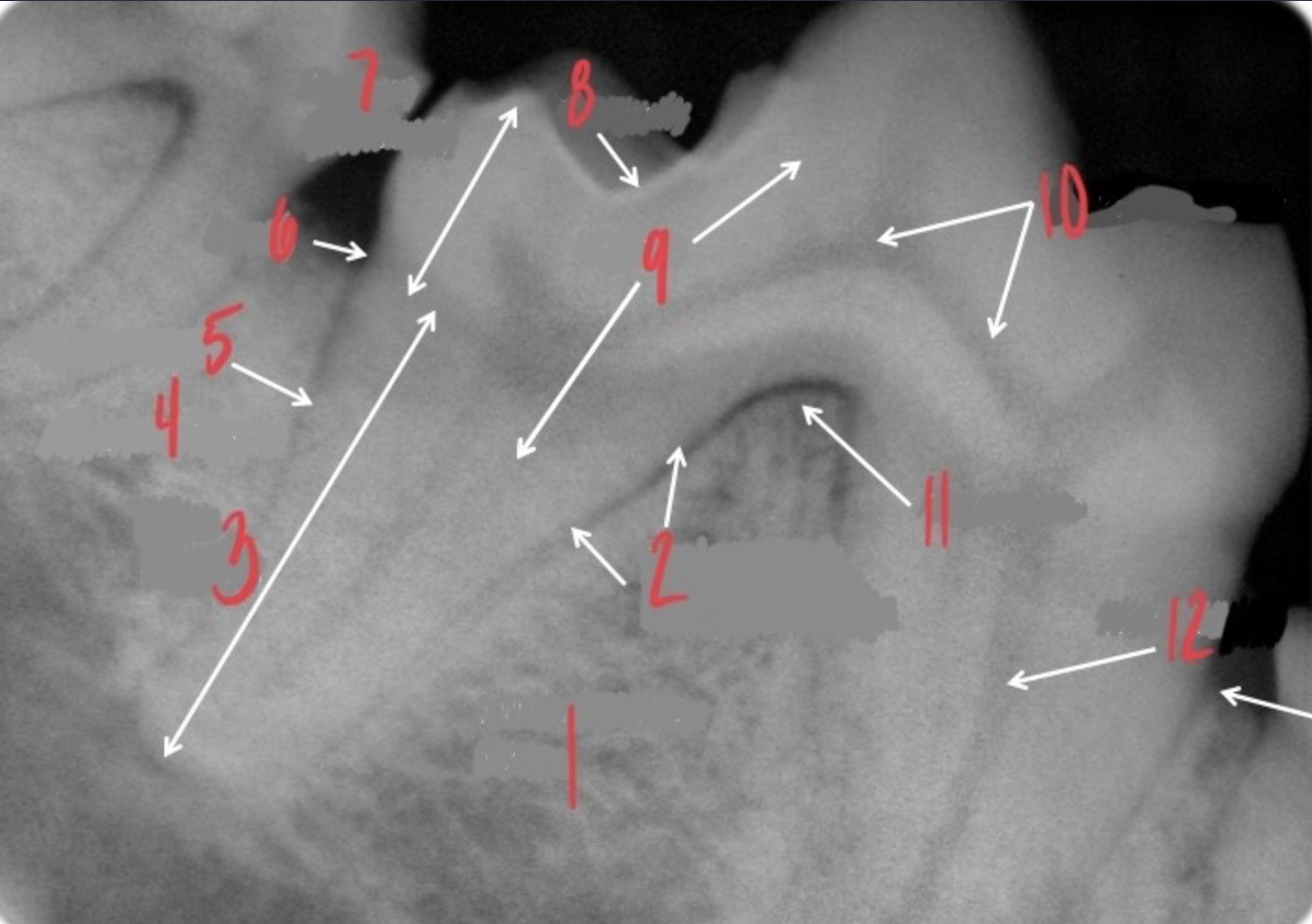
Periodontal ligament space
What is 2?
Root (apical)
What is 3?
Alveolar bone
What is 4?
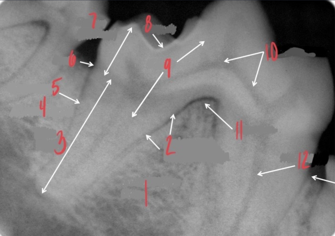
Cementum
What is 5?
Neck
What is 6?
Crown (coronal)
What is 7?
Enamel
What is 8?
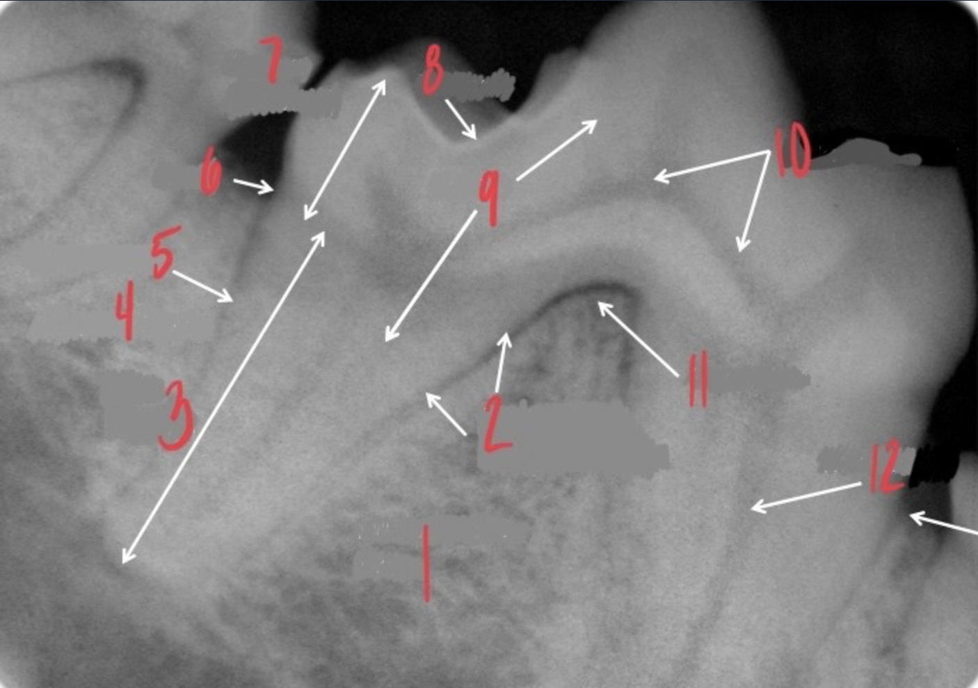
Dentin
What is 9?
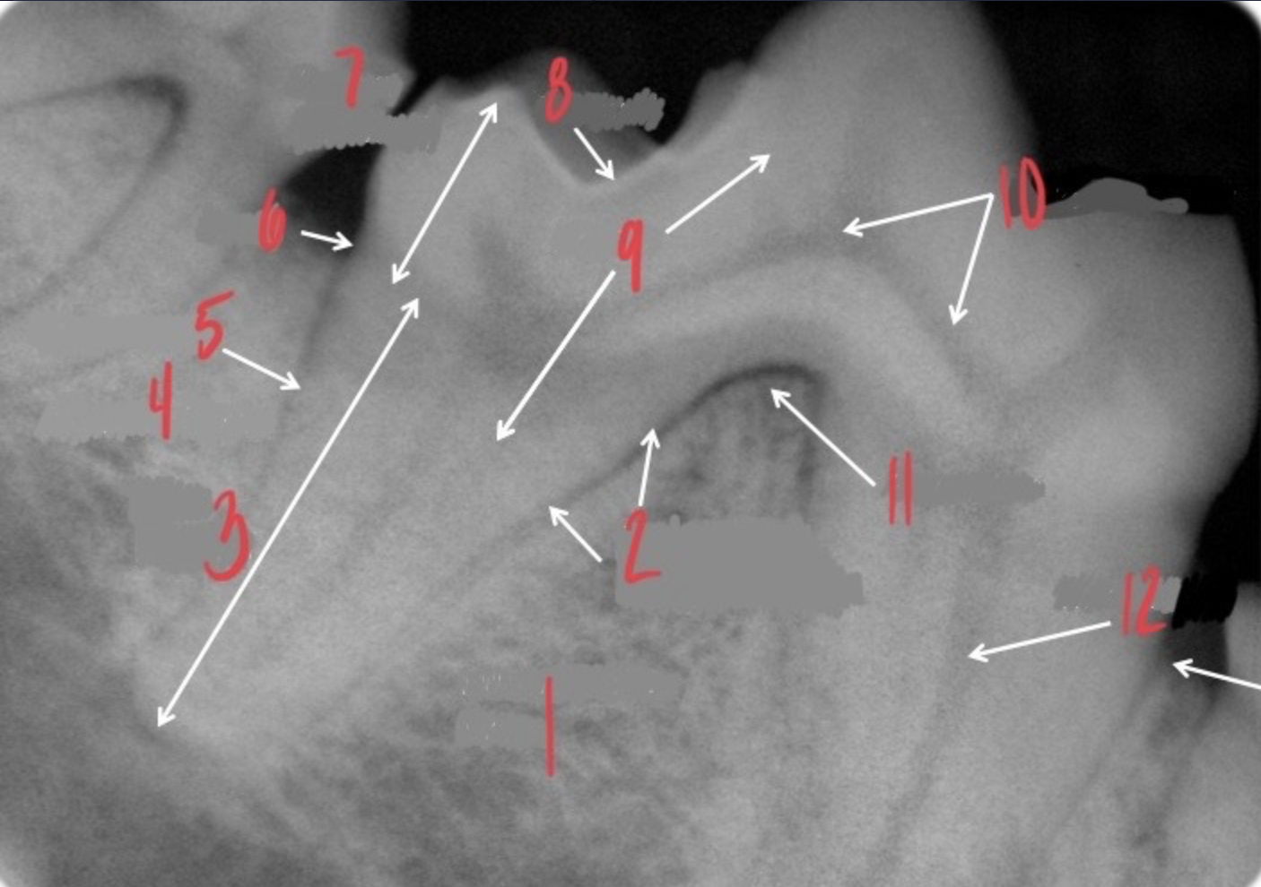
Pulp chamber
What is 10?
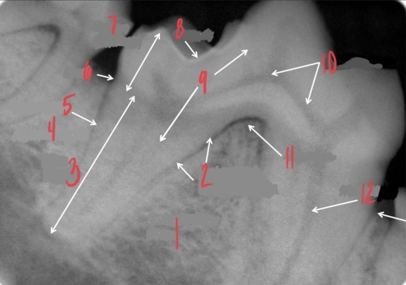
Furcation
What is 11?
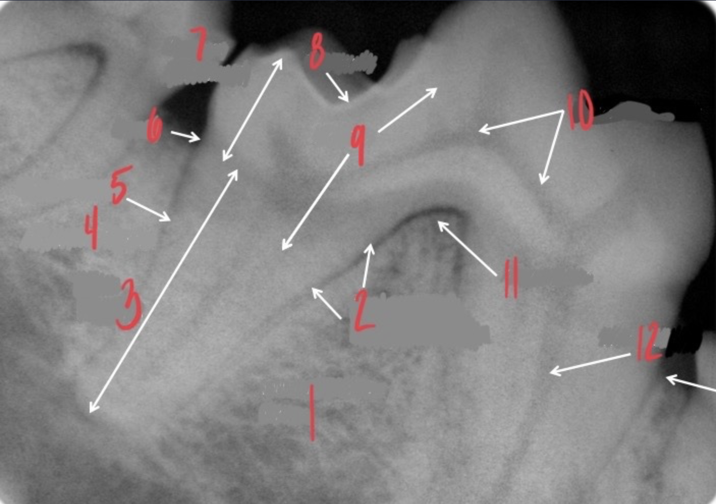
Root canal
What is 12?
304 on the left side = mandible
Facing the dog
What arcade and number is 2?
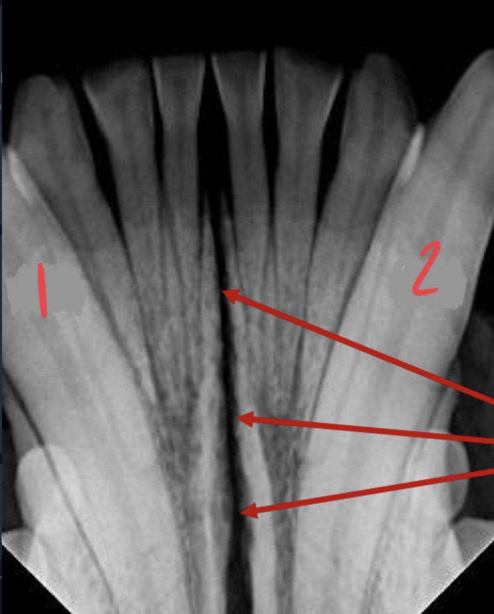
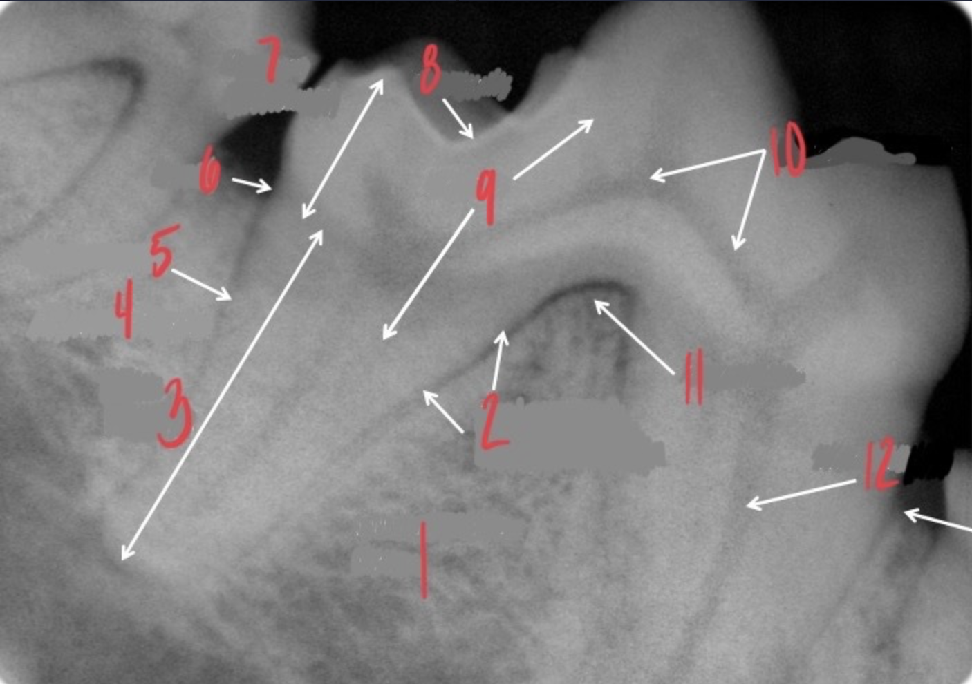
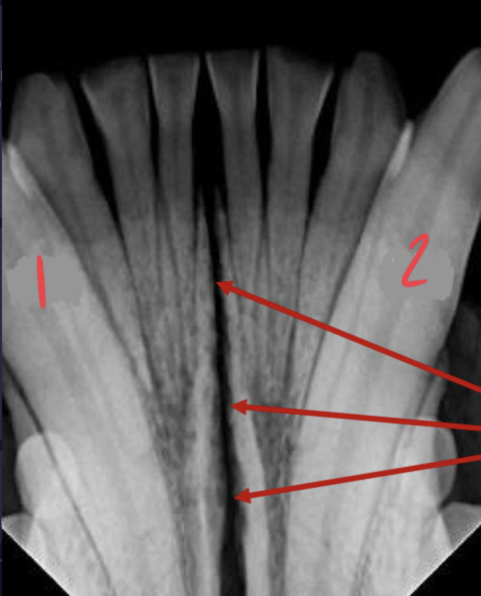
Mandibular symphysis
What are the arrows pointing to?
404 on the right side = mandible
FACING the dog
What arcade and number is 1?
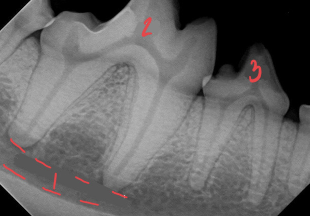
Mandibular canal
What are the dotted lines of 3 supposed to represent?
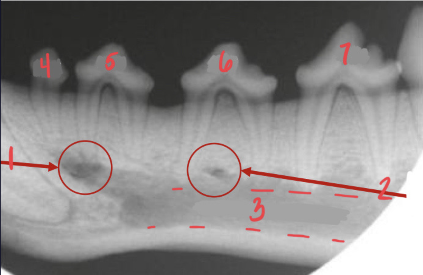
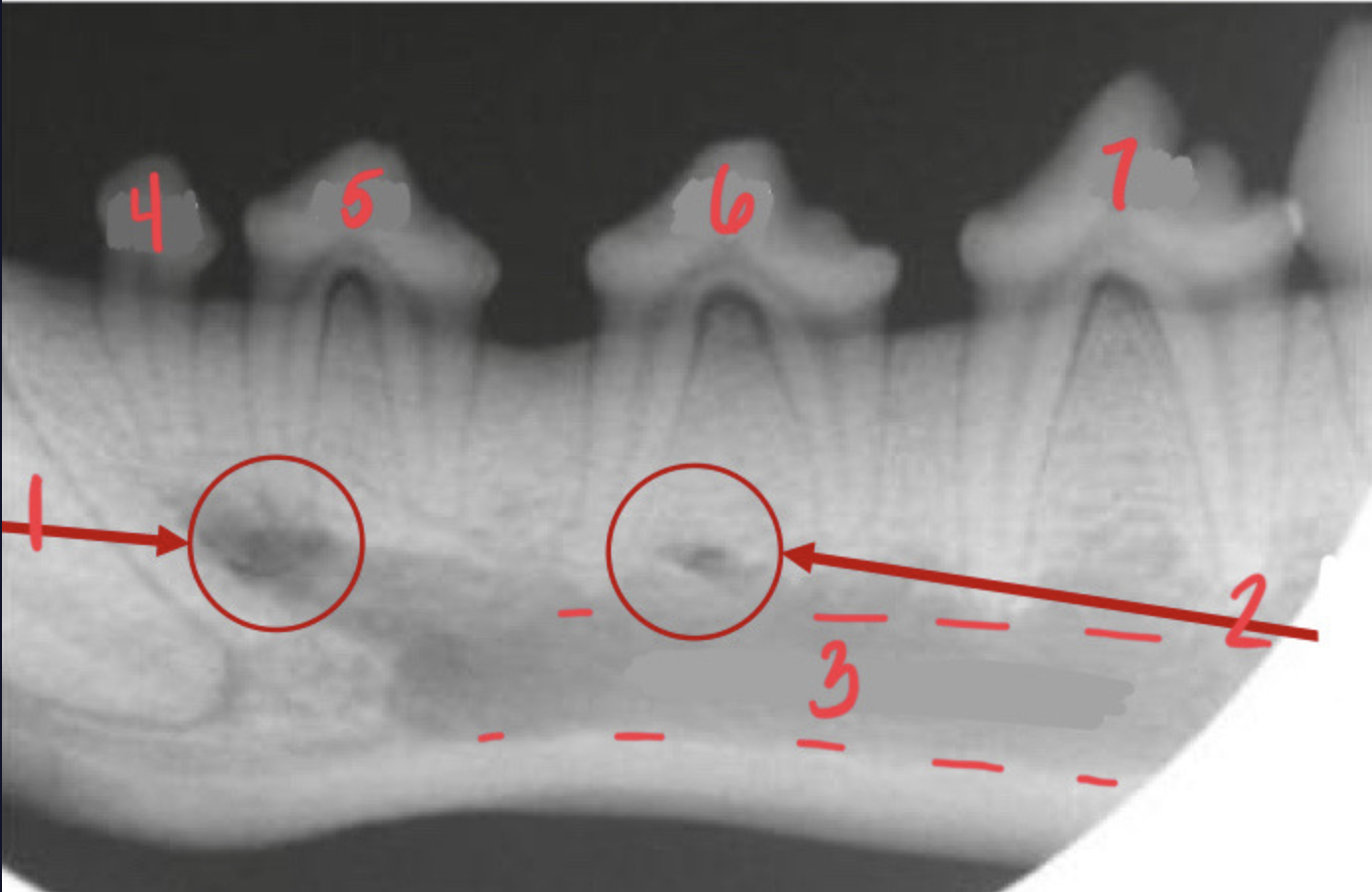
Middle mental foramen (on the mandible)
What is 1?
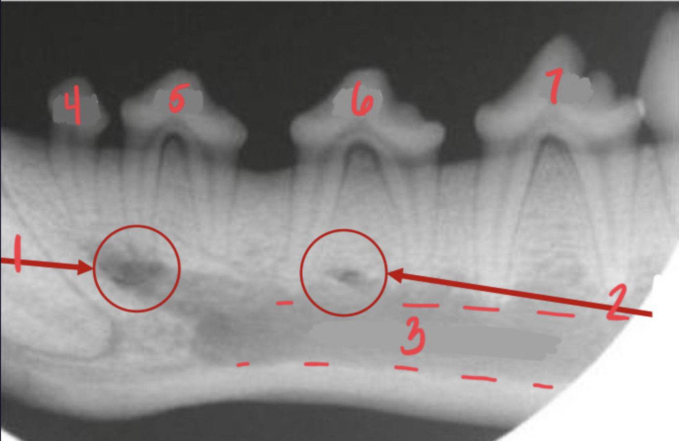
Caudal mental foramen
What is 2?
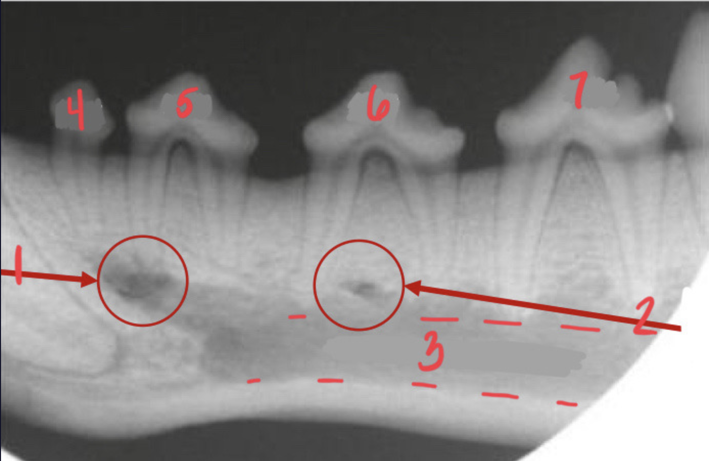
305, first premolar with 1 root = mandible
What is 4? What arcade are you in?
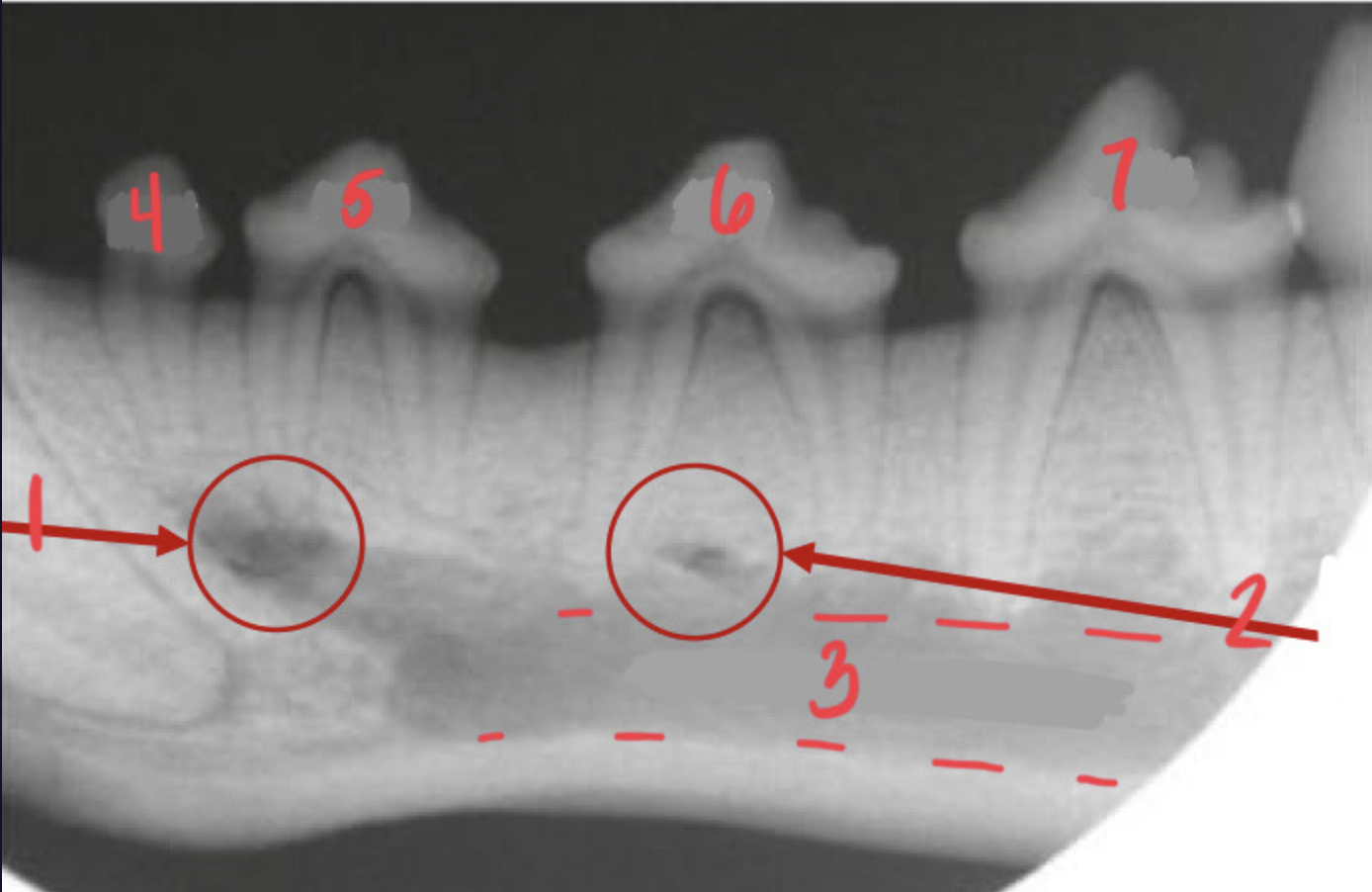
306 = mandible
What is 5? What arcade are you in?
307 = mandible
What is 6? What arcade are you in?
308 = mandible
What is 7? What arcade are you in?
Mandibular canal
What is 1?
409 = mandible. to the right is rostral meaning this is the right side of the mouth
What is 2? What arcade are you in? Which way is rostral?
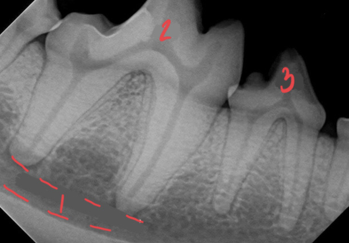
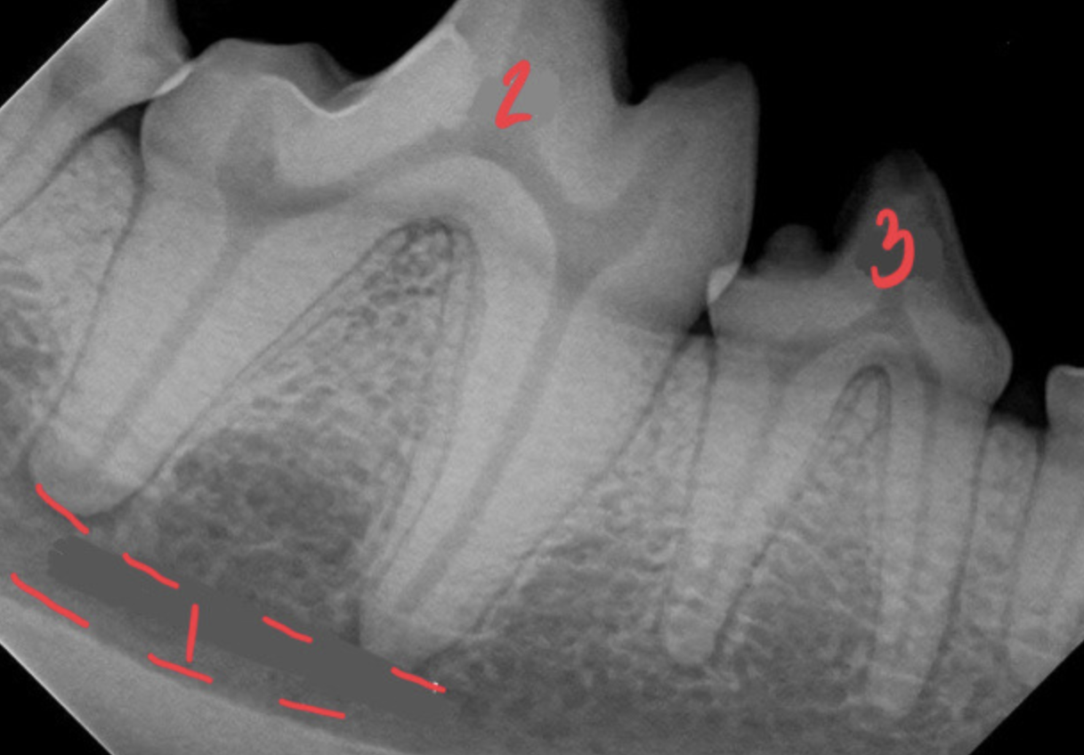
408 = mandible. to the right is rostral meaning this is the right side of the mouth (the low to high peaks point right)
What is 3? What arcade are you in? Which way is rostral?
Mandibular canal
What is 1?
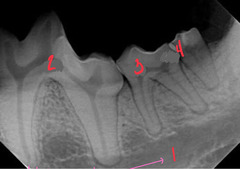
309 = mandible. To the left is rostral meaning this is the left side of the face
What is 2? Which arcade are you in? Which way is rostral?
310 = mandible. the left is rostral meaning left side of the face
What is 3? Which arcade are you in? Which way is rostral?
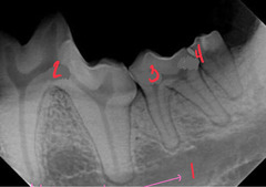
311 = mandible. the left is rostral
What is 4? Which arcade are you in? Which way is rostral?
Palatine fissures which are only present in the maxilla
What are the arrows at 1 pointing to?
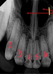
103 = right side maxilla
What is 2? What arcade are you in?
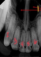
102 = right side maxilla
What is 3? What arcade are you in?
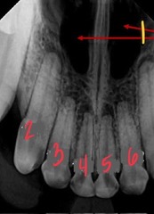
101 = right side maxilla
What is 4? What arcade are you in?
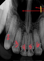
201 = left side maxilla
What is 5? What arcade are you in?
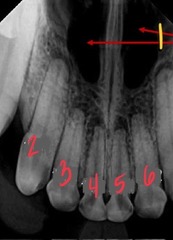
202 = left side maxilla
What is 6? What arcade are you in?
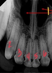
Nasal surface of the alveolar process of the maxilla
What are the arrows of 4 pointing to?
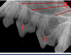
205 = maxilla's 1st premolar with 1 root (to the left is rostral, meaning youre on left side of the face)
What is 1? What arcade are you in?
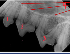
206 = maxilla
What is 2? What arcade are you in?
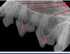
207 = maxilla. to the left is rostral meaning this is the left side of the face
What is 3? What arcade are you in? Which direction is rostral?
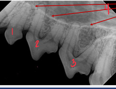
208, the 4th maxillary premolar. the arrows are pointing to the large 3 roots of the tooth. this means it has to be the maxilla because the mandible doesn't have any 3 rooted teeth
What is 1? What are the arrows pointing to?
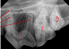
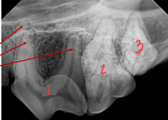
209 = maxilla
What is 2? What arcade are you in?
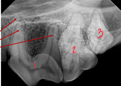
210 = maxilla
What is 3? What arcade are you in?
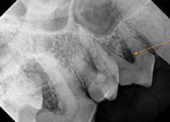
Furcation in maxilla
What is this pointing to?
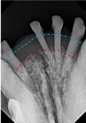
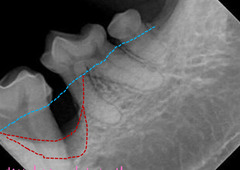
Mandible. There lines represent the original alveolar margin in blue and alveolar margin after periodontal disease attachment loss in red
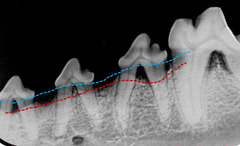
horizontal bone loss
What are these lines supposed to represent and why do we care? Where are you?
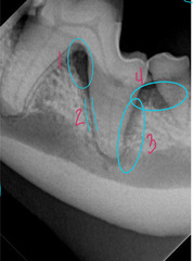
Furcation
What is 1?
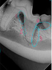
Normal periodontal ligament space
What is 2?
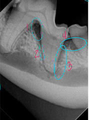
Some vertical bone loss
What is 3?
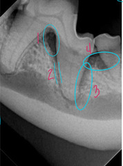
Horizontal bone loss
What is 4?
Horizontal
What pattern of bone loss is this?
Vertical
What pattern of bone loss is this?
The tooth on the left of the image is abnormal (404). Enlarged pulp chamber and root canal shows a diseased fragile tooth. There is also loss of lamina dura
secondary dentin is not filling in = dead tooth
Which tooth is normal, which is diseased? How do you know?
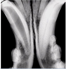
Endodontic disease = enlarged pulp cavity with periapical lucency (309)
Furcation with horizontal bone loss
What is happening here?
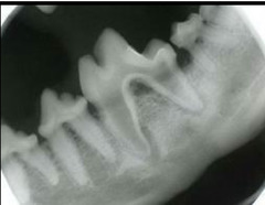
NORMAL lamina dura = the normal bright white line below the canine tells you the darker space between the tooth and the line is actually NOT apical bone loss and is completely normal
Describe what you are seeing here
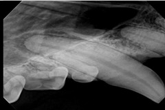
The right side of the image is healthy. You can see the bright white lamina dura and healthy periodontal ligament between the tooth and dura.
The left side of the image shows the periodontal ligament breaking apart because there is NO normal lamina dura, meaning the dark space is all apical bone loss
Which side is normal? Abnormal? Describe what you are looking at
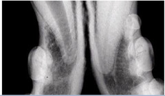
Periapical lucency
What are the arrows pointing to?
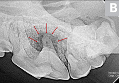
stage 1, 2, 3, 4a, 4b, 4c, and 5
Name the tooth resorption stages
Cats
Tooth resorption is more common in *cats or dogs*?
Stage 2
Which tooth resorption stage?
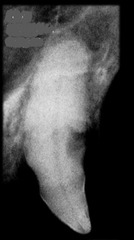
Stage 3
Which tooth resorption stage?
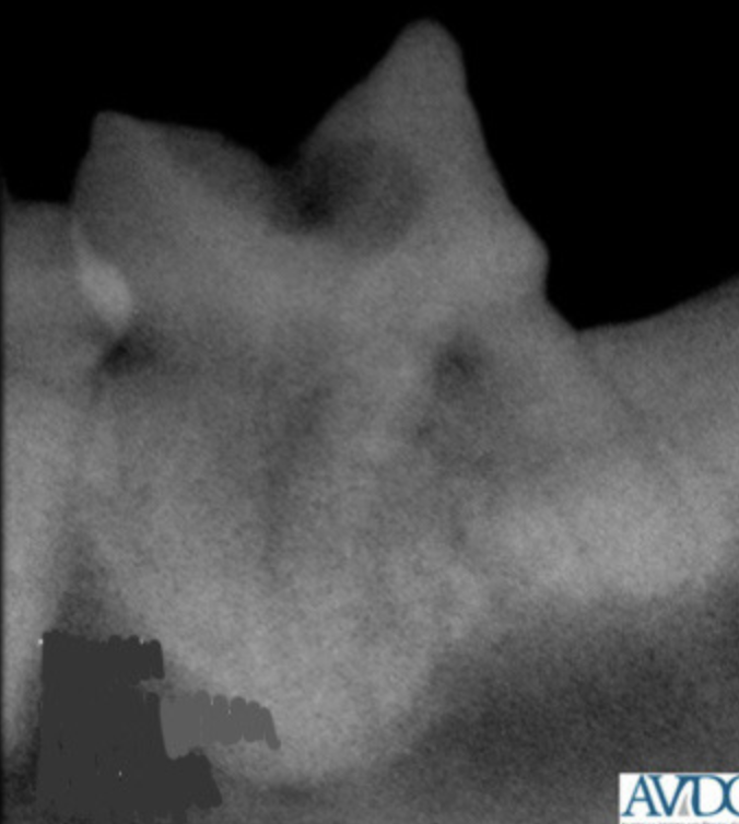
Stage 4a
Which tooth resorption stage?
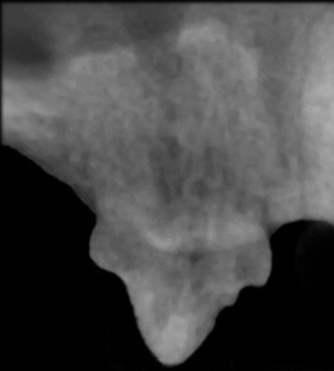
Stage 1: periodontal ligament space is still apparent but you should extract the tooth with open surgical technique
Which tooth resorption stage? What is happening and what should you do?
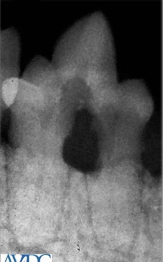
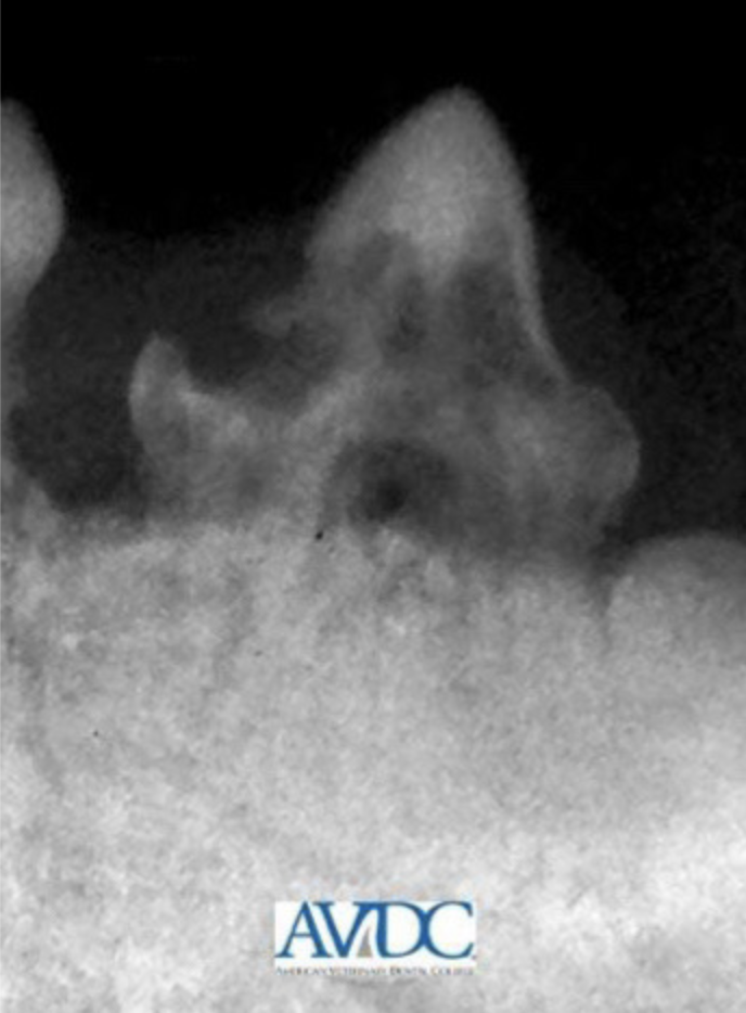
Stage 2: there is a lytic area on the top of the tooth and decrease radiopacity of part of the tooth, narrowing of the periodontal ligament in some areas.
Crown amputation only with intentional root retention indicated (aka leave roots behind because the cat will resorb them and has already started)
Which tooth resorption stage? What is happening and what should you do?
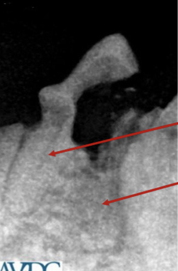
Stage 3: for the top arrow, extract the root segment, but for the bottom arrow crown amputation only for this root segment. There has been lytic activity in this tooth, there is no normal structure, and the periodontal ligament is gone
Note: stage 3 is a combination of type 1 and 2
Which tooth resorption stage? What is happening and what should you do?
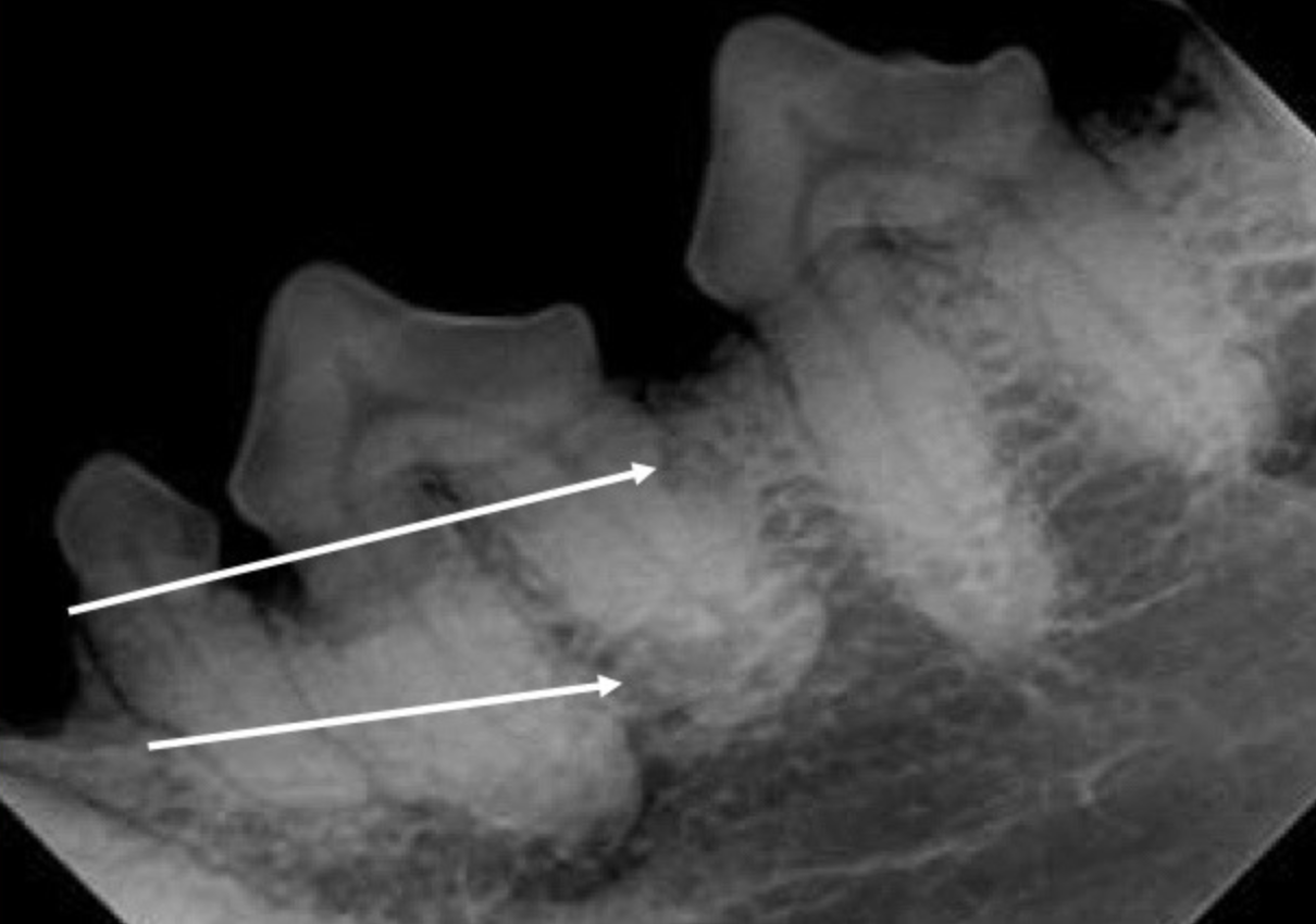
Mandibular premolars often starting in the roots
Tooth resorption is less common in dogs. If it does occur, where does it happen?
Tooth resorption
What is happening here in this dog?
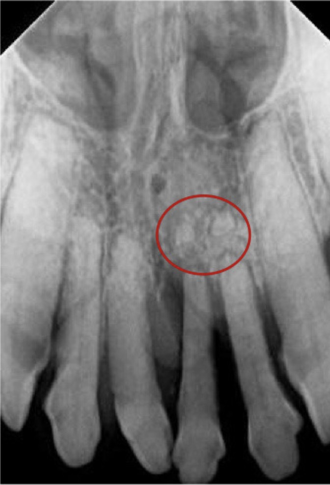
Fractured root tips
What is happening here?
Roots
If you CANNOT safely remove the roots = REFER
What do you have to make sure you DO NOT LEAVE BEHIND when doing dental extractions?
Dilacerated mesial root (it is a fracture risk if you try and remove). Occurs when small breed dogs have large teeth in relation to the size of their mandibles
REFER
What is this? Why does it commonly occur?
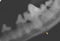
The tooth has a fracture (around the neck i think) and apical lucency
Describe this image
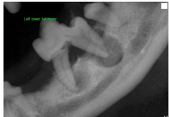
Roots are left behind
What is happening in this post extraction radiograph?