Pelvic Inflammatory Disease and Infertility chapt. 21 PENNY
1/59
There's no tags or description
Looks like no tags are added yet.
Name | Mastery | Learn | Test | Matching | Spaced | Call with Kai |
|---|
No analytics yet
Send a link to your students to track their progress
60 Terms
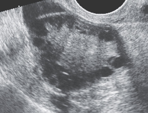
1. The 24-year-old patient in Figure 21-25 presented with hirsutism and obesity. What is the most likely clinical diagnosis based on these sonographic findings?
a. Asherman syndrome
b. PCOS
c. Endometriosis
d. Fitz-Hugh–Curtis syndrome
b. PCOS
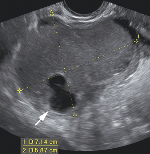
2. The patient in Figure 21-26 complained of dysmenorrhea and infertility. The structure between the calipers was noted in the right adnexa. What is the most likely diagnosis?
a. Brenner tumor
b. PCOS
c. Endometrioma
d. Endometrioid
c. Endometrioma
3. What is the term for inflammation of connective tissue adjacent to the uterus?
a. Myometritis
b. Salpingitis
c. PID
d. Parametritis
d. Parametritis
4. Which of the following is not true concerning PID?
a. PID often results in pelvic adhesions.
b. PID often results in dyspareunia.
c. PID often results in endometritis.
d. PID often results in leukopenia.
d. PID often results in leukopenia.
5. Which of the following would be the likely cause of the production of artifactual echoes being produced from the endometrium in a patient with known PID?
a. Dilated blood vessels
b. Pneumouterus
c. Endometriosis
d. Pyosalpinx
b. Pneumouterus
6. What is inflammation of the ovary?
a. Oophoritis
b. Ovaritis
c. Oogenesis
d. Parametritis
a. Oophoritis
7. Which of the following may require hysteroscopic uterine septoplasty in the case of infertility?
a. Endometriosis
b. Asherman syndrome
c. PCOS
d. Septate uterus
d. Septate uterus
8. What is the most common location of endometriosis?
a. Ovary
b. Uterus
c. Fallopian tube
d. Bowel
a. Ovary
9. Which of the following is an endocrinologic ovarian disorder?
a. Asherman syndrome
b. Ovulation induction
c. Endometriosis
d. PCOS
d. PCOS

10. The patient in Figure 21-27 is undergoing ovulation induction. What type of ovarian cysts are noted in this image?
a. Corpus luteal cysts
b. Polycystic ovarian cysts
c. Theca lutein cysts
d. Nabothian cysts
c. Theca lutein cysts
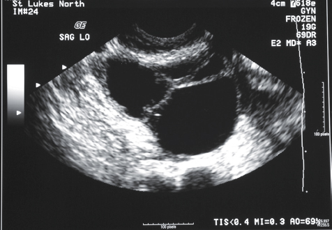
11. The patient in Figure 21-27 is also suffering from electrolyte imbalance, oliguria, and nausea. The patient also has ascites. What other sonographic finding is most likely present?
a. Pleural effusion
b. Hepatic periportal cuffing
c. Pancreatic enlargement
d. Subhepatic adhesions
a. Pleural effusion
12. Which of the following may present like gallbladder disease in the patient suffering from PID?
a. Ovarian torsion
b. PCOS
c. Fitz-Hugh–Curtis syndrome
d. Endometriosis
c. Fitz-Hugh–Curtis syndrome
13. The ovarian volume for the diagnosis of PCOS should not exceed:
a. 4 mL.
b. 5 mL.
c. 8 mL.
d. 10 mL.
d. 10 mL.
14. Which of the following would least likely cause of the development of intrafallopian tube synechiae?
a. Long-standing PID
b. Endometriosis
c. Tubal surgery
d. Sonohysterography
d. Sonohysterography
15. Which of the following is not a way sonography assists in reproductive technology?
a. Provides a definitive diagnosis for infertility
b. Provides a way to monitor ovulation
c. Provides imaging assistance during follicular aspiration
d. Provides imaging assistance during oocyte retrieval
a. Provides a definitive diagnosis for infertility
16. What blood disorder is associated with OHS?
a. Sickle cell disease
b. Hyperlipidemia
c. Thromboembolism
d. Polycythemia
c. Thromboembolism
17. What is inflammation of the muscular part of the uterus?
a. Perimetritis
b. Myometritis
c. Cervicitis
d. Endometritis
b. Myometritis
18. Which of the following would not appear as a “T” shaped IUD with sonography?
a. Mirena
b. Liletta
c. Lippes loop
d. Kyleena
c. Lippes loop
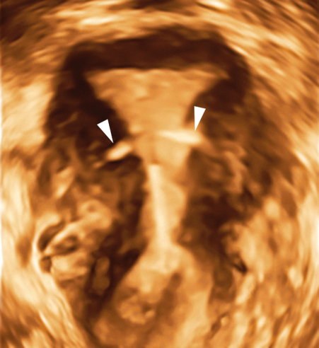
19. The patient in Figure 21-28 presented with a lost IUD string and vaginal bleeding. What is the most likely diagnosis?
a. The IUD is located within the endometrium.
b. The IUD is located within the myometrium.
c. The IUD is located within both the endometrium and the myometrium.
d. The IUD is located within the endometrium, myometrium, and through the perimetrium.
c. The IUD is located within both the endometrium and the myometrium.

20. What would the patient also likely suffer from in Figure 21-28?
a. Uterine synechiae
b. PID
c. Endometriosis
d. Uterine cramping
d. Uterine cramping
21. What is the radiographic procedure used to evaluate the patency of the fallopian tubes?
a. Sonohysterography
b. Hysterosalpingography
c. Hysteroscopy
d. Hysteroscopic fallopian septoplasty
b. Hysterosalpingography
22. The sonographic finding of a tubular, simple-appearing, anechoic structure within the adnexa is most consistent with:
a. dyspareunia.
b. hematometra.
c. hydrosalpinx.
d. endometritis.
c. hydrosalpinx.
23. All of the following are considered risk factors for PID except:
a. IUD.
b. multiple sexual partners.
c. post childbirth.
d. uterine leiomyoma.
d. uterine leiomyoma.
24. Which of the following would be the least likely clinical finding for a patient with endometriosis?
a. Pelvic pain
b. Dysmenorrhea
c. Painful bowel movements
d. Hyperandrogenism
d. Hyperandrogenism
25. Which of the following is not a potential cause of PID?
a. Intrauterine contraception use
b. Postabortion
c. Chlamydia
d. Pyelonephritis
d. Pyelonephritis
26. A patient presents to the sonography department with a fever, chills, and vaginal discharge. Sonographically, what findings would you most likely not encounter?
a. Cul-de-sac fluid
b. Uterine adhesions
c. Dilated uterine tubes
d. Ill-defined uterine border
b. Uterine adhesions
27. A 26-year-old patient presents to the sonography department with a history of infertility and oligomenorrhea. Sonographically, you discover that the ovaries are enlarged and contain multiple, small follicles along their periphery, with prominent echogenic stromal elements. What is the most likely diagnosis?
a. Ovarian torsion
b. OHS
c. PID
d. PCOS
d. PCOS
28. The most common initial clinical presentation of PID is:
a. endometritis.
b. tubo-ovarian abscess.
c. vaginitis.
d. pyosalpinx.
c. vaginitis.
29. Sonographic findings of the endometrium in a patient with a history of PID, fever, and elevated white blood cell count would include all of the following except:
a. ring-down artifact posterior to the endometrium.
b. thin, hyperechoic endometrium.
c. endometrial fluid.
d. thickened, irregular endometrium.
b. thin, hyperechoic endometrium
0. What is another name for an endometrioma?
a. Dermoid
b. Teratoma
c. Chocolate cyst
d. String of pearl
c. Chocolate cyst
31. Fitz-Hugh–Curtis syndrome could be described as:
a. clinical findings of gallbladder disease as a result of PID.
b. the presence of uterine fibroids and adenomyosis in the gravid uterus.
c. coexisting intrauterine and extrauterine pregnancies.
d. the presence of pyosalpinx, hydrosalpinx, and endometritis.
a. clinical findings of gallbladder disease as a result of PID.
32. All of the following statements concerning PID are true except:
a. PID is typically a unilateral condition.
b. PID can be caused by douching.
c. PID can lead to a tubo-ovarian abscess.
d. dyspareunia is a clinical finding in acute PID.
a. PID is typically a unilateral condition.
33. A patient presents to the sonography department with complaints of infertility and painful menstrual cycles. Sonographically, you discover a cystic mass on the ovary consisting low-level echoes. Based on the clinical and sonographic findings, what is the most likely diagnosis?
a. Cystic teratoma
b. Endometrioma
c. PID
d. OHS
b. Endometrioma
34. The development of adhesions between the liver and the diaphragm as a result of PID is termed:
a. Fitz-Hugh–Curtis syndrome.
b. Dandy–Walker syndrome.
c. Stein–Leventhal syndrome.
d. Asherman syndrome.
a. Fitz-Hugh–Curtis syndrome.
35. Assisted reproductive therapy can result in all of the following except:
a. heterotopic pregnancy.
b. multiple gestations.
c. OHS.
d. Asherman syndrome.
d. Asherman syndrome
36. PCOS may also be referred to as:
a. Fitz-Hugh–Curtis syndrome.
b. Plateau syndrome.
c. Stein–Leventhal syndrome.
d. Asherman syndrome.
c. Stein–Leventhal syndrome.
37. PID can lead to all of the following except:
a. infertility.
b. polycystic ovarian disease.
c. ectopic pregnancy.
d. scar formation in the fallopian tubes.
b. polycystic ovarian disease.
38. What term is used to describe painful intercourse?
a. Dyspareunia
b. Dysuria
c. Dysmenorrhea
d. Dysconception
a. Dyspareunia
39. The presence of functional, ectopic endometrial tissue outside the uterus is termed:
a. adenomyosis.
b. Asherman syndrome.
c. Fitz-Hugh–Curtis syndrome.
d. endometriosis.
d. endometriosis.
40. All of the following are sonographic findings of a tubo-ovarian abscess except:
a. the presence of 10 or more small cysts along the periphery of the ovaries.
b. cul-de-sac fluid.
c. thickened, irregular endometrium.
d. fusion of the pelvic organs as a conglomerated mass.
a. the presence of 10 or more small cysts along the periphery of the ovaries.
41. A patient presents to the sonography department with a history of chlamydia and suspected PID. Which of the following would be indicative of the typical sonographic findings of PID?
a. Enlarged cervix, thin endometrium, and theca lutein cysts
b. Atrophic uterus, free fluid, and small ovaries
c. Bilateral, cystic enlargement of the ovaries with no detectable flow
d. Thickened irregular endometrium, cul-de-sac fluid, and complex adnexal masses
d. Thickened irregular endometrium, cul-de-sac fluid, and complex adnexal masses
42. Causes of female infertility include all of the following except:
a. previous IUD use.
b. PCOS.
c. Asherman syndrome.
d. endometriosis.
a. previous IUD use.
43. Infertility is defined as:
a. the inability to conceive a child after 2 years of unprotected intercourse.
b. the inability to conceive a child after 5 years of unprotected intercourse.
c. the inability to conceive a child after 1 year of unprotected intercourse.
d. the inability to conceive a child after 3 months of unprotected intercourse.
c. the inability to conceive a child after 1 year of unprotected intercourse.
44. A 25-year-old patient presents to the sonography department complaining of pelvic pain, dyspareunia, and oligomenorrhea. An ovarian mass, thought to be a chocolate cyst, is noted during the examination. Which of the following is consistent with the sonographic appearance of a chocolate cyst?
a. Simple-appearing anechoic mass
b. Echogenic mass with posterior shadowing
c. Cystic mass with low-level echoes
d. Anechoic mass with posterior shadowing
c. Cystic mass with low-level echoes
45. Amenorrhea, hirsutism, and obesity describe the clinical features of:
a. Fitz-Hugh–Curtis syndrome.
b. Stein–Leventhal syndrome.
c. Asherman syndrome.
d. endometriosis.
b. Stein–Leventhal syndrome.
46. The sonographic evidence of a hyperemic fallopian tube is consistent with:
a. pyosalpinx.
b. hydrosalpinx.
c. endometritis.
d. salpingitis.
d. salpingitis.
47. The sonographic “string of pearls” sign is indicative of:
a. PCOS.
b. tubo-ovarian disease.
c. PID.
d. OHS.
a. PCOS.
48. Complex-appearing fluid within the fallopian tubes seen with PID is most likely:
a. pyosalpinx.
b. pyometra.
c. hydrosalpinx.
d. hematometra
a. pyosalpinx.
49. Sonographic findings of OHS include all of the following except:
a. cystic enlargement of the ovaries.
b. ascites.
c. pleural effusions.
d. oliguria.
d. oliguria.
50. The development of adhesions within the uterine cavity is termed:
a. Fitz-Hugh–Curtis syndrome.
b. Dandy–Walker syndrome.
c. Stein–Leventhal syndrome.
d. Asherman syndrome.
d. Asherman syndrome.
51. OHS can cause multiple large follicles to develop on the ovaries termed:
a. theca lutein cysts.
b. chocolate cysts.
c. corpus luteum cysts.
d. dermoid cysts.
a. theca lutein cysts.
52. What is another name for adhesions within the endometrial cavity?
a. Endometritis
b. Synechiae
c. Septation
d. Mural nodules
b. Synechiae
53. A female patient presents to the sonography department with a clinical history of Clomid treatment. (infertility medication) She is complaining of nausea, vomiting, and abdominal distension. What circumstance is most likely causing her clinical symptoms?
a. Stein–Leventhal syndrome
b. Polycystic ovarian disease
c. Fitz-Hugh–Curtis syndrome
d. OHS
d. OHS
54. A 35-year-old patient presents to the sonography department with a history of tubal ligation and positive pregnancy test. What condition should be highly suspected?
a. Asherman syndrome
b. Polycystic ovarian disease
c. Endometriosis
d. Ectopic pregnancy
d. Ectopic pregnancy
55. Patients with OHS are at increased risk for:
a. ovarian torsion.
b. chlamydia.
c. gonorrhea.
d. vaginitis.
a. ovarian torsion.
56. Which of the following would be described as functional cysts that are found in the presence of elevated levels of hCG?
a. Theca lutein cysts
b. Chocolate cysts
c. Corpus luteum cysts
d. Endometrial cysts
a. Theca lutein cysts
57. The presence of pus within the uterus defines:
a. pyosalpinx.
b. pyometra.
c. pyocolpos.
d. pyomyoma.
b. pyometra.
58. The occurrence of having both intrauterine and extrauterine pregnancies at the same time describes:
a. PID.
b. ectopic pregnancy.
c. heterotopic pregnancy.
d. molar pregnancy.
c. heterotopic pregnancy.
59. Excessive hair growth in women in areas where hair growth is normally negligible would be seen with:
a. ectopic pregnancy.
b. Fitz-Hugh–Curtis syndrome.
c. Asherman syndrome.
d. Stein–Leventhal syndrome.
d. Stein–Leventhal syndrome.
60. What form of permanent birth control would be seen sonographically as echogenic, linear structures within the lumen of both isthmic portions of the fallopian tubes?
a. Essure devices.
b. ParaGard.
c. Lippes loop.
d. Mirena.
a. Essure devices.