W2: Principles and actions of eye movements
1/44
There's no tags or description
Looks like no tags are added yet.
Name | Mastery | Learn | Test | Matching | Spaced |
|---|
No study sessions yet.
45 Terms
5 main types of eye movement
Saccades
Smooth pursuit
Vergence
Optokinetic
Vestibular
Identify and define the 3 main categories of ocular positions
Primary position: eyes looking straight ahead with the head vertical
Secondary positions: any vertical or horizontal movement from the primary position
Tertiary positions: oblique movement of the eyes, a combination vertical and horizontal movement
Ocular movement occurs along the _________.
Equator
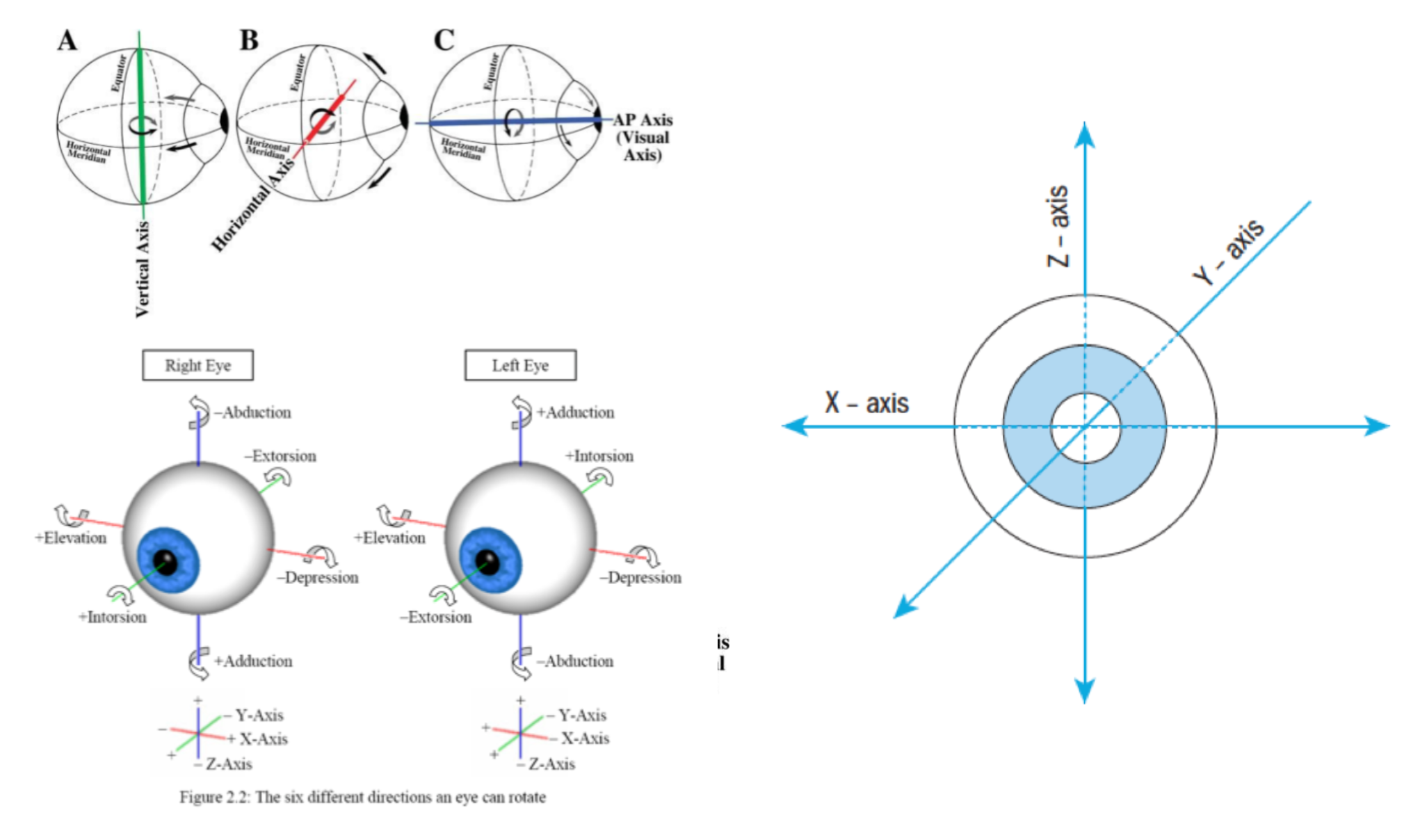
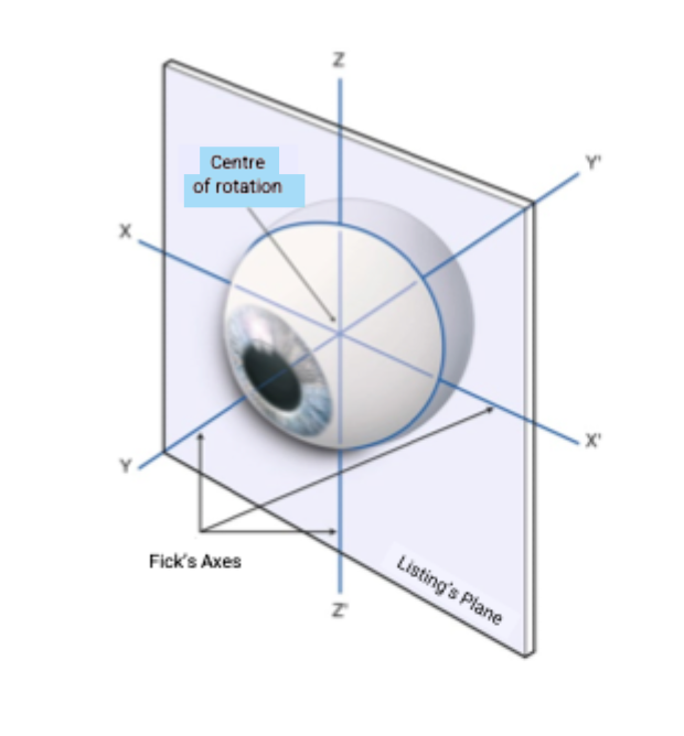
Describe Axes of Rotation (Fick’s axes)
Eye movements occur along 3 axes.
X axis - horizontal
Y axis - antero-posterior
Z axis - vertical
The different directions of eye movements occuring within the axes are:
X axis - vertical eye movement
Y axis - torsion
Z axis - horizontal eye movement
Describe Listing’s plane
An imaginary plane passing through the rotational centre of the eye, including the X and Z axes of Fick. Any movement into a tertiary position involves this plane.
Define ductions and identify the actions.
Monocular movements of the eye.
Adduction
Abduction
Supraduction
Infraduction
Incycloduction
Exocycloduction
Define adduction/adduction and identify the type of action.
Ductions
Adduction - towards the nose
Abduction - towards the temple
Define supraduction/infraduction and identify the type of action.
Duction
Supraduction - upwards
Infraduction - downwards
Define incycloduction/exocycloduction and identify the type of action.
Version
Incycloduction - rotation inwards (torsion) around anteroposterior axis
Exocycloduction - rotation outwards (torsion) around anteroposterior axis
Define versions and identify the actions.
Binocular movements where the eyes are moving in the same direction - CONJUGATE
Dextroversion
Laevoversion
Supraversion
Infraversion
Dextrocycloversion
Laevocycloversion
Define dextroversion/laevoversion and identify the type of action.
Version
Dextroversion - both eyes to the right
Laevoversion - both eyes to the left
Define supraversion/infraversion and identify the type of action.
Version
Supraversion - both eyes upwards
Infraversion - both eyes downwards
Define dextrocycloversion/laevocycloversion and identify the type of action.
Version
Dextrocycloversion - torsional rotation to right
Laevocycloversion - torsional rotation to left
Define vergences and identify the actions.
Binocular movements where the eyes are moving in the opposite direction - DISGUGATE
Convergence
Divergence
Supravergence
Incyclovergence
Exocyclovergence
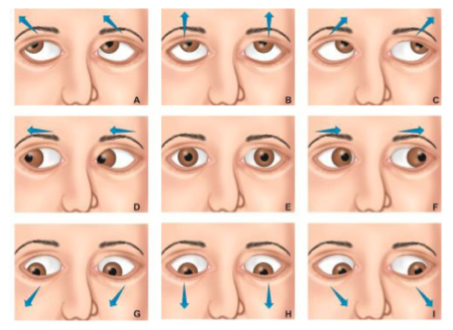
Identify the Cardinal Positions of Gaze
Primary position
Elevation
Depression
Laevoversion
Dextroversion
Dextroelevation
Laevoelevation
Dextrodepression
Laevodepression
Key components to the mechanics of ocular movements include:
Centre of rotation
Tangential point
Arc of contact
Pulleys
Muscle plane
Describe the centre of rotation. Where is it located?
The eye is a sphere in a socket that rotates freely around this centre of rotation.
Located 13.5mm behind cornea

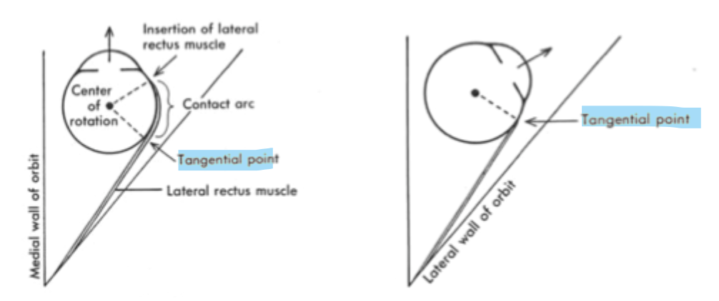
Describe the Tangential Point.
It is the first point of contact of the muscle onto the globe, described at the ‘effective‘ insertion of the muscle.
The position of this point varies when the muscle contracts/relaxes and the globe rotates
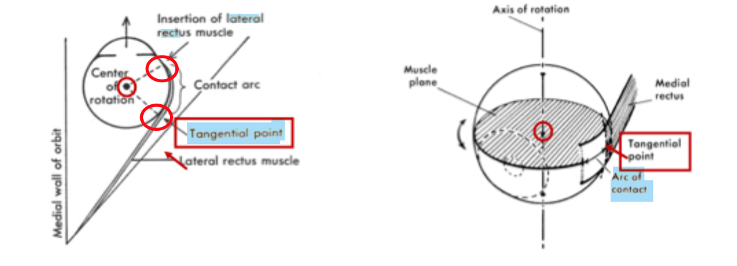
Describe the Arc of Contact.
The area between the anatomical insertion and point of first contact (tangential point). The muscle lies in direct contact with the globe.
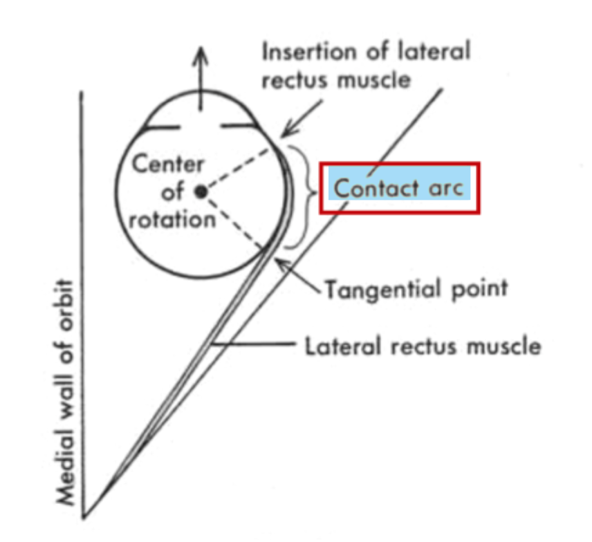
What happens to the Tangential Point when the muscle contracts?
As muscle contracts, the muscle unrolls from the globe. This causes the Tangential Point to move to Insertion point.
Describe extraocular muscle pulleys and their significance in eye movement.
They are connective tissue sleeves enveloping the rectus muscles.
Considered to function as the mechanical origin of the muscle
Stabilises rotation of the globe during complex eye movements
Prevents excessive retraction of the globe
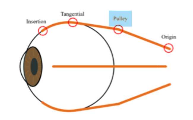
Identify the mechanical origin of extraocular muscles.
Muscle pulleys
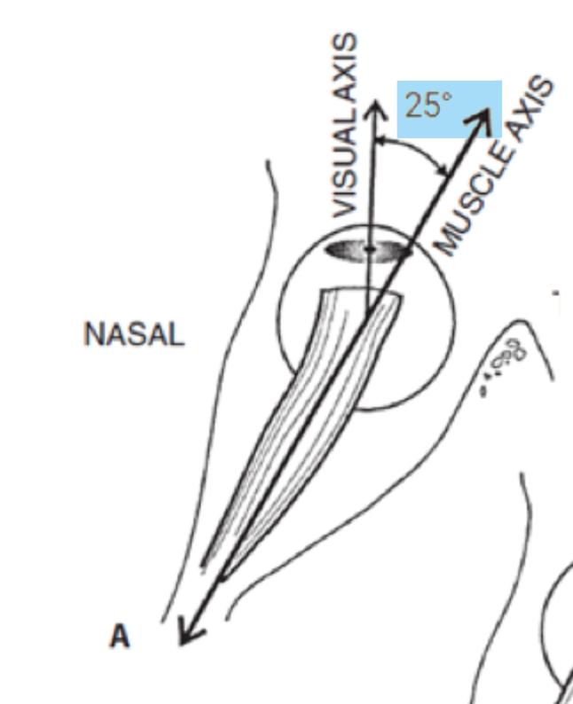
Muscle plane
Determine by a line drawn between the anatomical insertion and tangential point when the eye is in primary position.
Visual axis
An imaginary line extending from the viewed object through the centre of the pupil to the fovea
Relationship between muscle plane and visual axis
They are positioned 25º to each other
Primary action
Occurs when the muscle plane and visual axis coincide and muscle contracts
Primary action of medial rectus and position of eye
MP coincides with VA when eye is in primary position
Primary action is adduction
Primary action of lateral rectus and position of eye
MP coincides with VA when eye is in primary position
Primary action is abduction
Primary action of superior rectus and position of eye
Primary action is elevation
MP coincides with VA when eye is in 25º abduction
Primary action of inferior rectus and position of eye
Primary action is depression
MP coincides with VA when eye is in 25º abduction
Primary action of superior oblique and position of eye
Primary action is depression
MP coincides with VA when eye is in 55º adduction
Primary action of inferior oblique and position of eye
Primary action is elevation
MP coincides with VA when eye is in 50º abduction
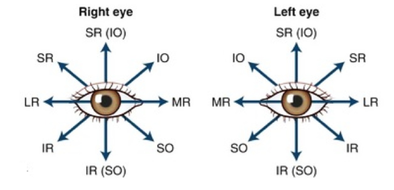
Draw the diagram relating to the fields of action.
Secondary actions
Occur when the visual axes and muscle plane are at 90º to one other
Secondary action of medial/lateral rectus and position of eye
Neither have a secondary action because the muscle planes coincide with the visual axes in primary position.
Secondary action of superior rectus and position of eye
Secondary action is intorsion and adduction
Occurs when eye adducts 65º
Secondary action of inferior rectus and position of eye
Secondary action is extortion and adduction
Occurs when eye adducts 65º
Secondary action of superior oblique and position of eye
Secondary action is intorsion and abduction
Occurs when eye abducts 35º
Secondary action of inferior oblique and position of eye
Secondary action is extorsion and abduction
Occurs when eye abducts 49º
Synergists
Two muscles working together to produce the same movement
Antagonists
Two muscles working against one another
When the agonist relaxes, then by reciprocal innervation, its antagonist relaxes
Yoke muscles
Contralateral synergists - the opposing muscles required to move both eyes into a particular position of gaze
Hering’s Law
Equal and simultaneous innervation flows to yoke muscles (contralateral synergists)
Whenever the impulse for a particular movement is sent out, corresponding muscles in either eyes recieve equal innervation to contract or relax
Sherrington’s Law
Whenever a change occurs in the innervation to an EOM, a proportionate but opposite change occurs in the antagonist.
Whenever an agonist receives the impulse to contract, the antagonist receives an equal impulse to relax and lengthen