Brain Structure: Developmental Perspective (2)
 Call Kai
 Learn
Learn Practice Test
 Spaced Repetition
 Match
Match Flashcards
 Knowt Play
1/86

There's no tags or description
Looks like no tags are added yet.
Name | Mastery | Learn | Test | Matching | Spaced | Call with Kai | Chat |
|---|
No analytics yet
Send a link to your students to track their progress
87 Terms
neural plate/tube becomes…
CNS
4 divisions of CNS that develop
prosencephalon, mesencephalon, rhombencephalon, caudal neural tube
prosencephalon becomes…
forebrain of diencephalon and telencephalon
mesencephalon becomes…
midbrain
rhombencephalon becomes…
hindbrain of cerebellum and brainstem (pons, medulla)
caudal neural tube becomes…
spinal cord
4 steps of CNS development and generally what happens
1) induction on neural plate → flat tissue becomes neural plate
2) neurulation → formation of neural tube from neural plate tissue due to induction signals (partly from notocord)
3) neural tube patterning → regions of brain begin to form due to morphogens
4) proliferation, differentiation, and migration → regions grow and produce neurons, which then differentiate and travel to correct location
PNS forms from…
neural crest → produces glial cells & PNS neurons, glial cells wrap around neurons
3 germinal layers of embryos & what they will become
inner: endoderm → internal organs
mesoderm → bones & muscle
outer: ectoderm → skin & nervous system
germinal layer from which the CNS & PNS
dorsal ectoderm layer → neural plate → neural ectoderm → CNS
most dorsal part of neural folds/closing neural tube → neural crest → PNS (& cell bodies for other tissues)
two steps of neurulation and generally what happens
1) primary neurulation → neural plate becomes neural tube, which forms brain and spinal cord up to lumbar level
2) secondary neurulation → caudal eminence forms, sacral & coccygeal (lower) spinal cord segments form
4 steps of primary neurulation
1) elongation
2) folding
3) convergence
4) closure
elongation in primary neurulation
neural plate cells proliferate and grow; neural plate starts to bend at medial hinge and form neural groove
medial hinge point
where neural plate starts to bend in primary neurulation, forming the neural groove and allowing vertical growth
folding in primary neurulation
neural folds (vertical halves of neural plate) elevate and approach the neural crest (border tissue continuous with ectoderm), folding towards the dorsal midline
convergence in primary neurulation
dorsolateral hinge points (DLHP) let neural folds bend inwards to converage at the top of the newly formed tube
closure in primary neurulation
neural folds fuse and the neural crest laminates, connecting the folded neural plate into the neural tube
how does closure in primary neurulation work?
neural folds first connect at closure points, then closure extends from those like a zipper
the anterior neuropore closes before posterior neuropore
~ time after conception for primary neurulation folding
18 days
~ time after conception for primary neurulation closure, anterior & posterior neuropores
anterior: 24 days
posterior: 26-28 days
anencephaly definition & how it occurs
embryo lacks a brain; occurs if anterior neuropore doesn’t close in primary neurulation
spina bifida definition & how it occurs
spine condition where the spinal cord never fully closes; occurs if posterior neuropore doesn’t close in primary neurulation
neural tube defects, like anecephaly or spina bifida, are known as…
dysraphic defects
secondary neurulation forms
caudal eminence, which fuses with the neural tube at the end (AKA tail bud but we don’t get tails)
saccral & coccygeal spinal cord segments → below lumbar region that primary neurulation stops at
defects in secondary neurulation will cause…
loss of leg/feet sensation, loss of bladder control (lower spinal cord is responsible for these functions)
~ time after conception that secondary neurulation occurs
5-6 weeks
2 steps of neural tube patterning, generally what happens
1) dorsal-ventral & rostro-caudal patterning (which coincides with neurulation) due to morphogen gradients
2) segmentation of neural tube (morphogens, organizers)
morphogens are…
signaling molecules released from different tissues that spread/diffuse, creating a concentration gradient. exposure to various amounts of gradients determines the identity/fate of cells, which helps create brain regions of specific types of progenitors → neurons from various neural tube territories
2 most important dorsal-ventral patterning morphogens & their effects
SHH (sonic hedgehog), ventralizing effect
BMPs (bone morphogenic proteins), dorsalizing effect
SHH stimulates creation of…
ventral neural tube → Basal plate → motor
BMPs stimulates creation of…
dorsal neural tube → Alar plate → sensory
what is the sulcus limitans & why does it form
structure forming between Basal and Alar plates where the two morphogen-caused gradients balance/cancel out, since SHH and BMPs are antagonistic signals and cannot overlap
rostral end of neural tube forms…
entire encephalon (brain)
caudal end of neural tube forms…
spinal cord
2 main steps of rostro-caudal patterning differentiation
1) 3 primary vesicles form in anterior neural tube
2) 5 secondary vesicles form
primary vesicles in rostro-caudal patterning, what they become
prosencephalon (forebrain)
mesencephalon (midbrain)
rhombencephalon (hindbrain)
~ time after conception that primary vesicles form in rostro-caudal patterning
4 weeks
secondary vesicles in rostro-caudal patterning, where they come from, what they become
prosencephalon → 2 telencephalic vesicles, diencephalon, also optic vesicles (optic stalk & cups, will become optic nerve and retina)
mesencephalon stays as-is
rhombencephalon → metencephalon (→ pons, cerebellum) & myelencephalon (→ medulla)
morphogens in rostro-caudal patterning & impact
larger variety of morphogen gradients that define distinct territories of gene expression, which become different vesicles
some morphogens: FGFs, Wnt, Retinoic Acid
~ time after conception of rostro-caudal secondary vesicle formation
8 weeks
what proliferation, differentiation, and migration mean generally
proliferation: progenitors divide and create more progenitors
differentiation: progenitors become different types of neurons upon terminal division (diff progenitors impacted by morphogens = diff neurons)
migration: different neurons migrate to appropriate final locations
where does progenitor proliferation occur
in germinal zone, creating the neuroepithelium
form neural tube wall and line neural canal (tube lumen)
terminal division, what happens
progenitors divide a last time into a specific type of neuron (determined by morphogen type and ‘dosage’ that that progenitor was exposed to), which will not re-enter the mitotic cycle and will migrate from germinal zone
how do newly formed neurons migrate out of germinal zone
radial glia progenitors become radial scaffold upon which neurons can travel
pathway of morphogen → different brain structures
morphogen type & ‘dosage’ → differently expressed transcription factors in progenitors → different progenitor fates → different types of cells/neurons → different distinct brain structures
2 important dorso-ventral neuron lineages → CNS
dorsalizing effect of morphogens (BMPs) → Alar plate formation → Alar progenitors → neurons for sensory structures
ventralizing effect of morphogens (SHH) → Basal plate formation → neurons for effector/motor structures
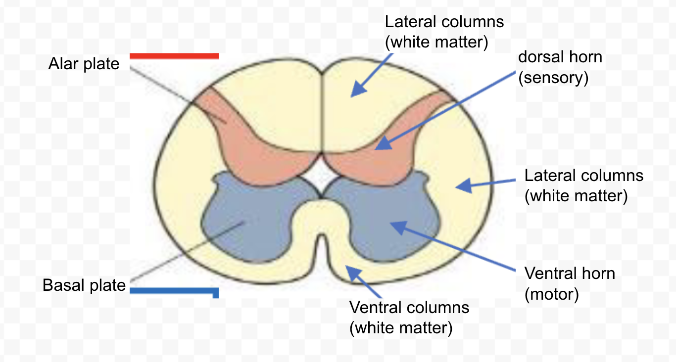
spinal cord gray matter final structures
dorsal horns, ventral horns, made of neurons derived from Alar & Basal plates
spinal cord white matter final structures
surrounds gray matter, made of fibers, includes dorsal, lateral, and ventral columns
derivation of dorsal horns
Alar plate → sensory neurons → dorsal horns (gray matter)
derivation of ventral horns
Basal plate → motor neurons → ventral horns (gray matter)
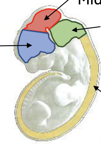
embryonic CNS structures (side view)
blue: forebrain
red: midbrain
green: hindbrain
yellow: caudal neural tube
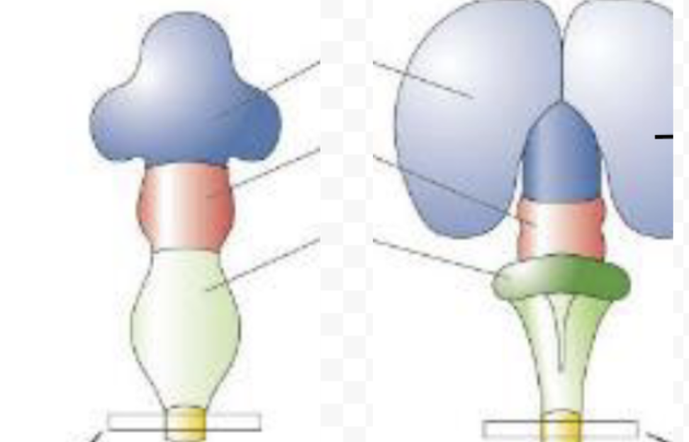
embryonic & postnatal CNS structures (front view)
blue: forebrain
red: midbrain
green: hindbrain
yellow: caudal neural tube
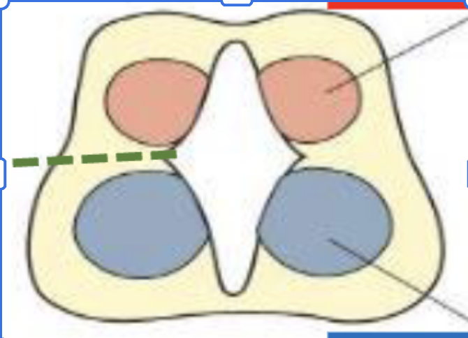
embryonic spinal cord cross section, label
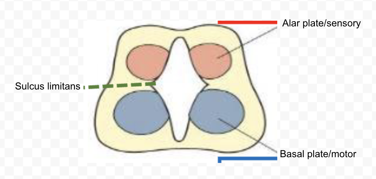
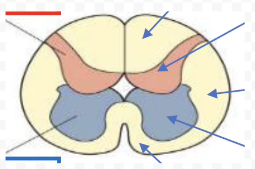
developed/postnatal spinal cord cross section, label
derived/differentiated hindbrain structure
rhombencephalon, including medulla, pons, cerebellum
maintains tube-like shape (along with spinal cord)
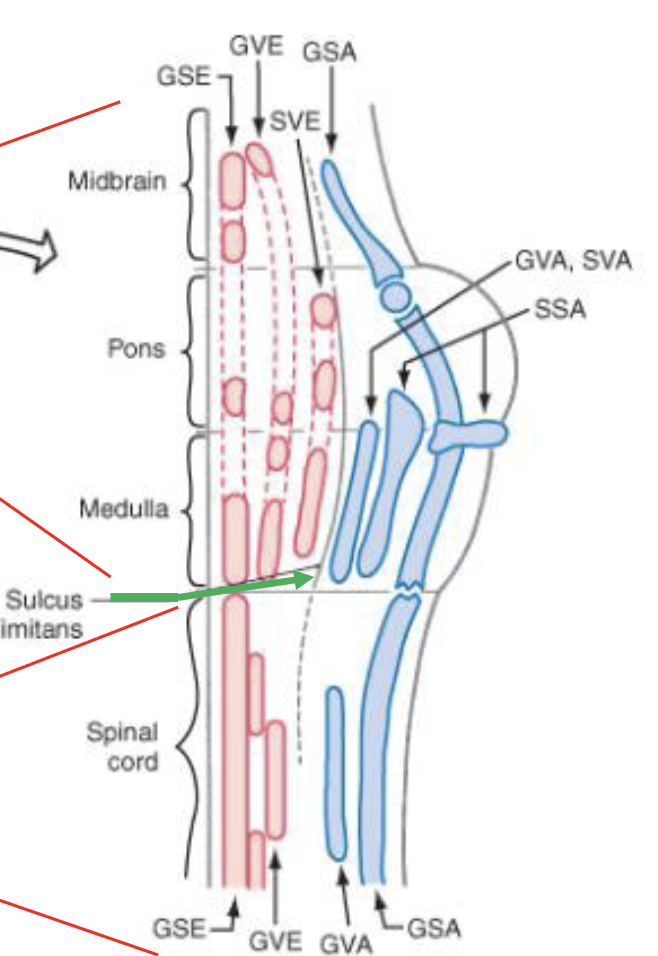
alar plate is dorsal to sulcus limitans, basal plate is ventral
alar plate derivatives are…
afferent/sensory
basal plate derivatives are…
efferent/motor
alar plate derivatives present in developed/differentiated hindbrain
special somatic afferent (SSA)
general visceral afferent (GVA)
special visceral afferent (SVA)
general somatic afferent (GSA)
basal plate derivatives in developed/differentiated hindbrain
general somatic efferent (GSE)
general visceral efferent (GVE)
special visceral efferent (SVE)
alar plate derivatives in developed/differentiated spinal cord
general visceral afferent (GVA)
general somatic afferent (GSA)
basal plate derivatives in developed/differentiated spinal cord
general somatic efferent (GSE)
general visceral efferent (GVE)
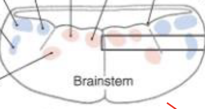
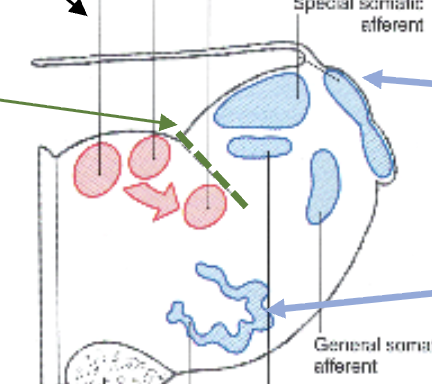
developed hindbrain cross section (dorsal - ventral), label
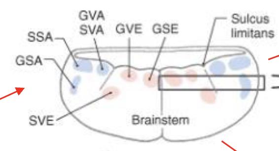

developed hindbrain & spinal cord cross sections (rostro-caudal) label
the myencephalon differentiates into…
caudal rhombencephalon (medulla)
mature medullas have…
ascending and descending axon bundles, alar and basal plate derivatives, pyramids of medulla
function of asc/desc axon bundles in mature medullas
connect spinal cord and other CNS structures
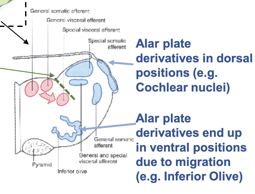
alar plate derivatives in mature medulla
somatic sensory nuclei, visceral sensory nuclei
basal plate derivatives in mature medulla
visceral motor nuclei
what happens with/what are pyramids of mature medulla
motor axons desc from cerebral cortex and cross midline (decussate) in pyramidal decussation (which is border bw spinal cord and medulla)
labeled mature medulla (myelencephalon diff)
the metencephalon differentiates into…
rostral rhombencephalon
characteristics of mature rhombencephalon
pons, cerebellum, rhombic lips
rhombic lips location, become
dorso-lateral position of metencephalon, grow dorsally to form cerebellum
motor axons from cortex synapse in __ and project to __
pons, cerebellum
alar plate derivatives in mature rhombencephalon
sensory nuclei of cranial nerves in cerebellum
basal plate derivatives in mature rhombencephalon
motor nuclei of cranial nerves in pons
why is it difficult to distinguish alar and basal plate derivatives in the mature prosencephalon, especially telencephalon & current hypothesis
very complex morphogen gradients and extensive cell migrations; very active research field → seems that almost all telencephalic structures are consistent with being alar plate derivatives
alar plate derivatives in diencephalon
alar plate deriv → thalamus
basal plate derivatives in diencephalon
basal plate deriv → hypothalamus
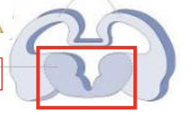
di/telencephalon label
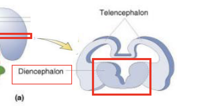
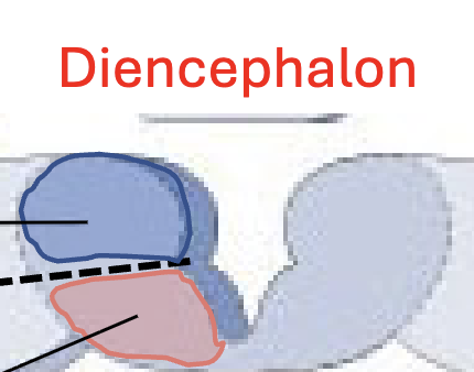
label

telencephalic vesicles, form
medial pallium → hippocampus
dorsal pallium → cerebral cortex
lateral pallium → amygdala, piriform cortex
ventral pallium → amygdala
LGE, MGE → subpallium → basal ganglia
dorsal pallium becomes
cerebral cortex (telencephalic diff)
dorsal pallium growth compared to other telencephalic vesicle sections?
disproportionate → expands until all available space in the skull is occupied, folding in on itself to pack more cortical tissue into limited space
telencephalic growth direction, shape
all directions but primarily dorsally & caudally, becomes a C shape due to bending and expansion of dorsal pallium within limited skull space
ganglionic eminence becomes, location
basal ganglia, stays in telencephalon core
telencephalon hollow space becomes
lateral ventricles (part of ventricular system