Medstudy 2
1/43
There's no tags or description
Looks like no tags are added yet.
Name | Mastery | Learn | Test | Matching | Spaced | Call with Kai |
|---|
No analytics yet
Send a link to your students to track their progress
44 Terms
The presence of a single central maxillary incisor should prompt additional investigation for
GH deficiency and hypopituitarism.
eruption of the teeth is often delayed in GH deficiency.
GH deficiency
Often AGA at birth.
Slowed lineargrowth may not present till 1-2y
Over time, they often appear “cherubic” (Kewpie doll-like) because of an excess of body fat, especially around the trunk and extremities, and deficiency of muscle mass
round, short, broad face; a depressed nasal bridge; small nose; well-developed nasolabial folds; and underdeveloped mandible and chin
Low IGF-1 and IGF-BP3
Congenital GH deficiency
May have apnea, and/or severe hypoglycemia associated with seizures
May have microphallus.
Herpes Esophagitis
Usually from reactivation and not primary
Usually Imunocompetent hosts
Often brought out by high doses of orticosteroids
pp
odynophagia and/or dysphagia, fever, and retrosternal chest pain.
Coexistent herpes labialis or oropharyngeal ulcers may be present
Early on, endoscopy may demonstrate individual vesicular lesions; lesions typically quickly coalesce to form well-circumscribed “volcano-like" ulcers with normal-appearing intervening mucosa.
Tx
If immunocompetent
short course (7–10 days) of oral acyclovir
If immunocomp
14–21 days of oral acyclovir
Barrett esophagus EGD findings
typically demonstrates long segments of columnar epithelium that extend above the esophagogastric junction. Columnar epithelium has a reddish color and velvet-like texture, whereas squamous epithelium has a more pale, glossy appearance. The squamocolumnar junction forms a visible line (the Z-line).
Candida EGD findings
white mucosal plaque-like lesions.
Caudal regression syndrome
200x more freq in diabetic mom
pp
incomplete to absent development of the sacrum and, to a lesser extent, the lumbar vertebrae.
spinal cord dysfunction → varying degrees of neurologic impairment of bowel and lower extremity function
Growth impairment of the lower extremities
developmental deformities
Perinaud occuloglandular syndrome
most common of the atypical presentations of catscratch disease caused by Bartonella henselae
occurs after direct inoculation of the eye with the hands after contact with a cat
pp
u/l conjunctival erythema w/o assoc discharge or significant pain +
prominent preauricular lymphadenpathy
Conjunctival granulomas are often identified at the site of inoculation.
Tx
symptomatic care and observation
At what age is it mandated by law for kids to wear a life jacket?
13y
describe traumatic myositis ossificans
Mature peripheral ossification with a distinct margin surrounding a radiolucent center of immature osteoid and primitive mesenchymal tissue
typically occurs in active adolescents after trauma
Hyperimmunoglobulin D syndrome
autorecess
presents by 1y of age
pp
fevers that generally last 3–7 days every 1–2 months.
abd pain
nondestructive large-joint arthritis
a diffuse nonmigratory erythematous macular rash, lymphadenopathy, headaches, oral/vaginal ulcers, and splenomegaly during febrile episodes.
IgD is often elevated (> 100 IU/mL) but not always, and IgA is also elevated in most cases
Cryopyrin=associated periodic fever syndromes CAPS
a group of autoinflammatory diseases with autosomal dominant inheritance. There are 3 periodic fever syndromes caused by mutations in the NLRP3 gene: familial cold autoinflammatory syndrome (FCAS), Muckle-Wells syndrome (MWS), and neonatal-onset multisystem inflammatory disease (NOMID). These disorders have overlapping clinical presentations that include fever, acute-phase inflammation, a neutrophilic urticarial skin rash, and joint involvement. There are distinguishing features as well, including sensorineural hearing loss in MWS and CNS involvement in NOMID.
For the QRS complex to be considered normal:
duration of QRS should not exceed more than 10ms longer than normal for that age group.
The R wave in V1 should also not exceed 15mm in infants less than 1 year or 10 mm if greater than 1 year.
Roseola pp
pp
Palpebral/periorbital edema (Berliner sign)
rash following defervescence
rash first appears on the trunk and quickly spreads to involve the extremities, face, and neck
nonpruritic. discrete, pinkish-red blanching macules and papules, sometimes associated with a peripheral halo of vasoconstriction.
enanthem consisting of erythematous papules involving the soft palate and uvula (Nagayama spots)
irritability, anorexia, diarrhea, upper respiratory symptoms, and suboccipital, postauricular and cervical lymphadenopathy.
Human herpesviruses 6 and 7 (especially human herpesvirus 6) are the major causes of roseola, which peaks at 6–15 months of age and is rare after 36 months of age
Gaucher disease
autorecess
lipid storage disease
pp
splenomegaly, hepatomegaly, skeletal abnormalities, and blood manifestations
Types 2 and 3 also demonstrate neurological symptoms
Type 2 is typically fatal by 2 years of age;
Erlenmeyer flask–like appearance of the femur on x-ray (don’t know why)
Dx
confirmed by measurement of glucocerebrosidase activity in peripheral leukocytes.
LEOPARD syndrome
(lentigines, electrocardiographic conduction abnormalities, ocular hypertelorism, pulmonic stenosis (a.k.a. pulmonary stenosis), abnormalities of the genitalia, restricted growth, and deafness). This is caused by mutations in the PTPN11 gene.
positive pathergy test
The phenomenon of developing a small, red papule, pustule, or ulceration at the site of needle insertion 1–2 days after the procedure
helpful in diagnosing Behçet disease
negative pathergy test does not rule out Behçet disease.
Pathergy is also associated with pyoderma gangrenosum
Familial mediterranean fever
usually < 10y
pp
fever from several hours to 5 days. predictable cycles that are patient specific. can be 3-5d q month, or just several times per year.
abd pain with the fever
Pleuritis, pericarditis, and scrotal swelling
An erysipelas-like rash can appear around the ankles
Arthritis, arthralgia, and myalgia are common.
Auspitz sign refers to
the appearance of small bleeding points after removal of a psoriatic scale in a patient with psoriasis
Bucket-handle deformity
describes a ridge of tissue, sometimes described as a bridge of skin, due to a fistulous tract extending to the perineal region in infants with a low imperforate anus.
hereditary hemorrhagic telangiectasia (HHT; a.k.a. Osler-Weber-Rendu syndrome)
autodom
pp
recurrent epistaxis and other bleeding diathesis
vascular malformations in the lungs, CNS, liver, and GI tract.
Nighttime epistaxis is typical and should raise the index of suspicion for this disorder.
Telangiectasias
most often found on the lips and oral cavity, palms, fingers, under the nails, soles, and ears (may not become apparent until adolescence or even later, making dx difficult).
Arteriovenous malformations may lead to pulmonary hemorrhage or intracranial bleeds; gastrointestinal bleeding can lead to significant anemia.
Eccentric lytic destruction and expansion of the metaphysis surrounded by a thin rim of sclerotic bone describes the radiographic findings of what?
aneurysmal bone cyst
Kobner
Darier
Auspitz
Kobner - defined as cutaneous hypersensitivity following superficial trauma (sJIA)
Darier - swelling, erythema, and pruritis of the skin in patients with mastocytosis or urticaria pigmentosa
Auspitz - Punctate bleeding when scales are scraped off in psoriasis
Water heater should be set to what?
120F or less
Branchial cleft cyst
congenital epithelial cyst
May form anywhere along 1st thru 4th branchial clefts (2nd most common)
These cysts are most often located along the lower half of the anterior margin of the sternocleidomastoid muscle, although they may also be preauricular or located at the mandibular angle.
Oftentimes, they go unnoticed until secondarily infected when they enlarge, become painful, and drain purulent material through a pore or fistula onto the cutaneous surface.
Recurrent infection is common without surgical removal or marsupialization.
dermatofibroma
a benign, well-defined, nodular connective tissue growth firmly fixed to the skin but easily movable over the subcutaneous fat.
dermoid cyst
a painless, firm lesion containing a greasy keratinous material, which may be attached to underlying structures. Dermoid cysts in the skin and subcutis occur mostly on the face, neck, and scalp.
Epidermal cyst
firm, slightly compressible, nontender nodules most common on the face, scalp, nape of the neck, and back.
Noonan syndrome
triangular shaped face
downslanting palpebral fissures
hypertelorism
short, webbed neck
joit laxity
pectus deformity
PS
bleeding diatheses
most frequent abnormality is Factor 11 deficiency.
Combined coagulation disorders and thrombocytopenia may also be identified
Excessive bruising; recurrent epistaxis; prolonged bleeding following injury, childbirth, or surgery; and menorrhagia are common complaints
short stature
congenital heart defects, most commonly pulmonary valve stenosis
pectus excavatum
webbed neck
low-set ears
hypertelorism
lymphedema
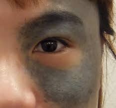
Nevus of ota
aka oculodermal melanocytosis
bluish-gray, irregular, patchy pigmentation on the face in an ophthalmic and/or maxillary distribution along the trigeminal nerve
In addition to pigmentation of the sclera, cornea, retina, and/or optic disc, ocular abnormalities that can be associated with nevus of Ota include hemangiomas of the optic disc and glaucoma, which have been reported in up to 10% of all affected individuals.
does not resolve with time
more common among females of Asian (particularly Japanese) descent and in Black individuals
Malignant transformation is very rare, although it may occur
nevus of Ito
similar to ota, but of the shoulders, sides of neck, upper arms, and/or scapular region.
Kasabach-Merritt phenomenon
aka hemangioma thrombocytopenia syndrome
When a large hemangioma is complicated by localized intravascular coagulation and hypofibrinogenemia
Risk of DIC
Miroangiopathic hemolytic anemia an elevated fibringen/fibrin degradatin products.
Tx Propranolol for large ones. Maybe corticosteroids
Nonresponders may benefit from interferon-α, irradiation, embolization, sclerotherapy, laser therapy, and/or surgery.
Bronchogenic cysts
Bronchogenic cysts are caused by abnormal budding of the tracheal diverticulum of the foregut before 16 weeks gestation. They can cause recurrent infections, pneumothorax (from cyst rupture), and can undergo malignant transformation. Diagnosis is by CT or MRI and treatment involves excision.
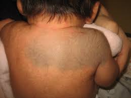
Chilblains
Chilblains are symmetrically distributed dark pinkish or violaceous nodules, plaques, or blisters located on exposed extremities (fingers, toes, nose, or ears) following exposure to cold, damp weather. They can be idiopathic or secondary to an underlying connective tissue disease.
Charcot-Marie-Tooth disease
inherited nuropathy
progressive distal ascending weakness manifesting as foot drop and pes cavus (high-arched feet).
The classic “stork leg” deformity results from distal calf muscle atrophy.
Deep tendon reflexes are lost.
Upper extremities are eventually involved in later stages of the disease process.
DOES NOT not involve cardiomyopathy, eye issues, or upgoing toes/Babinski sign.
Juvenile spinal muscular atrophy
presents between 2 and 17 years of age with proximal muscle weakness. Pyramidal signs are absent and there is no involvement of the visual or cardiac systems.
Rash from neonatal lupus
discoid lesions, periorbital erythema, annular lesions, scaly atrophic patches, and/or telangiectasia.
Lemierre disease
Suppurative thrombophlebitis of the jugular vein
Due to extension of a recent preceding oropharyngeal or dental infection to the lateral pharyngeal space.
Septic pulmonary emboli commonly occur in conjunction with the localized head and neck manifestations.
Fever, rigors, and respiratory symptoms are typically associated with dysphagia, trismus, pain, decreased range of motion of the neck, and tonsillar/peritonsillar swelling, as well as tenderness, swelling, and/or induration overlying the jugular vein, angle of the jaw, or sternocleidomastoid muscle.
lung abscesses and/or empyema may further complicate the disorder.
Although less common, septic arthritis and/or osteomyelitis may also occur
Normal oropharyngeal flora cause suppurative thrombophlebitis of the jugular vein, the most common pathogen being the anaerobe Fusobacterium necrophorum.
Bcxs often positive
Lemierre syndrome may also complicate infectious mononucleosis
Tx
surgical drainage and β-lactamase stable antimicrobials for 3–6 weeks.
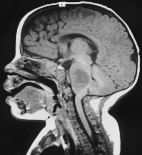
Diffuse intrinsic pontine glioma (DIPG)
Are enmeshed with the pons (portion of the brainstem) and are unresectable.
Radiation and chemotherapy have little or no effect.
The survival rate is < 5% and is measured in months from the time of diagnosis.
Langerhans cell biopsy, dx confirmed with:
stains for CD1a, CD207, and S100
Electron microscopy is no longer used in common clinical practice
Howell-Jolly bodies
nuclear remnants seen as red blood cell inclusions after splenectomy
Atlantoaxial instability ss
neck pain
radicular pain
torticollis
gait abnormalities
loss of bowel or bladder control
weakness
numbness
pasticity (unusual ‘tightness’ of certain muscles)
change in muscle tone
gait difficulties
hyperreflexia
change in bowel or bladder function
Ramsay Hunt syndrome
Caused by herpes zoster involving the geniculate ganglion.
pp
Ear pain with tinnitus and vertigo
Vesicles and crusted vesicles with erythema can be seen on the tongue, in the ear canal, on the pinna, soft palate ← these are if the genniculate ganglion is involved which it usually is i think.
TM normal
unilateral ear pain (which often radiates outward into the pinna), vertigo, ataxia, tinnitus, and ipsilateral hearing loss
Facial nerve palsy
usually reaches maximal severity within a week of onset of symptoms.
Tx
Systemic corticosteroids and oral acyclovir
Prog
50% do not recover facial nerve function