Central Visual Pathways and the Perceived Visual Field (Week 2, Mod 8)
1/11
There's no tags or description
Looks like no tags are added yet.
Name | Mastery | Learn | Test | Matching | Spaced |
|---|
No study sessions yet.
12 Terms
What are 4 ways we can assess vision in the veterinary world? Keep in mind the limitations that we have.
1) Observing the patient in an unfamiliar environment
If blind, may bump into things
2) Ability to track
Cotton ball test - drop a cotton ball on either side of the animal just at its peripheral… animal should turn to look at the object
3) Visual placing
Dog being picked up and brought forward to a table… should reach out to step onto the table on its own
4) Menace response
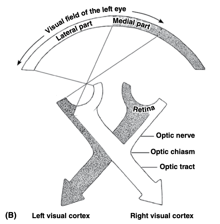
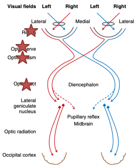
What are the 7 general steps of the visual pathway? KNOW THIS
Retina →
Optic nerve →
Optic chiasm →
Optic TRACT →
Lateral geniculate nucleus (of the thalamus) →
Optic radiation →
Occipital cortex
What is the “visual field”? How does it differ between predators and prey?
Visual field: The total area in which objects can be seen in the peripheral vision as you focus your eyes on a central point
Predators:
Eyes forward, towards front of the skull
Shorter snout
= BINOCULAR vision
Prey:
Eyes on the sides of the face; have their own visual fields, only become binocular directly in front of the snout
Long snout
Allows for better peripheral vision
Describe the variation of the level of CROSSOVER at the optic chiasm between species… is there more crossover in animals with increased binocular vision?
In predatory animals, the degree of crossover of visual information from the eyes to the lateral geniculate nucleus is LESS than prey animals:
Humans: 50%
Cats: 65%
Dogs: 57%
Horse: 80%
Birds and fish: 100%
Is INVERSELY correlated to the size of the binocular visual field
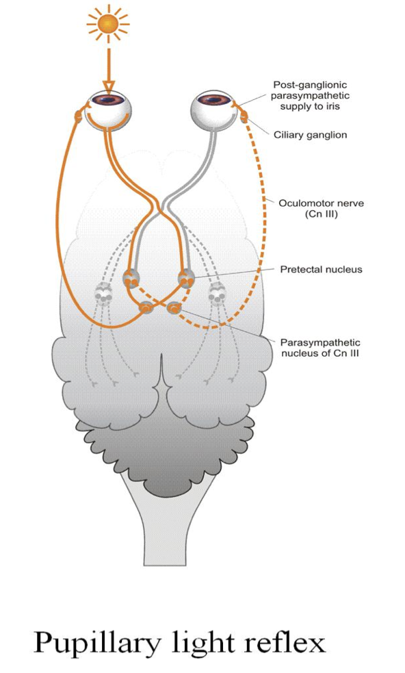
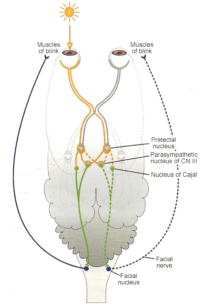
Describe the pupillary light reflex test… what parts of the visual pathway is this testing?
PLR = shining a bright light in ONE eye… should see BOTH pupils contract
Tests the function of the Optic Nerve (CN II) → optic chiasm → optic tract → Pretectal nucleus → Parasympathetic nucleus of Oculomotor nerve (CN III) pathway
** CN III = oculomotor nerve = constriction of the pupil
What is the menace response? What parts of the visual pathway is this testing?
The menace response is a reaction where a hand is moved in front of the eye suddenly, and the animal blinks in response
Is a RESPONSE, not a reflex, so will have cortical involvement and will therefore be more complicated
Pathway it testsL
Optic nerve (CN II) → optic chiasm → optic tract → lateral geniculate nucleus → optic cortex → motor cortex → pontine nucleus → cerebellum → FACIAL nerve nuclei (CN VII)

What is the major difference between the menace response and the pupillary right reflex? Why might we use the both of these during a visual assessment?
Both evaluate the retina, the optic nerve, and the optic chiasm
BUT the PLR test doesn’t asses the cortex portion of the visual pathway… simply tests the REFLEX itself
Combining the two tests in a visual assessment can tell us what kind of blindness a patient may have (essentially, whether the damage is localized to the eye or brain)
What is central (cortical) blindness? Where would you find a lesion in the visual pathway with this kind of blindness? What are the clinical signs?
Is blindness without apparent lesions of the eyes
Involves the PROCESSING system, rather than the hardware
Will see a lesion in:
Lateral geniculate nucleus
Optic radiation
Occipital cortex
Clinical signs:
NORMAL PLR
ABSENT menace response
Blind

What is peripheral (subcortical) blindness? Where would you find a lesion in the visual pathway with this kind of blindness? What are the clinical signs?
Involved the collection / distribution system
Will see lesions in the:
Eye
Optic nerves
Optic chiasm
Optic tract
Clinical signs:
ABSENT PLR
ABSENT menace response
Blind
What is the dazzle reflex? What parts of the visual pathway is this testing?
Is very similar to the PLR test, but triggers FACIAL NERVE CN VII to produce a BLINK REFLEX
Instead of Oculomotor nerve CN III, which is used for pupil constriction
Should see BILATERAL blinking
Light should be very bright for the reflex
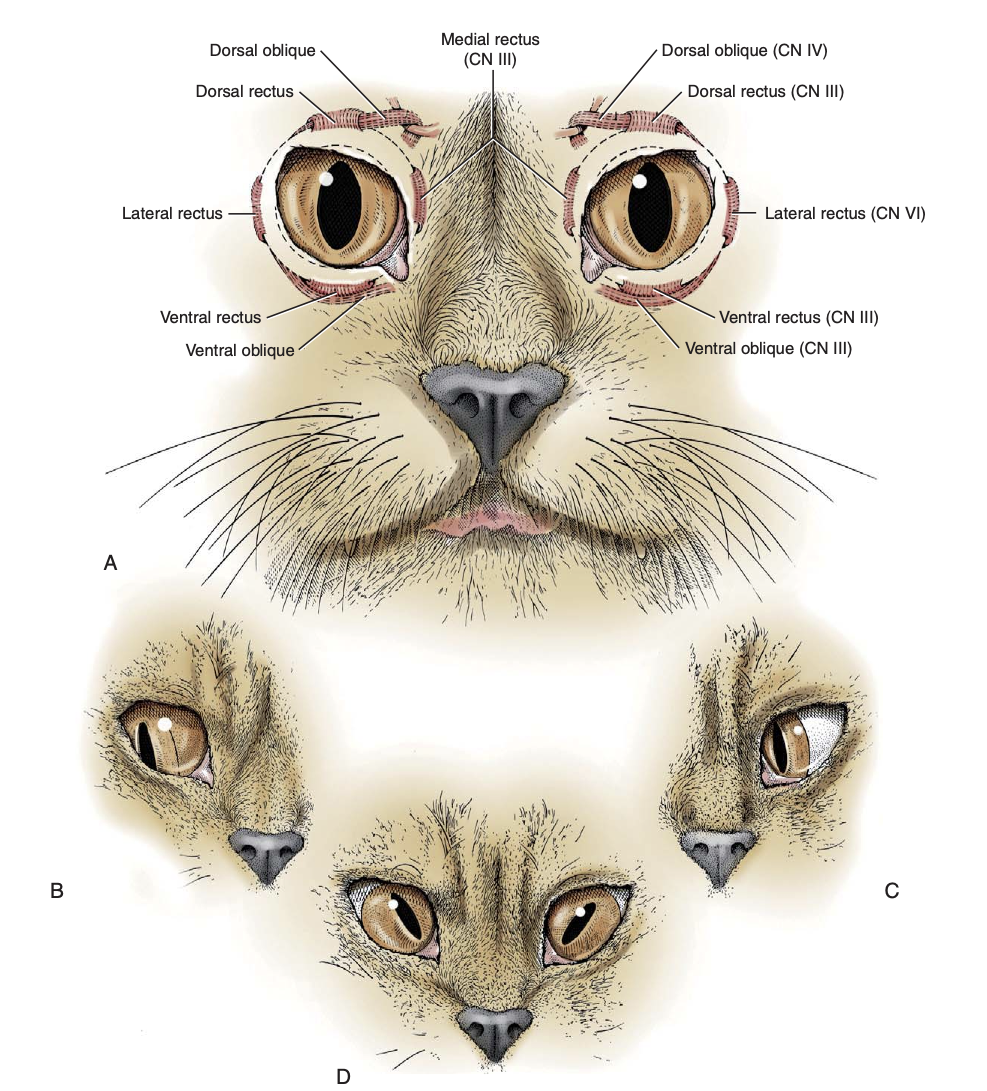
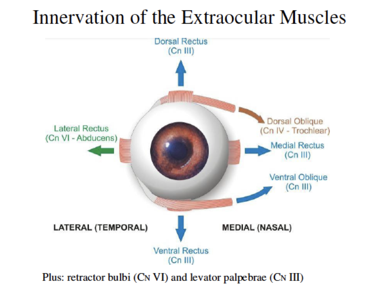
Associate the extraocular muscles with the nerve that innervates them… might be easier to remember them this way
Oculomotor nerve (CN III)
Dorsal Rectus
Medial Rectus
Ventral OBLIQUE
Ventral Rectus
Think of it this way: oculomotor = eye movement… so the oculomotor nerve innervates the MOST extraocular muscles
But doesn’t move laterally because ABDUCTING is the NEXT nerve’s job…
Abducens nerve (CN VI)
LATERAL Rectus
Retractor bulbi muscle
Trochlear nerve (CN IV)
Dorsal Oblique
Think of it as the muscle that turns the bottom portion of the eye towards the trochlea…
What is strabismus clinically? What would happen to the eye if there was a lesion at CN III? CN VI? CN IV?
Strabismus is ABNORMAL positioning of the eye
Lesion at CN III → eye moves ventrolaterally because the lateral rectus and other ventral muscles are the ONLY ones working still
Lesion at CN IV → eye moves dorsomedially… dorsal oblique muscles have stopped working, so no longer turning the bottom portion of the eye toward the trochlea
Makes pupil tilt
Visible in the cat, NOT noticeable in the dog
Lesion at CN VI → eye moves medially, lateral rectus no longer functioning