Chapter 26: Blood Supply, Stroke, Fluid Dynamics, and Intracranial Pressure Objectives
1/14
There's no tags or description
Looks like no tags are added yet.
Name | Mastery | Learn | Test | Matching | Spaced | Call with Kai |
|---|
No analytics yet
Send a link to your students to track their progress
15 Terms
Blood supply to brain
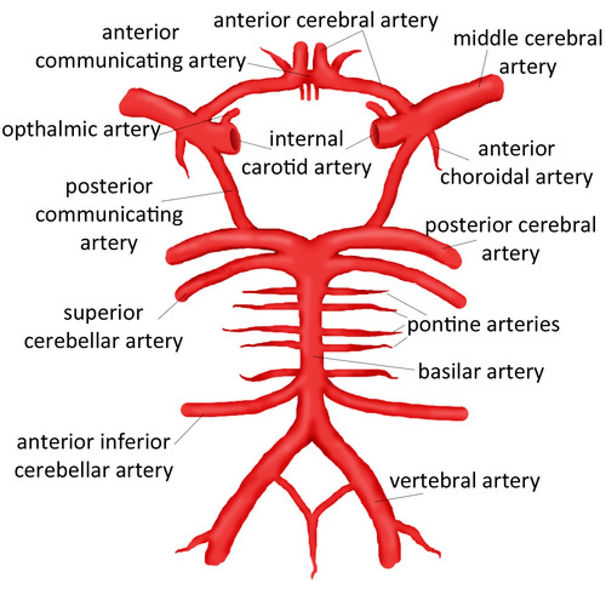
-circle of Willis: 9 arteries that form a hexagon
-8 paired arteries and the anterior common artery
-internal carotid aa. supply blood to most of the cerebrum
-vertebral aa provide blood to occipital and inf. temporal lobes and brainstem/cerebellar region
-basilar a. and its branches supply pons and most of cerebellum. Becomes posterior cerebral aa btwn pons and midbrain

1. Describe the circle of Willis and name the three major cerebral arteries (p. 9).
-9 aa creating a hexagon
-allows collateral circulation
-fed by internal carotid and basilar aa.
-major terminal aa supply specific regions and overlap in WATERSHED areas
-large aa: 2 anterior cerebral aa (branches of int. carotid aa, TERMINAL BRANCH), the internal carotid aa, 2 posterior cerebral aa (branches of basilar a, TERMINAL BRANCH).
-Anterior communicating a. (unpaired): joins the anterior cerebral aa. together.
-2 Posterior communicating aa: link each internal carotid a. with a posterior cerebral a.
Arterial supply to cortical and subcortical structures
-Three major terminal branches:
-middle cerebral a. (MCA)-not direct part of circle of willis
-anterior cerebral a. (ACA)
-posterior cerebral a. (PCA)
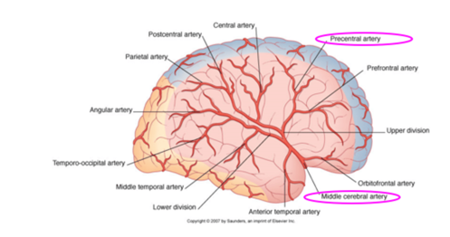
Middle cerebral a. (MCA)
-Main continuation of internal carotid a.
-rec'v 60-80% of blood flow
-supplies: frontal, parietal, temporal, optic tract/radiation, basal ganglia, internal capsule, thalamus
-main areas of blockage: base/trunk of a.
-area in pink in picture
Anterior cerebral a. (ACA)
-branch of internal carotid a.
-passes above optic chiasm to supply medial aspect of hemisphere
-forms arch around corpus callosum: internal capsule, frontal and parietal lobes
-area in blue in picture
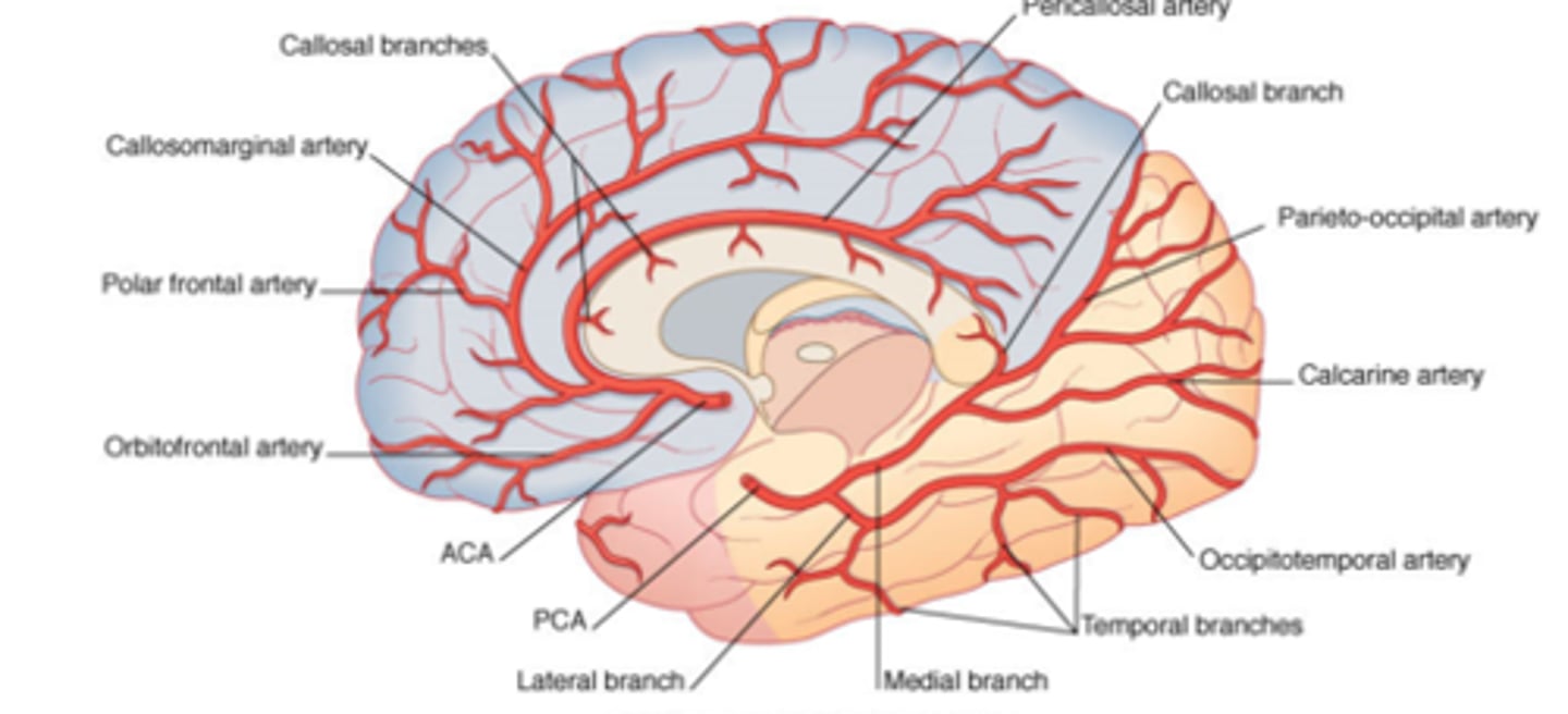
Posterior cerebral a. (PCA)
-curves around brainstem: occipital lobe, lower temporal lobe, choroid plexus
-yellow area in picture

2. Compare and contrast TIA, completed stroke (aka stroke), infarction, and hemorrhage.
-Transient ischemic attack (TIA): mini stroke, stroke symptoms occur and resolve within 24 hrs, 15% of people who have them will have a large stroke within a year
-Complete stroke: Neurological deficits from vascular disorders that persist for longer than 1 day and are stable (not progressing or improving)
-Infarction: embolus lodges in vessel and occludes blood flow (cause 80% of strokes). Onset may be fast or may worsen over days
-Hemorrhage: deprives downstream vessels of blood and extravascular blood exerts pressure on surrounding brain. Generally present with worse deficits within hours of onset, then improvement occurs as edema decreases
3. Identify the most commonly occluded cerebral artery.
- >90% of anterior circulation ischemic strokes affect the middle cerebral a (MCA)
4. Define arteriovenous malformation and aneurysm.
-arteriovenous malformation: developmental abnormalities with arteries connected to veins by abnormal, thin-walled vessels larger than capillaries. Don't cause symptoms until they rupture. Then bleeding causes dysfunction due to lack of blood the area.
-Aneurysm: dilation of the wall of an artery or vein. Saccular aneurysm most common.
-berry aneurysm: type of saccular aneurysm. Small sac protruding off of cerebral a. and has thin connection with the artery.
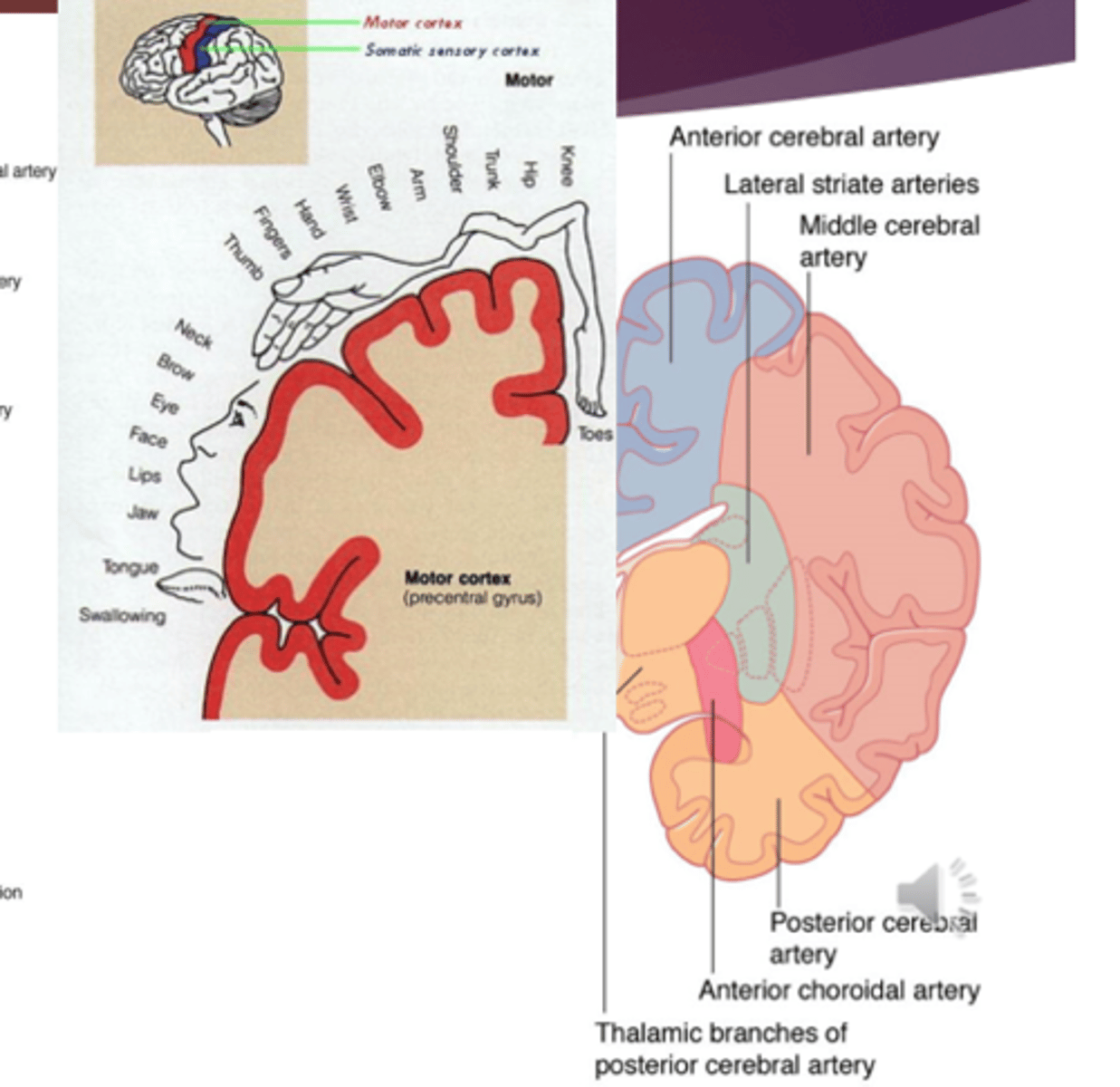
5. Use the motor and sensory homunculi to explain motor and sensory impairments listed in Figure 26.10.
-Next cards
Middle cerebral a. infarction
-Somatosensory: hemisensory loss affecting face and upper limb more than lower limb
-Motor: face and upper limb more impaired than lower limb
-special senses and autonomic function: homonymous hemianopia
-emotions and behavior: R. hemisphere: easily distracted, poor judgement, impulsiveness. L hemisphere: right hemiplegia, apraxia, compulsiveness, overly cautious
-cognition, lang, and memory: L MCA: aphasia. R MCA: difficulty understanding spatial relationships, neglect, impairment of nonverbal comm., dressing apraxia, constructional apraxia
-other: none

Anterior cerebral a. infarction
somatosensory: loss of fine sensation in lower limb
-motor: hemiplegia (lower limb more than upper limb and face)
-special senses and autonomic function: no changes
-emotions and behavior: flat affect, impulsiveness, perseveration, confusion, motor inactivity
-cognition, language, memory: difficulty with divergent thinking
-other: urinary incontinence
Posterior cerebral a. (PCA) infartcion
-Somatosensory: hemisensory loss: slow nociception
-Motor: If near origin of a., eye mvmt probs: vertical gaze palsy, oculomotor n. palsy, loss of medial deviation of eye with preserved convergence, vertical skew deviation of eyes. Rarely hemiparesis.
-special senses and autonomic function: homonymous hemianopia, cortical blindness, hallucinations, lack of depth perception, visual agnosia, limbs may show vasomotor and/or trophic changes.
-emotions and behavior: none
-cognition, lang, and memory: difficulty reading, memory loss
-other: none

5. Describe the blood-brain barrier.
-specialized permeability barrier btwn capillary endothelium of CNS and extracellular space
-formed by tight junctions btwn capillary endothelial cells
-excludes large molecules
-prevents pathogens, drugs, protein antibodies from accessing the brain
6. Describe the four types of brain herniations listed in the chapter.
-cingulate herniation: when a mass in one hemisphere displaces the cingulate cortex under the falx cerebri.
-may not cause symptoms
-if anterior cerebral a. is compressed it could cause motor deficits affecting lower limb
-uncal herniation: a space-occupying lesion in the temporal lobe displaces the uncus medially (like subdural hematoma) forcing uncus in to the tentorium cerebri. This compresses the midbrain which interferes with function of the oculomotor n. and consciousness
-central herniation: space-occupying lesion in the cerebrum exerts pressure on the diencephalon, moving it, the midbrain, and pons inferiorly. This stretches branches of the basilar a. causing brainstem ischemia and edema. Bilateral paralysis, consciousness and oculomotor control are impaired
-Tonsillar herniation: pressure from uncal herniation, tumor in brainstem/cerebellar region, hemorrhage, or edema may force cerebellar tonsils through the foramen magnum. This compresses brainstem which interferes with vital signs, consciousness, and flow of CSF.