Cardiovascular Disease - Heart Failure & Cardiogenic Shock
 Call Kai
 Learn
Learn Practice Test
 Spaced Repetition
 Match
Match Flashcards
 Knowt Play
1/42

There's no tags or description
Looks like no tags are added yet.
Name | Mastery | Learn | Test | Matching | Spaced | Call with Kai | Chat |
|---|
No analytics yet
Send a link to your students to track their progress
43 Terms
Types of hemodynamic monitoring
Pulmonary artery catheter (PAC) or Swan-Ganz catheter
Central venous pressure catheter
Arterial artery catheter
Pulmonary Artery Catheter (PAC)
Used to:
Evaluate cardiac function
Help diagnose the cause (etiology) of shock
Evaluate response to:
Fluids
Medications/interventions
Monitor cardiac pressures
Pulmonary Artery Catheter (PAC) - Central Venous Pressure (CVP)
Normal: 2–6 mm Hg
Helps evaluate:
Fluid status
Right-sided heart function
↑ CVP → may indicate fluid overload or right-sided heart failure
Pulmonary Artery Catheter (PAC) - Pulmonary Artery Occlusion Pressure (PAOP)
Normal: 6–12 mm Hg
Also called pulmonary wedge pressure
Helps evaluate:
Fluid balance
Left ventricular function
↓ PAOP → may indicate low fluid volume
↑ PAOP → may indicate left ventricular dysfunction/left-sided heart failure
Pulmonary Wedge Pressure
How it is obtained:
Inflate the balloon briefly
Pressure appears on the monitor
Immediately deflate the balloon
Do NOT leave the balloon inflated.
Prolonged inflation can cause pulmonary artery rupture.
PAC or CVP Access Sites
Internal jugular vein **
External jugular vein
Subclavian vein
Femoral vein
Basilic vein
Central Venous Pressure (CVP)
Central line → enters the central venous circulation:
SVC
IVC
Right atrium (RA)
Useful for assessing fluid volume status and preload
Less precise/rapid than the pulmonary artery system
No valves between the vena cava and right atrium → pressure reflects CVP
Allows for rapid, high-volume fluid resuscitation
Allows for frequent blood draws
CVP can be monitored:
Continuously
Intermittently
Fluids can run through the port when a CVP reading is not being obtained
Central Line Uses
Administer medications
Administer fluids
Monitor CVP
Evaluate fluid volume status
Obtain blood samples
Provide continuous IV access
Catheter is inserted through a large vein and advanced toward the right atrium.
Arterial Line
Provides:
Continuous blood pressure monitoring
Arterial blood pressure readings
Access for arterial blood samples/ABGs
Arterial Line - Before Insertion
Perform an Allen's test
Checks circulation to the hand/fingers
Assess:
Capillary refill
Sensation
Skin color
Skin temperature
Pulses
Arterial Line - Catheter/Site Care
Maintain appropriate dressing and perform dressing changes as needed.
Immobilize the body part where the catheter is placed in a neutral position.
Prevent joint flexion or extension
Perform ROM of the hand/fingers
Do not hyperextend the wrist → risk of neuromuscular injury
Assess the site every 1 hour for:
Redness
Drainage
Bruising
Blanching
Pallor
Firmness
Swelling
Assess distal circulation:
Skin color
Temperature
Capillary refill time (CRT)
Distal pulses
Motor function
Sensory function
Removing an Arterial Catheter
Apply direct pressure for approximately 5 minutes
Helps prevent bleeding.
Complications from Heart Disease
Heart Failure
Cardiogenic Shock
Heart Failure (HF)
Inability of the heart to pump enough blood to meet the body's needs for oxygen and nutrients
A syndrome characterized by:
Fluid overload or
Inadequate tissue perfusion
Involves a problem with:
Systolic failure → problem with heart contraction/pumping
Diastolic failure → problem with heart filling
Some cases of heart failure are reversible
Most HF is a progressive, lifelong disorder
Managed with lifestyle changes and medications
HF Causes
CAD
Hypertension
Cardiomyopathy
Valvular disorders
Renal dysfunction
Heart Failure — Diagnosis: Echocardiogram
Evaluates cardiac function
Measures ejection fraction (EF)
EF <40% → impaired left ventricular systolic function
Heart Failure — Diagnosis: B-type Natriuretic Peptide (BNP)
Laboratory value associated with heart failure
Secreted by the ventricles
Released in response to increased pressure/stretch
↑ BNP → may indicate heart failure or worsening heart failure
BNP >100 → suggestive of heart failure
Heart Failure — Diagnosis: Symptomatology
Patient's signs and symptoms are also used when diagnosing HF
Heart Failure — Types & Classifications
Types
Acute HF → develops suddenly
Chronic HF → develops gradually and persists over time
Acute-on-chronic HF → sudden worsening of pre-existing chronic HF
Heart Failure Classifications
1. NYHA (New York Heart Association)
Class I–IV
Based on how much physical activity causes symptoms
2. ACC/AHA
Stage A–D
Based on the progression of heart failure and risk/structural disease
Left-Sided Heart Failure
LV failure → left ventricle cannot pump effectively
Blood backs up into the lungs
Causes pulmonary congestion
Left = Lungs
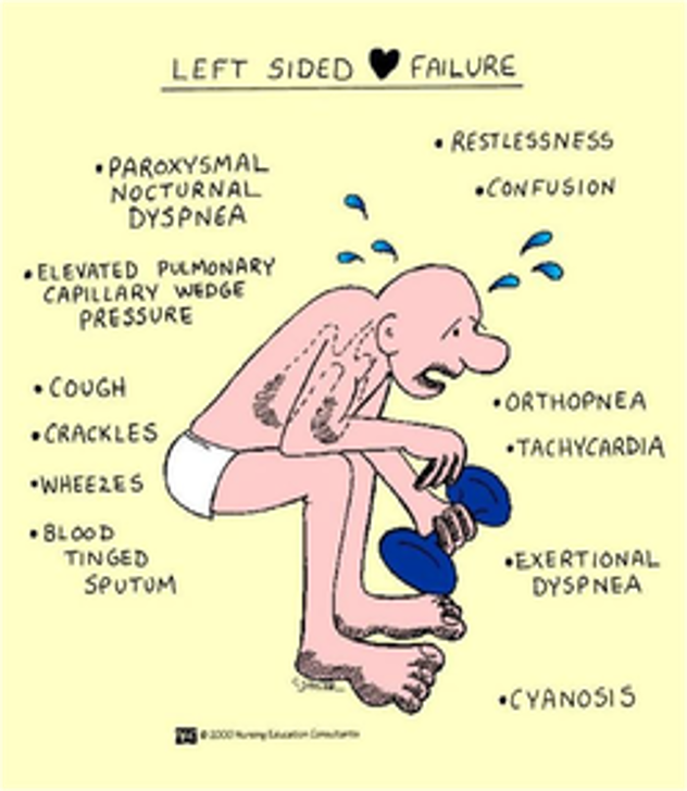
Left HF Clinical Manifestations:
Dyspnea / SOB → shortness of breath
DOE → dyspnea on exertion
Cough
Crackles
PND → paroxysmal nocturnal dyspnea (waking up at night short of breath)
↓ O₂ / hypoxemia
S3 heart sound
Right-Sided Heart Failure
RV failure → right ventricle cannot pump effectively
Blood backs up into the systemic circulation
Causes systemic congestion
Right = Rest of the body
Right HF Clinical Manifestations:
Peripheral edema → swelling, especially legs/feet
JVD → jugular venous distention
Hepatomegaly → enlarged liver
Ascites → fluid accumulation in the abdomen
Anorexia / nausea
Weakness
Weight gain → usually from fluid retention
Left vs Right HF

Left HF
Right HF

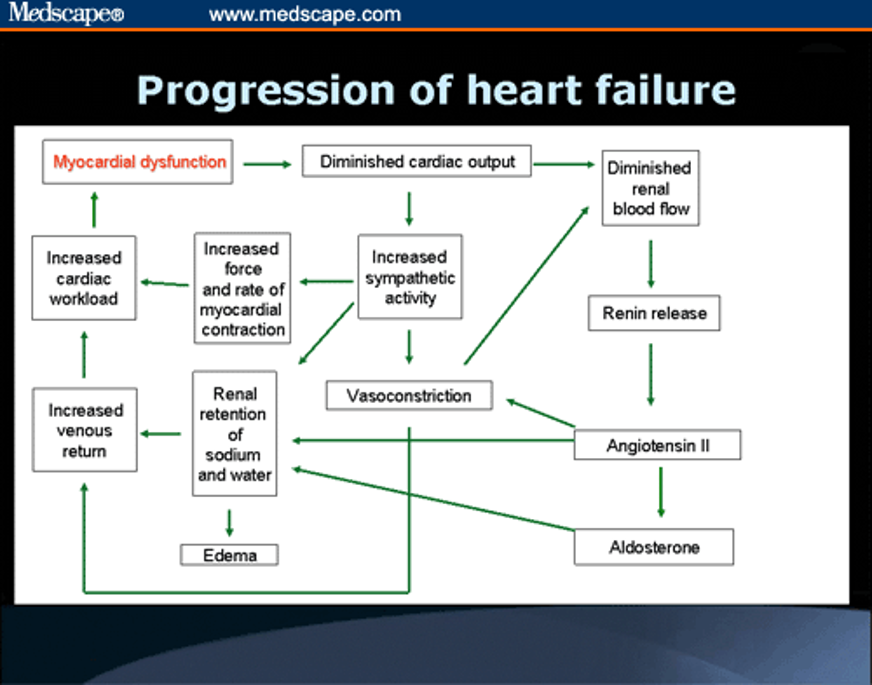
1. Myocardial dysfunction
→ Heart muscle is not working properly
→ ↓ Cardiac output
2. ↓ Cardiac output
→ Kidneys receive less blood
→ ↓ Renal blood flow
3. ↓ Renal blood flow
→ Renin release
→ ↑ Angiotensin II
4. Angiotensin II
→ Vasoconstriction → ↑ resistance/workload on the heart
→ Stimulates aldosterone
5. Aldosterone
→ Kidneys retain sodium + water
→ ↑ Blood volume
→ ↑ Venous return (preload)
→ Edema
6. ↓ Cardiac output also activates the sympathetic nervous system
→ ↑ Heart rate and force of contraction
→ Initially helps maintain cardiac output
→ But increases cardiac workload and oxygen demand
7. Increased blood volume + vasoconstriction + increased cardiac work
→ ↑ Cardiac workload
→ Further myocardial dysfunction
Heart Failure — Nursing Diagnoses
Activity intolerance and fatigue
Related to decreased cardiac output and reduced oxygen delivery
Excess fluid volume
Related to sodium and water retention
May cause edema, weight gain, crackles, and congestion
Anxiety
Related to symptoms, illness, and uncertainty about condition
Powerlessness
Related to the chronic nature of heart failure and changes in lifestyle
Noncompliance
Difficulty following the prescribed medications, diet, fluid restriction, or activity plan
Heart Failure — Nursing Management
Provide symptom relief
Monitor for dysrhythmias
Hemodynamic monitoring
Monitor fluid status
I&O
Daily weights
Assess for edema
Provide psychosocial support
Heart Failure — Medications
ACE inhibitors
↓ Blood pressure
↓ Afterload
↓ Cardiac workload
Help prevent/worsen remodeling
ARBs
Block the effects of angiotensin II
↓ Vasoconstriction
↓ Blood pressure
↓ Cardiac workload
Beta blockers
↓ Heart rate
↓ Contractility
↓ O₂ demand
Decrease cardiac workload
Diuretics
Remove excess fluid and sodium
↓ Fluid volume
↓ Edema and pulmonary congestion
↓ Preload
Digitalis (Digoxin)
Increases the force of cardiac contraction
Helps improve cardiac output
Slows heart rate
Heart Failure — IV Infusions
Dobutamine (Dobutrex)
Increases cardiac contractility
Helps increase cardiac output
Used for acute/severe heart failure when increased cardiac pumping is needed
Milrinone (Primacor)
Increases cardiac contractility
Causes vasodilation
↑ Cardiac output
↓ Cardiac workload/vascular resistance
ICD — Implantable Cardioverter-Defibrillator
Recommended for patients with heart failure who are at high risk for:
Ventricular tachycardia (VT)
Ventricular fibrillation (VF)
The ICD detects dangerous ventricular rhythms
It can deliver an electrical shock to terminate the rhythm
Helps prevent sudden cardiac death/cardiac arrest.
LifeVest
LifeVest = wearable cardioverter-defibrillator (WCD)
Used for high-risk heart failure patients who are at risk for dangerous ventricular dysrhythmias.
It is an external defibrillator worn by the patient.
Can serve as a temporary alternative/bridge to an ICD when an implanted device is not immediately available or appropriate.
The LifeVest can detect dangerous rhythms and deliver a shock to restore a normal rhythm.
Intra-Aortic Balloon Pump (IABP)
Circulatory assist device
↓ workload of the heart during contractions but does not perform the actual pumping work of the heart.
Consists of a catheter with a balloon at the distal end.
Balloon is positioned in the descending aorta.
Uses counterpulsation:
Balloon inflates and deflates according to the cardiac cycle
This helps augment the pumping action of the heart.
Synchronized with the ECG or arterial blood pressure (ABP) to identify systole and diastole and time balloon inflation/deflation.
Supports a failing LV by mechanically displacing blood within the aorta.
Effects of the IABP
Increases oxygen-rich blood supply to the myocardium
Balloon inflates during diastole
→ pushes blood toward the coronary arteries
→ ↑ coronary perfusion
Decreases myocardial oxygen demand
Balloon deflates just before systole
→ ↓ resistance/afterload against the LV
→ ↓ LV workload
→ ↓ myocardial oxygen demand
IABP: Common Uses
Treatment of pump failure:
Cardiogenic shock → heart cannot pump enough blood to meet the body's needs.
Intraoperative MI → MI occurring during surgery.
Low cardiac output after bypass surgery → heart is not pumping effectively after cardiopulmonary bypass.
Other uses:
Weaning from cardiopulmonary bypass → provides temporary LV support while the heart resumes effective pumping.
Cardiomyopathies → supports the heart when severe myocardial dysfunction causes inadequate cardiac output.
IABP: Complications
Infection
Risk from the catheter insertion site.
Ischemia
Reduced blood flow to the femoral, renal, visceral, or spinal arteries.
Can occur because the catheter/balloon obstructs arterial blood flow.
Embolic shower
Microscopic clots/debris can form on the balloon and travel through the bloodstream.
Can cause:
Peripheral thrombosis
Myocardial ischemia
Hemodynamic decompensation
Late pseudoaneurysm
Balloon/mechanical failure
Balloon can malfunction or rupture.
Balloon rupture requires removal of the IABP, with surgical/vascular expertise as needed.
Aortic dissection
Tear in the aortic wall.
Can lead to hemorrhage into the mediastinum.
Cerebral embolism
Embolic material can travel to the brain, particularly during insertion.
→ Possible neurologic injury/stroke.
Compartment syndrome
Increased pressure within a limb compartment can compromise blood flow and nerves.
Cardiogenic Shock
Life-threatening condition with a high mortality rate.
Occurs when the heart cannot pump enough blood to meet the body's needs.
Cardiogenic Shock - Causes
Myocardial infarction (MI)
Heart failure (HF)
Cardiac tamponade
Pulmonary embolism (PE)
Cardiomyopathy
Dysrhythmias/arrhythmias
Cardiogenic Shock: Pathophysiology
Heart is unable to pump effectively
↓ Cardiac output
↓ Tissue perfusion
↓ Oxygen delivery to tissues
Tissue hypoxia
Cells switch to anaerobic metabolism
↑ Lactic acid → metabolic acidosis
Poor perfusion causes organ dysfunction
Continued poor cardiac function → worsening shock
Compensation:
Sympathetic nervous system activates → ↑ HR and vasoconstriction
This temporarily helps maintain BP, but increases the heart’s workload and oxygen demand.
The heart becomes too weak to pump enough blood, often due to severe heart damage such as an MI.
Cardiac output decreases, so less blood reaches the body's tissues and organs.
Oxygen delivery decreases, causing tissue hypoxia.
Cells switch to anaerobic metabolism because they do not receive enough oxygen.
Lactic acid builds up, causing metabolic acidosis.
Continued poor perfusion causes organ dysfunction and eventually organ failure.
Cardiogenic Shock: Clinical Manifestations
symptoms of heart failure
shock state
hypoxia
Cardiogenic Shock: Clinical Manifestations
Decreased cardiac output (CO)
Low blood pressure → systolic BP <85 mmHg
Tachycardia → heart beats faster to try to maintain cardiac output
Rapid, weak/thready pulse
Acidosis → poor tissue perfusion causes lactic acid buildup
Poor peripheral perfusion
Cool, clammy skin
Decreased peripheral pulses
Kidney dysfunction
Renal insufficiency
Decreased urine output because the kidneys receive less blood flow
Pulmonary edema
Fluid backs up into the lungs because the failing LV cannot pump blood forward effectively.
↑ PAOP → indicates increased left-sided filling pressure
↑ CVP → may occur as heart failure progresses and right-sided pressures increase
Loss of consciousness
Severe decrease in brain perfusion/oxygen delivery can cause altered mental status or loss of consciousness.
Cardiogenic Shock: Treatment
Correct the underlying cause
Treat what caused the cardiogenic shock, such as MI, dysrhythmia, or heart failure.
Decrease the heart's workload
Reduce the heart's oxygen demand and workload.
Improve oxygenation and cardiac output
Diuretics → remove excess fluid and decrease pulmonary congestion.
Positive inotropes → increase the strength of heart contractions → ↑ cardiac output.
Vasopressors → increase vascular tone and help maintain blood pressure and organ perfusion when hypotension is severe.
Intra-aortic balloon pump (IABP)
Provides temporary support to the failing LV.
↓ myocardial workload and oxygen demand
↑ coronary perfusion.