psych cooked
1/21
There's no tags or description
Looks like no tags are added yet.
Name | Mastery | Learn | Test | Matching | Spaced |
|---|
No study sessions yet.
22 Terms
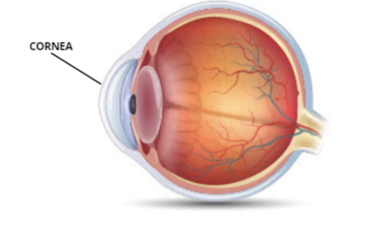
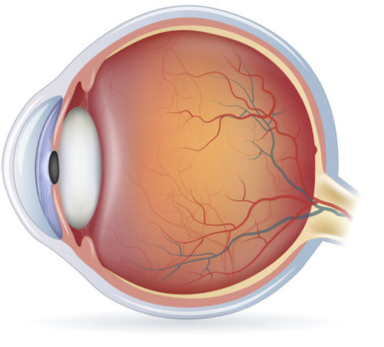
what does the Cornea do and where is it
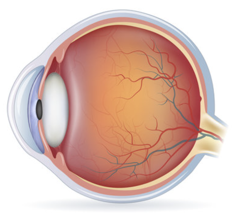
Clear outer layer that bends (refracts) incoming light to start focusing it
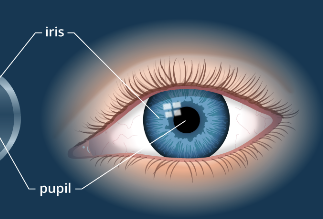
what does the iris do and where is it
Colored ring of muscle that controls how much light enters by changing the pupil’s size.
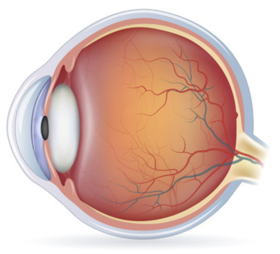
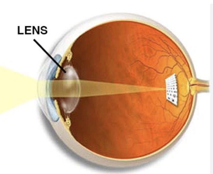
what does the lens do and where is it
Flexible structure that fine-tunes focus by changing shape (process called accommodation)
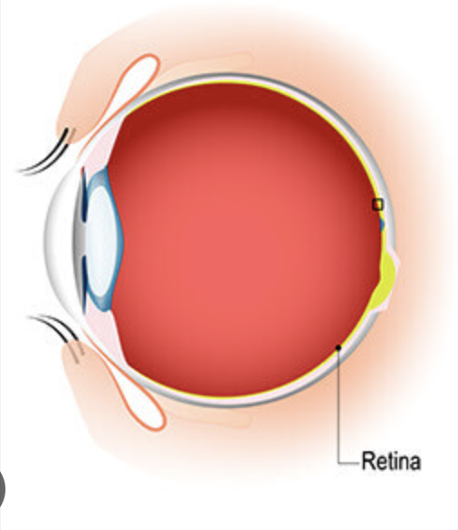
what is retina and where is it
Back layer of the eye that contains the photoreceptors (rods and cones); where light gets turned into neural signals (transduction).
what r rods and where r they
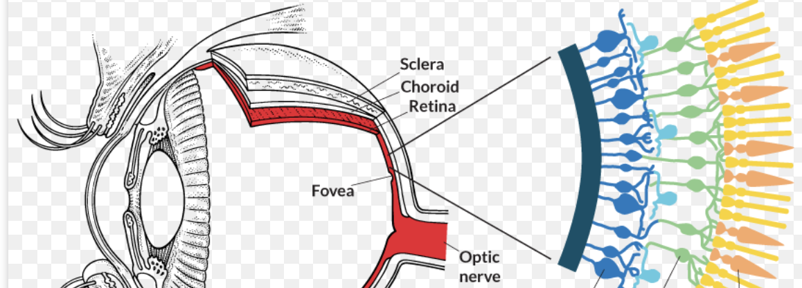
Photoreceptors that detect black, white, and gray — work best in dim light, help with night and peripheral vision.
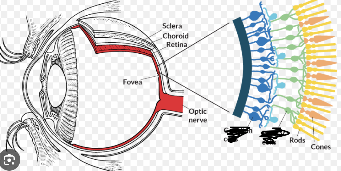
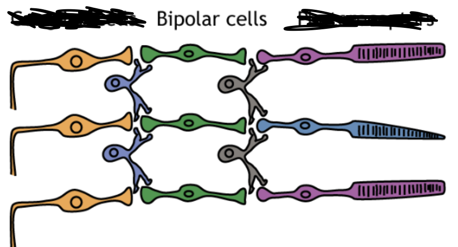
what are Bipolar Cells and where r they
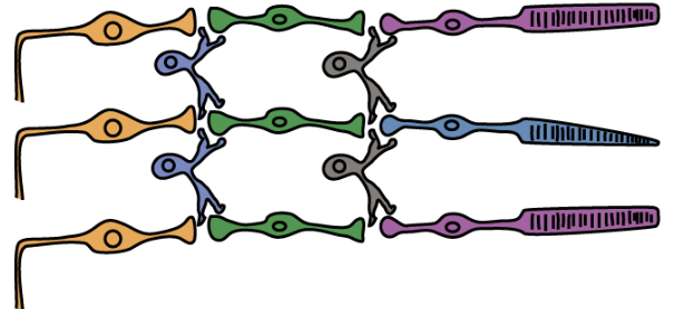
Middle layer of cells that receive signals from rods/cones and pass them to ganglion cells.
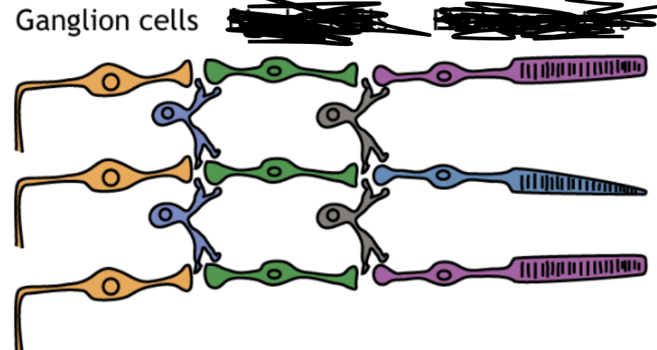
what are Ganglion Cells and where r they
Their axons bundle together to form the optic nerve — the final step before the brain.
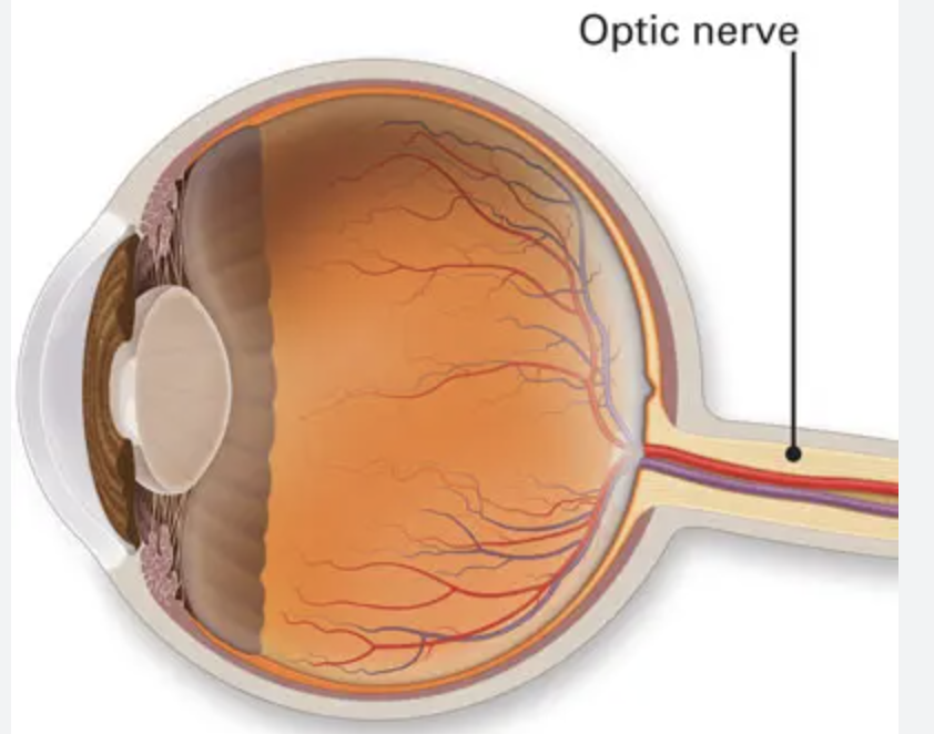
what is the Optic Nerve and where is it
Carries the visual information from the eye to the brain (specifically the occipital lobe).
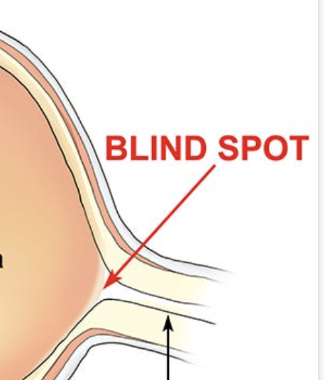
what is Blind Spot and where is it
The point where the optic nerve leaves the eye — no rods or cones there, so you can’t detect light in that small spot.
Negative Afterimages
When you stare at one color for a long time, then look away and see its opposite color — proof of the Opponent-Process Theory (red-green, blue-yellow, black-white).
Place Theory
We hear high-pitched sounds because different frequencies hit specific places on the basilar membrane inside the cochlea.
Frequency Theory
We hear low-pitched sounds because the entire basilar membrane vibrates at the same frequency as the sound wave.
Volley Principle:
For middle pitches — hair cells take turns firing rapidly in groups (a “volley”) so the brain can interpret higher frequencies than a single neuron could handle.
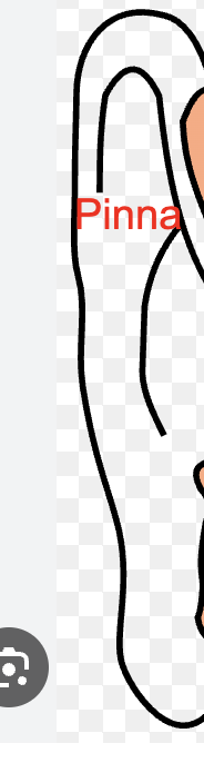
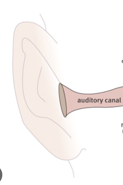
where is Pinna and what does it do
Visible outer ear — collects and funnels sound waves into the auditory canal
Auditory Canal where is it and what does it do
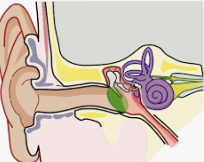
Tube that channels sound waves toward the eardrum.
Eardrum (Tympanic Membrane) and where is it
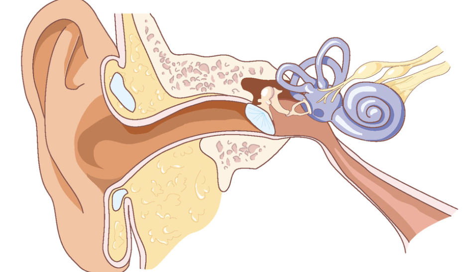
Vibrates when sound hits it — converts sound waves into mechanical vibrations
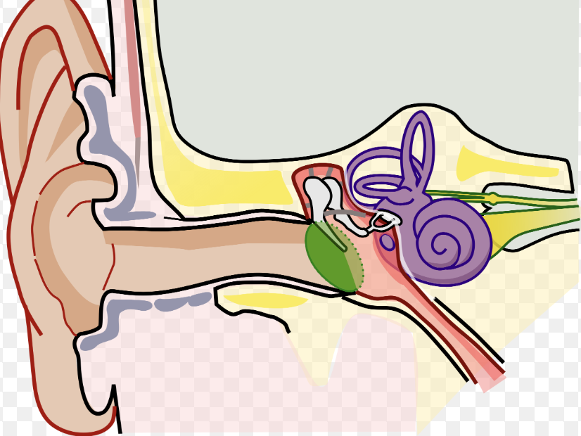
Malleus (Hammer), Incus (Anvil), Stapes (Stirrup)
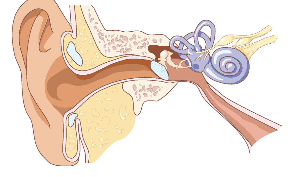
Three tiny bones (ossicles) in the middle ear that amplify and transmit vibrations from the eardrum to the inner ear.
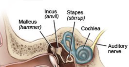
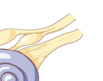
Cochlea
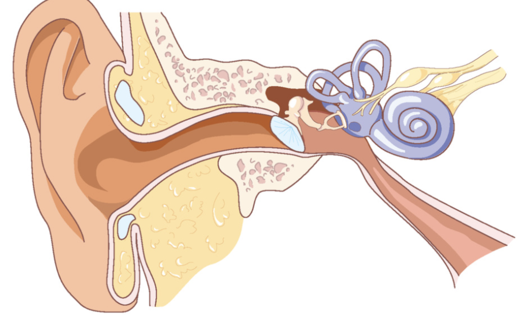
Snail-shaped structure in the inner ear filled with fluid and lined with the basilar membrane — where transduction happens (sound → neural signal).
Basilar Membrane
Inside the cochlea; contains hair cells that move when fluid vibrates — these hair cells trigger neural impulses.
Auditory Nerve:
Carries the electrical signals from the cochlea to the temporal lobe in the brain.
Vestibular Sense:
Sense of balance and spatial orientation — tells you if you’re upright, spinning, or falling.
Semicircular Canals:
Three curved, fluid-filled tubes in the inner ear that detect head movement and rotation — main part of vestibular sense.