Biology Lab Final Study
1/60
There's no tags or description
Looks like no tags are added yet.
Name | Mastery | Learn | Test | Matching | Spaced |
|---|
No study sessions yet.
61 Terms
CSE Journal citation
uthor(s). Date. Article title. Journal title. Volume(issue):location.
for example: Mazan MR, Hoffman AM. 2001. Effects of aerosolized albuterol on physiologic responses to exercise in standardbreds. Am J Vet Res. 62(11):1812–1817.
Parenthetical Citation CSE
(Author(s) Year).
if 3-10 authors: (Smart et al. 2003)
if 2 authors: (Mazan and Hoffman 2001)

DNA
genetic blueprint for all living organisms
some viruses have only RNA, not DNA.
is a virus a living organism?
not generally considered to be living organisms, since they do not have all of the characteristics of life
components of DNA
A five-carbon deoxyribose sugar, whose carbons are numbered 1′ to 5′.
the prime (′) symbol is used to differentiate the carbon in carbon atoms in deoxyribose from nitrogenous base
one of four nitrogenous bases covalently bound to carbon 1’ of deoxyribose
at least one phosphate groups covalently bound to carbon 5’ of deoxyribose (can be as many as 3 phosphate groups)
Types of nitrogenus bases
Adenine
Thymine
Cytosine
Guanine
what is pyrimidine and purine?
A and G = purine
C and T = Pyrimidine
Histones
positively charged protein which Dna wraps around to condense 2m of DNA into 5 nanometers .
name for eukaryotes
Chromatin
DNA in a highly condensed structure
What do we take advantage of to extract DNA for genetic analysis
cell membranes are made of lipids that can be disrupted by detergents
DNA interacting preteens have positive charges that can be neutralized by negatively-charged ions from salts
DNA has a negatively charged backbone that can attract specific dyes
double stranded DNA had 2 antiparrallel strands making it a polar molecule
how is DNA extracted
detergent lyses cell membranes and denatures proteins
salt (NaCl) helps separate DNA from histones through positive electrostatic interactions with the negatively charged DNA.
negatively charged chloride ions from favourable electrostatic interactions with positively charged histones.
In prescient of sodium DNA remains highly soluble in water since water and DNA are polar molecules.
To precipitate DNA, isopropanol is added to reduce solvent polarity, allowing long DNA strands to wind around glass rod for collection.
Why are we using DNa as source of DNA
Strawberries, like many crop plants, have been bred to have multiple copies of each chromosome also known as polyploidy
policy of commercial strains of strawberries:
octoploid and contain eight copies of each chromosome
larger amount of DNA gives us more DNA to extract and helps strawberries grow larger and taste sweeter
How can we tell that what we have extracted from the cells is really DNA?
DNA is invisible to naked eye.
specific dyes bind strongly to the negatively charged phosphates and hydrophobic bases of DNA.
what dye did we use and how does it work
fluorescent dye called SYBR Gold® (Invitrogen™)
gives off bright yellow-gold light when bound to DNA and exposed to blue light
dye interacts with both the negatively charged backbone and the closely stacked bases found in double-stranded DNA
single-stranded DNA does not bind strongly to the
dye because the bases are not stacked tightly in this form of DNA
Sodium Hydroxide
a strong base that removes some of the protons in the nitrogenous bases of DNA, thus disrupting the hydrogen bonding needed to stabilize the structure of double-stranded DNA
Confirming DNA property from dye and fluorescence
visually estimating the amount of fluorescent light given off when the dye is added to either water, DNA, or DNA denatured by the addition of a small amount of sodium hydroxide.
mutations
permanent changes to the genetic information of a cell
generally occur at a fairly low rate in most organisms
occur by many different mechanisms, one of which is exposure
of cells to ionizing radiation, such as ultraviolet light from the sun or artificial sources (e.g., fluorescent and halogen lights).
How does UV cause mutations
radiation can ionize water molecules to form free radicals, which
in turn react with the nitrogenous bases of DNA to change their chemical structure, altering their ability to form specific base pairs with other base
Etc about UV
Some exposure to UV radiation is normal, and organisms have evolved mechanisms to deal with the amount of free radicals generated by a limited degree of radiation exposure
high amounts of exposure cause more generation of free radicals than cells can control
when too much free radical image occurs then DNA damage is more likely to become a mutation, damaging health of cells
3 types of exposure to UV
Sunscreen
Foil
No foil
Bar chart
compare category or groups
Line graph
represent trends overtime or relationships between variables
plot points on a graph and connect them with lines making it easier to see change in value over time or discrete intervals
useful in tracking changes over time or identifying patterns or correlations
Scatter plot
display individual data pouts as dots on a graph with one variable plotted on the x axis and another on the y axis
used to visualize relationships b/w 2 variables and identify patterns or correlation between the,.
useful in identifying outliers, clusters, or trends
benefit from addition of a trend line
Box plots
concise summary for distribution of data set
highlight key stats amusers like median, quartiles, and range
effective for comparing spread and central tendency of multiple groups or categories within a data set
care good at seeing sim and diff between multiple groups or categories
Regarding the wild-type strain data set alone, describe the results from this
experiment. What conclusion can you draw with respect to your null hypothesis that sunscreen will have no protective benefit from UV radiation on yeast survival?
wild-type strain:
The plates with the highest UV exposure/ saran-wrapped plates, yielded the lowest average number of colonies
The sunscreen-protected plates yielded a higher average number of colonies
results reject the null-hypothesis that sunscreen will have no protective benefit for yeast survival.
Regarding the light-sensitive mutant strain data set alone, describe the results from this experiment. What conclusion can you draw with respect to your null hypothesis that sunscreen will have no protective benefit from UV radiation on yeast survival?
mutant strain
plates with the highest UV exposure/ saran-wrapped plates, yielded the lowest average number of colonies
The sunscreen-protected plates yielded a higher average number of colonies
results reject the null-hypothesis that sunscreen will have no protective benefit for yeast survival.
Compare the data between the wild-type yeast strain and the light-sensitive mutant strain and describe the results in your own words. Based on this data, what conclusions can you draw with regard to your second null hypothesis that there will be no difference between yeast strains in response to similar UV-blocking conditions?
mutant strain is not as efficient in its ability to resist/ survive damage caused by UV radiation causing cellular death compared to the wild-type strain (especially in "None" and SPF 15 groups)
reject their null hypothesis that wild-type and mutant strains will show no difference in response to similar UV-blocking conditions
What is the biological significance of your experimental results? Your response should be concise and touch on each exposure condition and yeast strain, and the functionality of DNA repair pathways. How do your results compare with literature on this topic? (Refer to the paper posted in Lab 4 folder: Alhamdy and Alsowayan, 2020)
Results suggest that any level of blocking (SPF 15, 50, or foil) is equally protective for wild- type cells, but light-sensitive mutant yeast only remain viable when UV is completely blocked by foil or SPF 50 sunscreen.
DNA repair is much more functional in sunscreen exposures of wild-type cells compared with mutants as observed with higher survival of wild-type
Lit comparison:
• Regular sunscreens use was shown to reduce the effect of UV exposure with similar results to ours (e.g. Alhamdy and Alsowayan, 2020)
The data presented in the graph is a compilation of data from all groups across all 33 lab sections. Compared with your own graph that you submitted, why are the SEM error bars smaller in this graph in Figure 1 above? What information can you infer from the error bars when trying to determine meaningful differences between samples? Give an example from the graph in Figure 1
Fewer samples in student graphs will result in larger SEM bars (OR more samples in compiled graph will give smaller SEM bars
Generally if error bars overlap with each other, there is no meaningful difference (and vice versa).
Can look at error bars between WT "none" vs WT Foil/sunscreens, or WT SPF15 vs Mut SPF15 (for differences) (1 for any example that works)
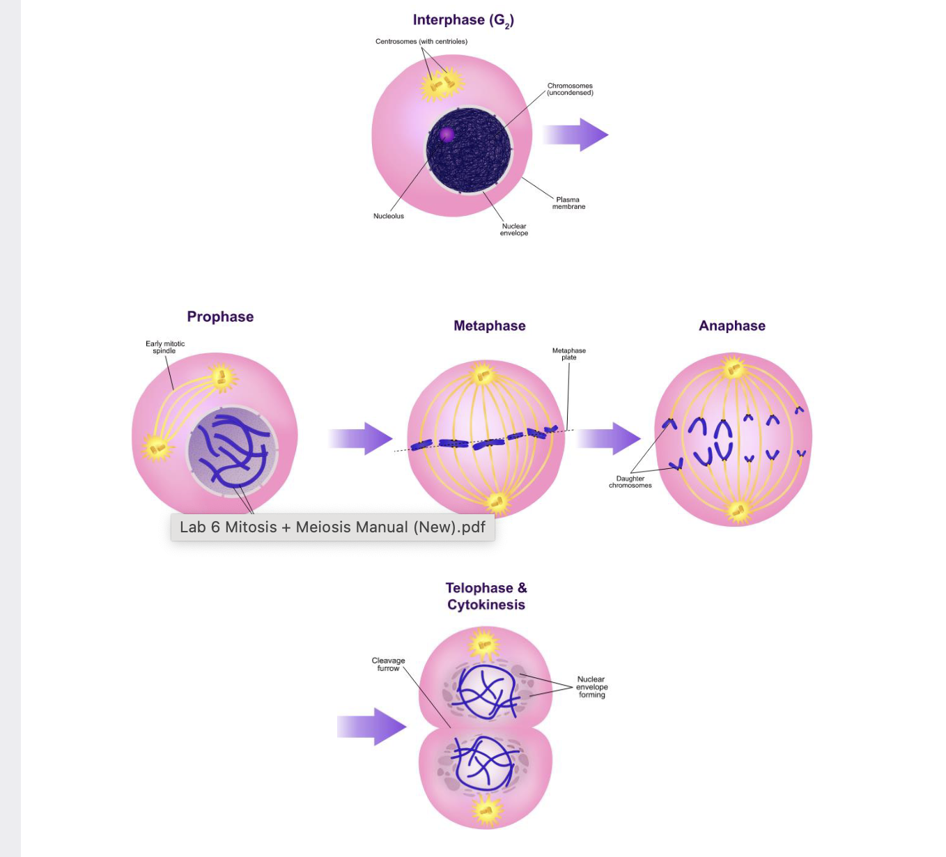
mitosis
divides genetic material equally between two daughter cells, giving each daughter cell the same genetic material as the mother cell
meiosis
divides genetic material among four daughter cells (gametes in animals and spores in plants or fungi), each of which contains half of the genetic material of
original germ cell
Mitosis Phases

Mitosis
one copy of each chromosome is given to each daughter cell, thus providing each daughter cell with a full complement of genetic material identical to that found in mother cell
occurs in all eukaryotic organisms. The process happens in stages that must be carried out very precisely, because mistakes would be catastrophic for daughter cells.
What happens when there is an error in mitosis
a daughter cell is given an incomplete complement of chromosomes or an excess copy of one or more chromosomes the daughter cells usually nonviable and will die.
extremely rare cases where such cells with defective complements of chromosomes somehow survive, such cells may behave in an abnormal fashion that may sometimes lead to disease states
such as cancer.
Meiosis
Meiosis is the process by which a germ cell divides its full genetic complement among four daughter cells, reducing its diploid chromosome set to haploid.
Purpose of Meiosis
Meiosis ensures genetic diversity by creating unique haploid cells, which combine during sexual reproduction to form a diploid zygote.
Meiosis in Animals
In animals, including humans, meiosis produces haploid gametes (sperm and egg cells) necessary for sexual reproduction.
Meiosis in Other Organisms
Meiosis doesn’t always produce gametes. In plants and fungi, it results in haploid spores instead.
Haploid cell
only one cope of each chromosome
diploid cell
2 copies of each chromosome
one copy of maternal and other is paternal
Homologous pair
also known as homologous chromosome or homologues
chromes set (n)
one representative chromosome from each of the pairs of homologous chromosomes in a diploid species.
diploid cells are 2n
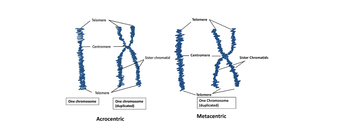
Acrocentric vs metacentric
used to describe structure of chromosomes
metacentric = has arms on either side of centromere equal in length
acrocentric = chromosome has one arm longer than other on one side of centromere than other side

Meiosis and diploid cells

Meiotic process and its functions
reduce diploid to haploid through segregation of alleles
through cross over and random movement of non homologous chromosomes producing new combo of genes or genotypes.
Etc about meiotic process
frequency of recombination in large sample of meiotic cells can by studied by recombinational analysis in Drosophila melanogaster, corn and other diploid organisms that are used as genetic tools.
genes
specific sequences of DNA that most often encode a protein whose expression
results in a specific phenotype
alleles
different DNA sequences of a gene
genotype
specific combination of alleles in an organism
genotype nomenclature
represents alleles of genes on different chromosomes. Alleles on homologous chromosomes are separated by a slash ( / ), while different genes are separated by a semicolon ( ; ).
Example of Genotype Representation
A male heterozygous for genes A and B is written as A/a; B/b; X/Y, where:
A/a and B/b show heterozygosity for genes A and B.
X/Y represents the sex chromosomes in males.
Role of Statistics in Research
Statistical tests help determine whether observed differences in data are meaningful or due to chance, aiding in hypothesis evaluation.
The Null Hypothesis
In statistical tests, the null hypothesis (H₀) assumes no significant difference between groups. A test determines whether to reject or fail to reject H₀.
The t-Test
t-test compares the means of two groups to assess statistical significance. A larger t-value increases the likelihood of a significant difference.
Interpreting the t-Test
calculated t-value is compared to a t-table based on degrees of freedom and p-value. If it meets or exceeds the critical t-value, the difference is statistically significant.
Understanding p-Value
p-value of 5% (p = 0.05) means that if the null hypothesis were true, we would observe a t-value this extreme in less than 5% of cases by chance.
Limitations of the t-Test
t-test can only compare two groups. Using multiple t-tests for more than two groups increases error risk—ANOVAshould be used instead.
Interpreting Non-Significant Results
If calculated t-value is smaller than the critical value, it doesn’t prove there’s no difference—just that the data failed to show a significant difference. A larger sample size may reveal one.
Statistical vs. Biological Significance
A statistically significant result does not always mean a biologically meaningful difference. Always consider real-world relevance before drawing conclusions.
t-test is based on mean and SEM for each of the two groups that you are comparing:
