Anterior abdominal wall and GI tract
1/87
There's no tags or description
Looks like no tags are added yet.
Name | Mastery | Learn | Test | Matching | Spaced | Call with Kai |
|---|
No analytics yet
Send a link to your students to track their progress
88 Terms
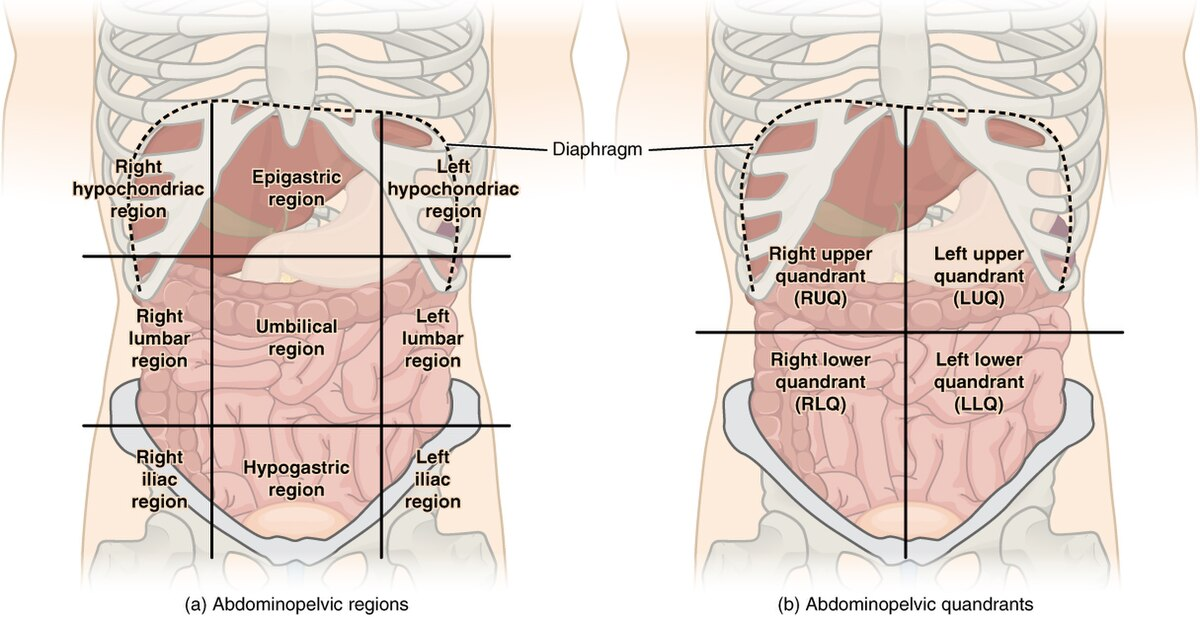
Recall the regions and quadrants of the abdomen.
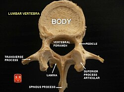
Describe the bones of the abdomen (lumbar vertebrae)
5 lumbar vertebrae forming lumbar spine: large, kidney-shaped vertebral bodies, a triangular vertebral foramen, short and sturdy spinous processes for muscle attachment, and thick pedicles and laminae that create a strong vertebral arch
Describe the arrangement of the muscles that make up the anterior and lateral abdominal wall and describe their actions.
All muscles aim to stabilize the vertebral column, move the trunk, protect abdominal organs, and aid in forceful expiration and activities that increase intra-abdominal pressure
vertical rectus abdominis muscles and three flat, stacked muscles: the external oblique, internal oblique, and transversus abdominis
Distinguish the major segments of the gastrointestinal (GI) tract and recall the key features of each segment.
mouth - ingestion
pharynx/oesophagus - propulsion
stomach - storage, mixing, digestion
small intestine - digestion + nutrient absorption
large intestine - water absorption + waste formation
rectum/anus - elimination
Describe the pattern of blood supply and venous drainage of the GI tract.
blood supply: by branches of the abdominal aorta: coeliac trunk (foregut), superior mesenteric artery (SMA, midgut) and inferior mesenteric artery (IMA, hindgut).
venous drainage:
SMV - midgut ie jejunum, ileum, cecum, appendix, ascending colon + first 2/3 of transverse colon
splenic vein - foregut ie stomach spleen and pancreas
IMV - hindgut, including the last 1/3 of transverse colon, descending colon, sigmoid colon, upper part of rectum
Which 2 main structures make up the abdomen?
abdominal wall and cavity
What is the abdominal wall made up of?
muscles make up both the anterior and posterior abdominal wall
Which viscera are found within the abdominal cavity?
GI tract (abdominal oesophagus, stomach, small intestine, large intestine, upper 1/3 rectum)
Accessory organs of digestion, (liver, gallbladder, pancreas)
Urinary system (kidneys, ureters)
Which vessels are found within the abdominal cavity?
arteries - abdominal aorta, veins - inferior vena cava, lymphatics etc
What qualifies an organ as being an “accessory“ organ of digestion?
no contact with food itself - will help store, secrete chime ie liquid food
What is the abdomen’s position relative to the thorax?
inferior, just below the diaphragm and xiphoid process
Describe the morphology of abdominal wall muscles
flat muscles surrounding abdominal cavity
What are the functions of abdominal wall muscles?
protect abdominal viscera (absence of bones means more protection is needed)
maintain posture and move the trunk
What are the 2 functional groups of abdominal wall muscles?
anterolateral muscles (lateral ie on left and right sides)
posterior muscles
What are the muscles of the abdominal wall from superficial to deep?
anterolateral wall with anterior and lateral muscles
external oblique muscles
rectus abdominis muscles
internal oblique muscles
transversus abdominis muscle
rectus sheath
What is the function of muscles in the abdominal anterolateral wall?
increase intra-abdominal pressure for defecation and childbirth eg
How are fibres within the anterolateral wall organised?
different fibre orientation = stronger meshwork of fibres
What are the 2 groups of muscles making up the anterolateral wall of the abdomen? How many muscles make up the two groups?
anterior muscles - 2 muscles on either side of the midline
lateral muscles - 3 muscles on either side of the abdomen
What connects the 2 groups of muscles of the anterolateral wall of the abdomen?
rectus sheath
What is the most superficial muscle of the anterolateral wall? How does its fibres run?
external oblique muscle
medially and inferiorly towards the midline (like hands in a pocket)
What forms the anterior wall of the rectus sheath?
external oblique muscle aponeurosis
What is aponeurosis?
a sheet of pearly white fibrous tissue that acts as a tendon in flat muscles, having a wide area of attachment - attaches muscle to bone
What do the muscles of the rectus abdominus run alongside?
the linea alba (white line)
What directions do the muscles of the rectus abdominus run?
long vertical muscles running superior to inferior
What are muscles of the rectus abdominus intersected by?
tendinous intersections
What two lines make up the “6 pack“ visible on some individuals?
linea alba (vertical) and tendinous intersections (horizontal) forming the pockets known as a 6 pack
What does the rectus abdominus muscle lie within?
rectus sheath
Which structure’s fibres run perpendicular to those of the external oblique muscle?
internal oblique muscle
Describe the orientation of the internal oblique muscle’s fibres
run medially and superiorly - perpendicular to those of the EOm
Where does the IOm aponeurosis divide? into which sections?
at rectus sheath
anterior ½ forms anterior wall, posterior ½ forms posterior wall
What is the deepest muscle?
transversus abdominis muscle
What separates the transversus abdominis muscle from the abdominal cavity?
transversalis fascia
How are the fibres of the transversus abdominis muscle orientated?
horizontally
What does the TAm aponeurosis form?
posterior wall of rectus sheath
What forms the anterior and posterior walls of the rectus sheath? What does the latter contain?
formed by aponeurosis of the 3 lateral muscles:
anterior made up of external oblique muscle aponeurosis + anterior ½ of the IOm
posterior made up of aponeurosis of posterior ½ of IOm and t transversus abdominis muscle
contains the rectus abdominis muscle
Define fascia
thin casing of connective tissue that surrounds and holds structures
What defines the midline of the rectus sheath?
linea alba
What is the goal of the gastrointestinal tract?
to process food and eliminate waste
What are secondary accessory organs of digestion?
salivary glands, liver, gallbladder, pancreas
What pathway does the GI tract follow?
continuous pathway from mouth to anus : Thoracic and abdominal oesophagus, stomach, small intestine, large intestine, upper 1/3 rectum
What does the blood supply for the GI tract come from?
from abdominal aorta: 3 branches coming anteriorly - coeliac trunk + superior mesenteric artery (SMA) + inferior mesenteric artery (IMA)
What are the 2 portions of the oesophagus?
thoracic
abdominal
What are the different parts of the stomach from superior to inferior?
from the oesophagus - cardia, fundus, body, pyloric region (antrum, canal and sphincter) + lesser and greater curvatures
What is the blood supply for the stomach?
coeliac trunk
Describe the anatomy of the thoracic portion of the oesophagus
superior and posterior mediastina, runs with vagal trunks, pierces diaphragm at oesophageal hiatus
Describe the anatomy of the abdominal portion of the oesophagus
very short, connects with stomach at cardiac sphincter
What is the blood supply to the small intestine ?
coeliac trunk + SMA
What are the 3 parts of the small intestine?
duodenum, jejunum, ileum
Which of the 3 parts of the small intestine is most distinct ? Why?
duodenum is very distinct with the major duodenal papilla
Which 2 parts of the small intestine are most similar ?
Jejunum and ileum share similarities but not the same! bc of different artery blood supply
What does the mesentery enclose? What does it attach these to?
small intestines and parts of colon (transverse/sigmoid)
posterior abdominal wall
What part of the small intestines is c shaped?
duodenum
What is the role of the major duodenal papilla?
nipple-like protrusion, external opening for the common bile duct and the pancreatic duct - aids in digestion, draining and controlling fluids from the bile and pancreatic ducts
Which structures are found within the mesentery?
blood vessels, nerves, lymphatic vessels, lymph nodes, and adipose (fatty) tissue
What is the blood supply to the small intestines?
coeliac trunk and superior mesenteric artery
What are different parts of the large intestines?
caecum, appendix, colon, rectum and anal canal
What is the colon divided into?
ascending, transverse, descending, and sigmoid colon
What are characteristic features of the large intestines?
epiploic appendices, taenae coli and haustrations
At what point does the small intestine become the big one?
at the ileocecal valve
What is the blood supply to the large intestines?
SMA + IMA
What do the embryological origins of the GI tract inform on? What are these 3 parts?
blood supply
foregut, midgut and hindgut
Which structures are found within the foregut?
From oesophagus to duodenum at the level of the major duodenal papilla (halfway along the duodenum)
What is the blood supply to the foregut?
coeliac trunk
Which structures are found within the midgut?
From major duodenal papilla to 2/3 of transverse colon
Which structures are found within the hindgut?
From last 1/3 of transverse colon to upper 1/3 rectum (+ anal canal)
What is the blood supply to the midgut?
SMA
What is the blood supply to the hindgut?
IMA
What are the 3 branches of the aorta supplying the GI tract?
first is celiac trunk, second is superior mesenteric artery, third is inferior mesenteric artery
At which level of the spine does the aorta bifurcate into L/R common iliac arteries?
L4 (lumbar 4)
At which level of the spine does the coeliac trunk come off the aorta?
thoracic 12, T12
At which level of the spine do SMA and IMA come off the aorta?
L1 and L3
At which level of the spine do renal and gonadal arteries come off the aorta?
renal off L1/L2, gonadal off L2
List the arteries that branch off the second branch of the abdominal aorta, the superior mesenteric artery
inferior pancreaticoduodenal
jejunal and ileal
ileocolic
right colic
middle colic
What does the inferior pancreaticoduodenal artery of the SMA of the abdominal aorta supply?
pancreas and last part of duodenum
What do the jejunal and ileal arteries of the SMA of the abdominal aorta supply?
jejunum and ileum
What does the ileocolic artery of the SMA of the abdominal aorta supply?
cecum (proximal ascending colon), ileum, and appendix
What does the right colic artery of the SMA of the abdominal aorta supply?
ascending colon
What does the middle colic artery of the SMA of the abdominal aorta supply?
ascending colon and 2/3 of transverse colon
What is the junction below the liver of the large intestine called? What supplies it?
hepatic flexure
middle colic artery
At which lumbar vertebrae is the midgut supplied?
L1 over the duodenum
At which lumbar vertebrae is the hindgut supplied?
L3, under duodenum
What are the 3 branches coming off the inferior mesenteric artery of the abdominal aorta?
left colic artery
sigmoid arteries
superior rectal artery
What does the left colic artery supply?
left side of colon ie last 1/3 transverse colon and descending colon
What do the sigmoid/sigmoidal arteries supply?
last part of descending colon and sigmoid colon
What does the superior rectal artery supply?
upper ⅓ of rectum
What separates the abdominal cavity from the thorax?
the diaphragm
What are the 3 lateral muscles of the anterolateral abdominal wall? the 2 vertical ones?
EOm, IOm and TAm
RAm