GAS Lectures
 Call Kai
 Learn
Learn Practice Test
 Spaced Repetition
 Match
Match Flashcards
 Knowt Play
1/195

There's no tags or description
Looks like no tags are added yet.
Name | Mastery | Learn | Test | Matching | Spaced | Call with Kai | Chat |
|---|
No analytics yet
Send a link to your students to track their progress
196 Terms
fibrous joints (synarthroses)
immovable joints such as sutures, gompohoses, synchondroses
cartilaginous joints (amphiarthroses)
slightly movable joints such as syndesmoses and symphyses (intervertebral)
synovial joints (diarthorses)
freely movable joints
uniaxial synovial joints
planar, pivot, hinge
biaxial synovial joints
ellipsoid, condyloid, saddle
multiaxial synovial joints
ball and socket
6 types of synovial joints
hinge, ball and socket, condyloid, pivot, planar, saddle
hinge joints
joints that open and close in one direction (knees and elbows)
ball and socket
rounded end of one bone fits into an indentation in another bone, can rotate and turn in almost any direction (shoulders and hips)
condyloid joints
made of two oval shaped bones that fit together, similar to ball and socket joints, except they can't rotate in a full 360 degrees (wrist joints and joints where toes meet the rest of your foot)
pivot joints
rotate in place without moving out of their original position (these joints in neck let the head move from one side to another)
planar joints
formed when 2 mostly flat bones come together, move by one piece of bone sliding over the other without rotating (carpal bones that join your wrist to your forearm and the joints between the vertebrae in your spine)
saddle joints
formed when two curved bones meet; can move in any direction, but can't twist or rotate (joint where your thumb joins your hand)
hyaline cartilage
smooth. most common. matrix closely packed with collage. tough but slightly flexible. chondrocytes arranged in small groups with solid and smooth matrix. allows tissues to slide/glide more easily (between ribs and sternum, nasal cartilage, articular cartilage)
fibrocartilage
toughest and durable. dense masses of white collagen fibers. provide support and rigidity (intervertebral discs, callus, public symphysis, junction where tendons insert to bone
elastic cartilage
flexible and provides support. chondrocytes located between a network of threadlike elastic fibers. provides support and maintains shape of earlobe, epiglottis and blood vessel walls
process of making cartilage
chondrogenesis
components of ECM that are important for cartilage formation
aggrecan and type 2 collagen
germ layer cartilage is formed from
mesoderm
zygapophyseal joint
joint between superior and inferior articular processes (symphyseal joints)
role of zygapophyseal joint
stabilizes and limits spinal rotation
anterior longitudinal ligament
stabilizes vertebral bodies and annulus fibrosus. limits spinal extension. (throat side)

posterior longitudinal ligament
stabilizes bodies and annulus fibrosus. limits spinal flexion
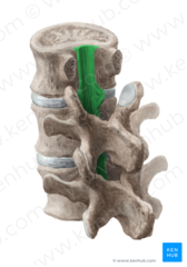
ligamentum flavum
stabilizes laminae. limits spinal flexion

interspinous ligament
stabilizes spinal process. limits spinal flexion
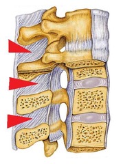
Supraspinous ligament
continuous with nuchal ligament above C7 (THICK) (posterior and back side)

Longitudinal vessels of spinal cord
1 anterior spinal artery & 2 posterior spinal arteries
Segmental spinal arteries (neck)
from vertebral and deep cervical arteries
Segmental spinal arteries (thorax)
from posterior intercostal arteries
Segmental spinal arteries (abdomen)
lumbar arteries
nerve plexus
network of nerves
what forms the nerve plexus
only ventral rami of spinal nerves
cervical plexus nerves
formed by ventral rami of C1-C4 nerves
cervical plexus innervation
skin (via cutaneous branches) & muscles of neck and shoulder (via ansa cervicalis)
Phrenic Nerve nerves (cervical plexus)
C3-5 (C3,4,5 Keeps you alive)
Phrenic nerve innervation (cervical plexus)
diaphragm, vital for respiration
Brachial plexus nerves
ventral rami of C5-T1
Brachial plexus innervation
innervates the upper limbs. lies in neck and armpit (axilla)
Musculocutaneous nerve of brachial plexus
flexor of arm (biceps etc)
Median nerve of brachial plexus
all flexors of forearm except 1.5 muscles
Ulnar nerve of brachial plexus
1.5 flexors of forearm (flexor carpi ulnas and medial 1/2 of flexor digitorum profundus)
Axillary nerve of brachial plexus
deltoid & teres minor
Radial nerve of brachial plexus
all extensors of arm and forearm
Lumbar Plexus nerves
ventral rami of L1-L4 nerves (lies in posterior abdominal wall)
2 main nerves of lumbar plexus
femoral nerve & obturator nerve
Femoral Nerve of lumbar plexus
innervates anterior muscles of thigh (quadriceps)
Obturator Nerve of lumbar plexus
innervates medial muscles of thigh (adductors)
Sacral Plexus nerves
ventral rami of L4-S4 (immediately caudal to lumbar plexus)
Main nerve in sacral plexus
sciatic nerve
Sciatic Nerve of sacral plexus
innervates muscles and skin of posterior thigh and most of the leg
thickest and longest nerve in the body
sciatic nerve
Herpes Zoster
virus that produces chickenpox. Sits dormant in cells of dorsal root ganglia. Once activated travels along neuronal bundles to areas supplied by the nerve (dermatome)
epaxial muscles
true back muscles (innervated by dorsal rami)
hypaxial muscles
superficial & intermediate back muscles & limb (innervated by ventral rami)
crossbar of H in gray matter
gray commissure (bundles of axons that cross from one side of CNS to another)
Where are ventral horns the largest?
in cervical and lumbar segments of the spinal cord due to innervation of upper and lower limbs
3 types of fibers in white matter
ascending, descending, commissural
Asending fibers
carry sensory information from sensory neurons of body up to the brain
Descending fibers
carry motor instructions form brain to spinal cord
Commissural fibers
carry information from one side of spinal cord to other
2 enlargements of spinal cord
cervical (C5-T1) & lumbosacral (L1-S3)
How many cervical spinal nerves are there?
8 (different than the 7 vertebrae in the cervical region)
How many coccygeal nerves are there?
1 (different than the 4 fused vertebrae in coccyx)
Where do spinal nerves lay?
in the intervertebral foramina
Where is CSF formed
choroid plexus
3 layers of meninges (superficial to deep)
Dura mater, arachnoid mater, pia mater
3 functions of the spinal cord
sensory and motor innervation, two way conduction pathways for signals between the brain and body, major center of reflexes
4 stages of development of spinal cord
neural plate --> neural fold --> neural tube --> spinal cord
What condenses into the neural plate?
neuroectoderm (derived from ectoderm)
Sulcus limitans
longitudinal groove formed by the neural tube that divides spinal cord into dorsal and ventral portions
Altar Plates
dorsal portions. location of sensory somatic and visceral input
Basal Plates
ventral portions. location of motor function output
Conus Medullaris definition & location
tapered end of spinal cord. L2
filum terminale
made of Pia mater. tapered end of conus medullaris. attaches to coccyx and anchors spinal cord
Cauda Equina
collection of descending nerve roots
Nucleus pulposus
pulpy interior of intervertebral discs. shock absorption. remnant of notochord. 80% water and loses water content with age
Annulus Fibrosus
fibrous outer ring. helps contain nucleus pulpous
Secondary curvatures
lordotic (cervical and lumbar)
Primary curvature
kyphotic (thoracic and sacral)
When does secondary curvature arise in babies
1 year
What kind of curvature do chimps have?
secondary curvature only in cervical region
hyperkyposis
hunchback. Cobb angle > 30. more common with age especially after 40
hyperlordosis
swayback. lordosis exceeding normal range. common in pregnant women
scoliosis
lateral curvature and rotation of vertebrae
types of scoliosis
congenital, degeneration, adolescent idiopathic (most common), neuromuscular
bones of shoulder and arm
clavicle, scapula, humerus
joints of shoulder and arm (3)
sternoclavicular, acromioclavicular, glenohumeral
sternoclavicular joint
saddle joint. medial clavicle articulates with manubrium of sternum
acromioclavicular joint
gliding joint. lateral clavicle articulates with acromion of scapular
glenohumeral joint (shoulder)
ball and socket joint. glenoid cavity of scapula articulates with head of humerus
acromion process of scapula
continuous with spine of scapula. articulates with clavicle
glenoid process of scapula
articulates with head of humerus of glenohumeral joint. splits into supraglenoid tubercle (origin of long head of bicep) & infraglenoid tubercle (origin of long head of tricep)
coracoid process of scapula
origins of coracobrachialis, short head of biceps brachii. insertion of pectoralis minor
4 ligaments supporting sternoclavicular joint
anterior sternoclavicular ligament, posterior sternoclavicular ligament, interclavicular ligament, costoclavicular ligament
acromioclavicular joint
plane gliding synovial joint with capsule
ligaments supporting acromioclavicular joints (2)
acromioclavicular ligament (reinforces joint capsule), coracoclavicular ligament (stabilizes distal clavicle and coracoid process), coracoclavicular ligament (conoid ligament -- medial and trapezoid ligament -- lateral)
glenohumeral joint
ball and socket synovial joint
functions of glenohumeral joint
abduction, adduction, flexion, extension, medial rotation, lateral rotation, circumduction
glenoid labrum
a fibrocartilaginous collar that deepens the glenoid cavity. continuous with tendon of long head of bicep brachii
transverse humeral ligament
within the intertubercular sulcus, stabilizes glenohumeral joint