A-Level Biology - 3.6.3 Skeletal muscles are stimulated to contract by nerves and act as effectors
 Call Kai
 Learn
Learn Practice Test
 Spaced Repetition
 Match
Match Flashcards
 Knowt Play
1/23

There's no tags or description
Looks like no tags are added yet.
Name | Mastery | Learn | Test | Matching | Spaced | Call with Kai | Chat |
|---|
No analytics yet
Send a link to your students to track their progress
24 Terms
How do muscles act?
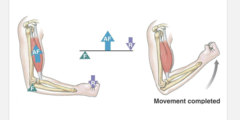
Muscles act in antagonistic pairs against an incompressible skeleton
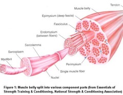
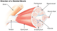
Describe the structure of skeletal muscle
made up of bundles of muscle cells (muscle fibres)
muscle fibres are elongated cell containing many nuclei
sarcoplasm contains myofibrils formed from contractile proteins
many mitochondria in the sarcoplasm produce ATP to release energy for muscle contraction
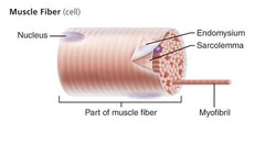
Name the subcellular structures of a muscle fibre
cell surface membrane = *sarcolemma*
cytoplasm = *sarcoplasm*
endoplasmic reticulum = *sarcoplasmic reticulum*
Describe the structure of a myofibril
protein filaments in myofibrils:
*myosin* (thick filaments)
*actin* (thin filaments)
Myofibrils can be divided into sections called *sarcomeres*
sarcomeres shorten during muscle contraction as myosin and actin filaments slide past each other
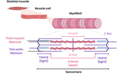
Name the features of a sarcomere
Z-line
M-line
A band
H zone
I band
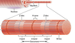
What is the Z-line?
The boundary between sarcomeres
Actin filaments attach here
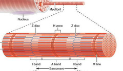
What is the M-line?
middle of sarcomere
Myosin filaments attach here
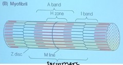
What is the A band?
region of the sarcomere across which myosin filaments extend
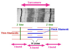
What is the H zone?
region containing only myosin filaments
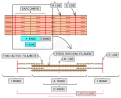
What is the I band?
region containing only actin filaments
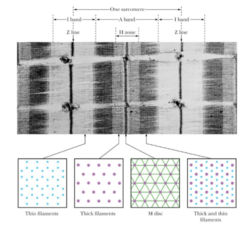
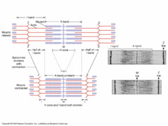
What happens to the sarcomere during contraction?
Z-lines get closer together
Myosin filaments don't change in length, so the A band remains same size
H zone shrinks during contraction as actin and myosin fires overlap more
I band shrinks during contraction as actin and myosin fires overlap more
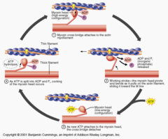
Describe the roles of calcium ions and ATP in the contraction of a myofibril
1) Sarcolemma is depolarised, so calcium ions diffuse into myofibrils from sarcoplasmic reticulum
2. Calcium ions cause movement of tropomyosin on actin
3) Causes exposure of the binding sites on the actin
4) Myosin heads attach to binding sites on actin (forms actinomyosin bridges)
5) Release of Pi and ADP from myosin heads cause them to bend, pulling actin molecules (power-stroke)
6) Attachment of a new ATP molecule to each myosin head causes myosin heads to detach from actin sites
7) Hydrolysis of ATP on myosin heads causes them to bend back to original position (re-cocked)
Describe what happens in a myofibril when the electrical impulse stops
contraction stops when the impulse stops: calcium ions actively transported back into sarcoplasmic reticulum
actin moves back and muscle can return to its relaxed length
What is the role of ATP in myosin contraction?
1) Reaction with ATP breaks binding of myosin to actin
2) Provides energy to move myosin head
What is phosphocreatine?
a molecule stored by muscles that can be used for the rapid production of ATP over short periods
Describe the role of ATP and phosphocreatine in muscle contraction
supply of ATP is required for muscle contraction:
-to break actinomyosin bridges
-to move the myosin head
-so actin filaments are moved inwards
-for active transport of calcium ions
a phosphate ion from phosphocreatine is transferred to ADP to rapidly generate ATP anaerobically
ADP + phosphocreatine → ATP + creatine
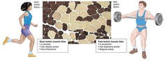
Name two types of muscle fibre
fast and slow skeletal muscle fibres
What are fast muscle fibres most suited to?
short bursts of high-intensity activity
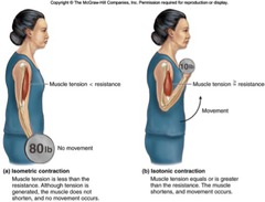
Give the properties of fast muscle fibres
higher concentration of glycogen and phosphocreatine
contract rapidly
ATP supplied mostly from anaerobic respiration
Fatigues rapidly due to high lactate production rate
fewer capillaries and mitochondria
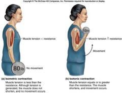
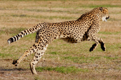
Give the location of fast muscle fibres
limbs of animals that flee predators or hunt prey at high speeds
human eyelid muscles
biceps and triceps in the upper arms of humans
What are slow muscle fibres most suited to?
sustained, low intensity activities
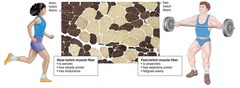
Give the properties of slow muscle fibres
lower concentration of glycogen and phosphocreatine
contract slower
ATP supplied mostly from aerobic respiration
Fatigues more slowly due to reduced lactate formation
more capillaries and mitochondria
Give the location of slow muscle fibres
limbs of animals that migrate or stalk prey over long distances
human back and leg muscles

Explain how the difference in glycogen concentrations is related to the different properties of fast and slow muscle fibres
1) Fast fibres used during short-term/intense exercise
2) Slow fibres used during longer-term exercise
3) Creatine used to form phosphocreatine, which combines with ADP to form ATP
4) Glucose stored as glycogen
5) Glycogen hydrolysed to glucose for respiration