Human Physiology Unit 2 Flashcards
1/610
There's no tags or description
Looks like no tags are added yet.
Name | Mastery | Learn | Test | Matching | Spaced |
|---|
No study sessions yet.
611 Terms
what kind of receptors do neuromuscular junctions contain?
nicotinic
Explain the somatic motor pathway
extends from CNS and releases ACh that binds to nicotinic receptor on target skeletal muscle
what are NaChR channels of skeletal muscke similar to?
NNACh receptors on neurons
what kind of channels are NaChR channels?
chemically-gated channels with 2 binding sites for ACh
are NaChR channels inhibitory or excitatory?
always excitatory and create muscle contractions
what is the synapse of a somatic motor neuron on a muscle fiber called?
neuromuscular junction (NMJ)
what does the NMJ consist of?
axon terminals, motor end plates, schwann cells, synaptic cleft, postsynaptic membrane
what do the motor end plates on muscle membrane in the NMJ contain?
high concentrations of ACh receptors and lies opposite to axon terminal that is modified into motor end plate
what is the synaptic cleft between the axon and muscle in the NMJ filled with?
fibrous matrix whose collagen fibers hold the axon terminal and motor end plate in proper alignment
what does the matrix in the synaptic cleft in the NMF contain?
acetylcholinesterase
acetylcholinesterase
enzyme that rapidly deactivates ACh by degrading it into acetyl and choline
what do nicotinic ACh receptor (nAChR) channels do in the active zone?
cluster together
what are the steps that lead to muscle contractions?
Action potentials arrive at axon terminal and open voltage-gated calcium channels in membrane
calcium diffuses into cell down its electrochemical gradient to trigger release of ACh containing synaptic vesicles
ACh diffuses across synaptic cleft and combines with NAChRs on skeletal muscle membrane
chemically gated ion channels with 2 binding sites for ACh
when ACh binds to receptor, channel gate opens and allows monovalent cations to flow through
in skeletal muscle, net entry into muscle fiber depolarizes it, triggering action potential that causes contraction of the skeletal muscle cell
what are the 3 types of muscle?
skeletal, cardiac, smooth
what kind of muscle is the skeletal muscle and what is it attached to?
striated muscle attached to bones of skeleton
what do skeletal muscles do?
control body movement and responsible for voluntary control
what neurons do skeletal muscles respond to?
somatic motor neurons
what kind of fibers are in skeletal muscles?
large, multinucleate cells that appear striped/striated under miscroscope
what kind of muscle is the cardiac muscle and what is it attached to?
striated muscle and found only in heart
what do cardiac muscles do?
move blood through circulatory system and responsible for involuntary control
what do cardiac muscle respond to?
autonomic innervation, spontaneous contraction, modulated by endocrine system
what kind of fibers are in cardiac muscle?
striated, smaller, branched, and cells are uninucleate and joined in series by junctions
what are intercalated discs
junctions that join cells in the fibers of cardiac muscle together in series
what kind of muscle is the smooth muscle and what is it attached to?
primary muscle of internal organs and tubes
what do smooth muscle do?
influence movement of material into, out of, and within body and responsible for involuntary control
what do smooth muscle respond to?
autonomic innervation, spontaneous contraction, modulated by endocrine system
what kind of fibers are in smooth muscle
small fibers that lack striations
in skeletal muscles, thick filament is…
actin
in skeletal muscles, thick filament is…
myosin
in thick filament, heavy chains are…
motor domain and have myosin ATPase
in thick filament, light chains are…
regulatory function
what are the regulatory proteins in skeletal muscle?
tropomyosin and troponin
what are the accessory proteins in skeletal muscle?
titin and nebulin
what do T-tubules do?
allow rapid AP movement from cell surface into interior of fiber
where do t-tubules transmit action potentials for synchronous contraction?
to regions near the sarcoplasmic reticulum
how do t-tubules help with synchronous contraction?
allow for coordinated calcium release throughout fiber and leads to uniform contraction
what are t-tubules?
extensions of cell membrane (sarcolemma) that associate with the ends (terminal cisternae) of the sarcoplasmic reticulum
myofibrils
muscle fiber contractile structures
sarcomere
contractile unit of microfibril
sarcomere: z discs
have filaments between them that have accessory proteins that link think filaments together
sarcomere: I band
made of thick filaments (actin)
sarcomere: A band
made of darker regions where both light and heavy vilaments overlap
sarcomere: H zone
clear band in middle of A band and has heavy filaments only
sarcomere: M line
proteins to which heavy filaments attach
what happens to sarcomeres overall during contraction?
shorten
in sarcomeres, what happens to actin and myosin during contraction?
don’t change length but slide past one another in an energy-dependent process
in sarcomeres, what happens to the H zone, I band, and A band during contraction?
H zone and I band shorten; A band remains constant
what are titin and nebulin together and what do they do?
proteins that ensure proper alignment of filaments in the sarcomere
what is titin responsible for individually?
stabilizes myosin
what is nebulin responsible for individually?
aligns actin
muscle tension
force created by muslces
load
weight/force that opposes contraction
relaxation
release of tension
what are the steps to skeletal muscle contraction
events occur at the NMJ
excitation-contraction (E-C) coupling
contraction-relaxation cycle
in regards to length, what happens to fibrils?
remain at a fixed length
what do myosin crossbridges do?
move actin filaments
myosin crossbridges: power stroke
myosin crossbridge swivels and pulls actin toward M line
myosin crossbridges: end of power stroke
myosin releases actin and resets and binds to another actin (heads are not released in unison)
true or false? the power stroke occurs only in one cycle
False; the powerstroke is repeated multiple times
myosin ATPase
energy places the myosin head into a “cocked" position”)
what happens when myosin comes in contact with actin
myosin ATPase initiates a pull
what does troponin do?
controls position of tropomyosin
what does troponin C do?
reversibly binds to calcium
what does tropomyosin do and why?
partially covers myosin binding site on actin to prevent myosin and actin from interacting
what are the steps of contraction when initiated by calcium signaling?
calcium released from terminal cisternae
calcium binds troponin
troponin pulls tropomyosin from myosin-binding sites on actin
myosin binds tightly to and moves actin
repeated as long as binding sites uncovered and ATP is available
where do crossbridges form and why?
forms between actin and myosin heads to trigger contraction
After a powerstroke, there is a rigor state where…
myosin is tightly bound to G-actin
steps of rigor state
ATP binds and myosin detaches (ATP decreases myosin affinity for actin)
ATP hydrolysis provides energy for myosin head to rotate and reattach to actin (head weakly binds to new actin)
powerstroke that begins in response to calcium binding to troponin (release of Pi allows head to swivel, pulling actin toward M line)
myosin releases ADP and makes room for next ATP
what does myosin ATPase do?
breaks down ATP to ADP and Pi
E-C coupling steps
ACh released from somatic motor neuron
ACh initiates AP in muscle fiber (binds to receptors on sarcolemma and causes depolarization of cell for an end plate potential and muscle AP)
muscle AP triggers calcium release from sarcoplasmic reticulum
calcium combines with troponin to initiate contraction
twitch
single contraction-relaxation cycle in skeletal muscle fiber
relaxation
when calcium is pumped back to sarcoplasmic reticulum using SERCA
timing of E-C coupling: latent period
short delay between muscle action potential and beginning of muscle tension development
what is central fatigue due to?
CNS
what is peripheral fatigue due to?
neuron/muscle
peripheral fatigue: what does submaximal exercise do?
extended submaximal exercise leads to depletion of glycogen stores
peripheral fatigue: what does short-duration maximal exertion do?
leads to increased levels of Pi which can slow Pi release from myosin and decrease calcium release
peripheral fatigue: what does maximal exercise do?
leads to ion imbalances because potassium leaves the muscle fiber, which leads to increased extracellular concentrations of potassium and altered membrane potential; also leads to changes in sodium/potassium pump activity
what are skeletal muscles classified by?
speed and fatigue resistance
skeletal muscle: slow-twitch fibers (ST / type I)
rely primarily on oxidative phosphorylation
skeletal muscle: fast-twitch fibers
develop tension faster (split ATP more rapidly), pump calcium into SR more rapidly
skeletal muscle: fast-twitch oxidative-glycolytic fiber (FOG / type IIA)
use oxidative and glycolytic metabolism
skeletal muscle: fast-twitch glycolytic fibers (FG / type IIB/X)
rely primarily on anaerobic glycolysis
what does myoglobin do?
binds oxygen in muscle so it is readily available for aerobic processes
oxidative fibers have what?
more myoglobin
sarcomeres contract with optimum force under what conditions?
if at optimum length (not too long or short) before contraction begins
what is tension directly proportional to?
number of crossbridges
the force of contraction increases with what?
summation — contractions are stronger when the muscle does not relax completely between action potentials
tetanus
maximal contraction
what is a motor unit?
1 motor neuron + all of the muscle fibers it innervates
what does contraction force depend on?
types and numbers of motor units
what does asynchronous recruitment of motor units help?
helps avoid fatigue
isotonic contractions
move loads
isotonic contractions: concentric action
shortening
isotonic contractions: eccentric action
lengthening
isometric contractions
force without movement
isometric contractions: what happens to sarcomeres?
shorten while elastic elements stretch, resulting in little change in overall length
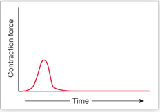
What does this graph show and give an example?
phasic smooth muscle that is usually relaxed; esophagus
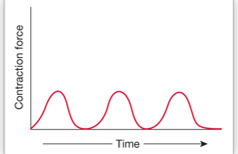
what does this graph show and give an example?
phasic smooth muscle that cycles between relaxation; intestine
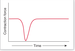
What does this graph show and give an example?
tonic smooth muscle that is usually contracted; sphincter that relaxes to allow material to pass