Biopsych Exam 1
 Call Kai
 Learn
Learn Practice Test
 Spaced Repetition
 Match
Match Flashcards
 Knowt Play
1/102

There's no tags or description
Looks like no tags are added yet.
Name | Mastery | Learn | Test | Matching | Spaced | Call with Kai | Chat |
|---|
No analytics yet
Send a link to your students to track their progress
103 Terms
Biopsychology
Seeks to understand behavior by studying the physiological processes that control it
Also referred to as physiological psychology, or behavioral neuroscience
How to we learn about brain functioning?
Animal models
Human brain injuries
Imaging of the brain
Genetic methods
Experimental Ablation
Removing or inactivating part of the brain
Lesion studies
Experiments in part of the brain is damaged and the animals behavior is observed
Discovers what functions are performed by different regions of the brain
Human Brain Injuries - Phineas Gage
Helped us to understand the role of the prefrontal cortex
Human Brain Injuries: H.M.
Helped us to understand the role of the hippocampus
Structural Brain Imaging
X-Ray
Computerized Tomography (CT)
Magnetic Resonance Imaging (MRI)
Still photo
Functioning Brain Imaging
Position emission tomography (PET)
Functional MRI (fMRI)
Mirco and macroelectrodes (EEGs)
Stimulating AND recording brain activity
Genetic Methods
All behavior is determined by interactions between and individual’s brain and their environment
Behavioral characteristics are often passed down
May play a role in physiological differences
Examples
Twin studies
Adoption studies
Genomic studies
Changes on perspectives of the brain
Historical: brain is static and does not change in adulthood
Now: neuroplasticity and neurogenesis
Neuroplasticity
The ability of brain areas to increase in organization, level of activation, and amount of connection to other areas based on use, practice, and experience
Neurogenesis
Involves the birth of totally new neurons
Neurons can be regenerated
New neurons appear throughout the lifespan
Evidence in hippocampus
Central Nervous System (CNS)
Brain and spinal cord
Encased in bone (skull and vertebral column)
Requires a large supply of blood and is chemically protected by the blood brain barrier
Peripheral Nervous System (PNS)
Nerves that rely informed between the CNS and the rest of the body
Cranial nerves, spinal nerves, and peripheral ganglia)
Neuron
Information-processing and information-transmitting element of the nervous systen
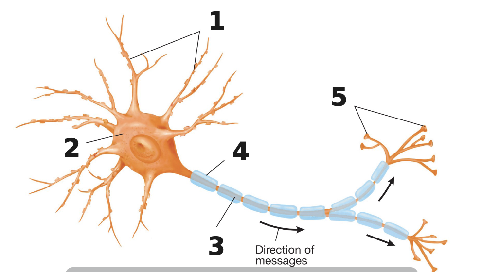
Label the neuron
Consists of dendrites, soma, axon, myelin shealth, axon terminal buttons
Soma (cell body)
contains organelles including the nucleus
Shape varies
Appears as GRAY MATTER in the brain and nerves
Dendrites
Branched, treelike structure
Receives info transmitted from other neurons
Axons
Long, thin, cylindrical structures
Carries info from the soma to the terminal buttons
The action potential is basic message
Myelin Sheath
Fatty substance that surrounds the axons and insulates them
Appears as white matter in the brain and nerves
Helps a message travel faster
Terminal buttons
Buds at the end of a branch of an axon
Form synapses with another neuron
Secrete chemicals called neurotransmitters
Axon Hillock
Where the action potential begins
Nucleus
Contains chromosomes and proteins
Mitochondria
Power plants; produces ATP
Membrane
Double layer of lipid (fat-like) molecules
Sensory neuron
IN PNS
Afferent - signals travel toward the CNS
Receives sensory info (light, sound, touch, taste, odor, contact with objects)
Motor neuron
In PNS
Efferent - signal travels away from the CNS
Control voluntary and involuntary motor behavior through contracting muscles/glands
Interneurons
Connections
Between sensory and motor neurons
In CNS

Label da diagram
Sensory, motor, and interneurons
Astrocytes
Supporting cell of the CNS
Support and repair
Nourishment
“neuron glue”
Phagocytosis
Oligodendrocytes
Supporting cells of the CNS
Multiple segments of myelin sheath
Insulation and support
Microglia
Supporting cell of the CNS
Immune defense
Phagocytosis
Schwann cell
Support cell for PNS
Support
One segment of myelin sheath
Action Potential
Rapid changes in electrical charge across the membrane of a neuron
Results in the release of neurotransmitters
Membrane Potential
Difference in charge (positive or negative) across the membrane of a neuron (inside or outside)
Extracellular fluid
Fluid surrounding the neuron
Contains ions
Chloride (Cl-) and Sodium (Na+) are of highest concentration
Intracellular fluid
Fluid inside the neuron
Potassium (K+) and Organic Anions (A-) are of highest concentration
What are the two forces that give rise to the membrane potential?
Diffusion
Electrostatic pressure
Diffusion
movement of molecules from regions of high concentration to low concentration
Electrostatic pressure
Force exerted by attraction or repulsion that moves ions from place to place
Cations are positive
Anions are negative
Anions repel each other; cations repel each other; anions and cations attract
Resting membrane potential
Axon at rest is negative inside the cell relative to the outside; neuron is polarized
-70 mV more negatively charged inside the axon in comparison to the outside
Sodium-Potassium Pump
Made of protein molecules embedded in the membrane
3 Na+ comes out of the neuron for every 2 K+ that is pumped in
Creates high concentration of sodium outside the cell and high concentration of potassium inside the cell
Uses 40% of the neurons metabolic resources (ATP is provided by mitochondria)
Without the pump, the resting potential would collapse, causing neurons and muscle cells to stop firing signals
Action potential
Consists of a series of changes in opening and closing of voltage gated ion channels along the axon of a neuron and the resulting redistribution of ions (and thus membrane potential)
What causes an action potential to occur?
Signals from other cells or sensory inputs cause positive ions (like sodium) to enter the cell
Begins when threshold of excitation is reached and the membrane potential moves closer to 0, becoming less negative
Threshold of excitation
-55mV
When reached, an action potential will fire and the voltage-gated ion channels open
Depolarization
The inside of the cell becomes less negative relative to the outside; moving the membrane potential closer to 0
Voltage-gated ion channels open, allowing positively charged sodium ions in
Triggers the rising phase of an action potential (electrical signal)
Repolarization
The cell returns its voltage back down to the negative resting state after depolarization
Sodium channels close and voltage-gated potassium channels open, allowing positive potassium ions to leave the cell
Resets the electrical charge inside the cell to the negative
Returns the neuron to resting potential
Hyperpolarization
The membrane potential becomes more negative than the normal resting potential
Potassium channels stay open a bit too long and potassium ions leave the cell
Creates a refractory period where its harder for the cell to fire another signal
Voltage-gated ion channels
Exist along the axon of a neuron and aid in the redistribution of ions and the membrane potential
Are only opened by changes in the membrane potential
2 kinds: voltage gated sodium channel (allows sodium ions to rush into cell) and voltage gated potassium channel (allows potassium ions to rush out of the cell)
All or nothing law
An action potential either occurs or does not occur
Once threshold of excitation is reached, an action potential will always occur
Synaptic Transmission
Primary means of communication between neurons

Structure of synapses
Pre and postsynaptic membranes
Face each other across the synapse
Synaptic vesicles
Made of membrane and filled with molecules
Neurotransmitters
Naturally occurring chemicals (endogenous), produced and released by neurons
Postsynaptic receptors
Specialized proteins that bind neurotransmitters released from presynaptic neurons, facilitating the transmission of neural signals
Synaptic Cleft
The space between terminal buttons of one neuron and dendritic or somatic membrane of another
Release of neurotransmitter
Action potential reaches terminal button
Vesicles (filled with neurotransmitters) on the presynaptic membrane open
Neurotransmitter is released into the synaptic cleft
Called Exocytosis
Molecules of neurotransmitters move from areas of high concentration to disperse across synapses (diffusion)
Exocytosis
Vesicles fuse with presynaptic membrane, open, and spill their contents into the synaptic cleft
Activation of receptors
NTs (either excitatory or inhibitory) fit the binding sites of receptors
This conveys the neural message from the presynaptic cell to the postsynaptic cell
Once binding occurs, the postsynaptic receptors open neurotransmitter-dependent ion channels
Movement of these ions changes local membrane potential (depends on what ion channel is opening)
Excitatory neurotransmitter
increase the likelihood that the neuron will fire an action potential
depolarization
Inhibitory neurotransmitter
decrease the likelihood that a neuron will fire an action potential
hyperpolarization
Termination of Postsynaptic potentials
Reuptake or Enzymatic deactivation
Reuptake
Extremely rapid removal of a neurotransmitter from the synaptic cleft by the terminal button
Enzymatic deactivation
Accomplished by enzyme that destroys molecules of the neurotransmitter
Ex: Acetylcholinesterase (AChE) deactivates acetylcholine (ACh)
Discovery of the Blood Brain Barrier
Paul Ehrlich’s experiment over 100 years ago
Injected dye into mice (dye injected in body stayed separate from the brain and vice versa)
Blood-Brain Barrier
Selectively permeable barrier produced by the cells in walls of brain’s capillaries
Let’s in only what is needed and keeps out what could be harmful
Maintains the right ionic balance within the brain and blocks substances that would disrupt neural functions
Some substances must be actively transported through the capillary walls by special proteins
Blood flow and the BBB
Brain receives up to 20% of blood flow from the heart
1 sec interruption = uses too much dissolved oxygen
6 sec interruption = unconsciousness
Few minutes = permanent brain damage
Cerebrovascular Disease or Stroke
Most common type of life-threatening injury to the brain in the U.S. and 5th most common cause of death
Happens when a blood clot or broken vessels presents blood from getting to the brain
Meninges
Protective sheaths around the brain and spinal cord
Dura Mater
Outermost meninges
Durable, thick, tough
Unstretchable
Arachnoid Membrane
Middle meninges
Soft and spongy with weblike tissue
Subarachnoid Space
Fluid-filled space that cushions the brain
Between arachnoid membrane and pia matter
Protects from head trauma
Reduces brain from 1400kg (3lb) to 80kg (1lb)
Contains cerebrospinal fluid (CSF)
Pia Matter
Innermost meninges
Follow every surface of the brain
Ventricular System
Consists of a series of 4 ventricles (hollow, interconnected chambers)
Produce and contain the cerebrospinal fluid, which provides support, nourishment, and removes waste
Cerebrum
Largest part of the brain (accounts for 4/5 total weight)
Divided into 2 symmetrical cerebral hemispheres
Made up of cerebral cortex, limbic system, and basal ganglia
Cerebral Cortex
surrounds the cerebral hemispheres
4 lobes
Consists mostly of glia, cell bodies, and dendrites
Grayish appearance called gray matter
Beneath are millions of axons that connect to other areas of the brain
White matter
Gray matter
Glia, cell bodies, and dendrites in the cerebral cortex
White Matter
Millions of axons that connect to other areas of the brain beneath the cerebral cortex
Large concentration of myelin gives it the opaque white color

Four Lobes of the brain (label)
Frontal
Parietal
Occipital
Temporal
Sensory cortices
Receive information from sensory organs; info sent to the contralateral hemisphere (except taste and smell)
Primary visual cortex
Receives and processes visual info
Located in occipital lobe
Primary auditory cortex
Receives and processes auditory information
Located in temporal lobe
Primary somatosensory cortex
Receives and process somatic info (detecting touch, body position and movement in space), pain, pressure, and temp
Located in parietal lobe
Motor cortex
Most directly involved in control of movement
Neurons in different parts of this are connect to muscles in different parts of the body
Contralateral
Located in front of the primary somatosensory cortex in the frontal lobe
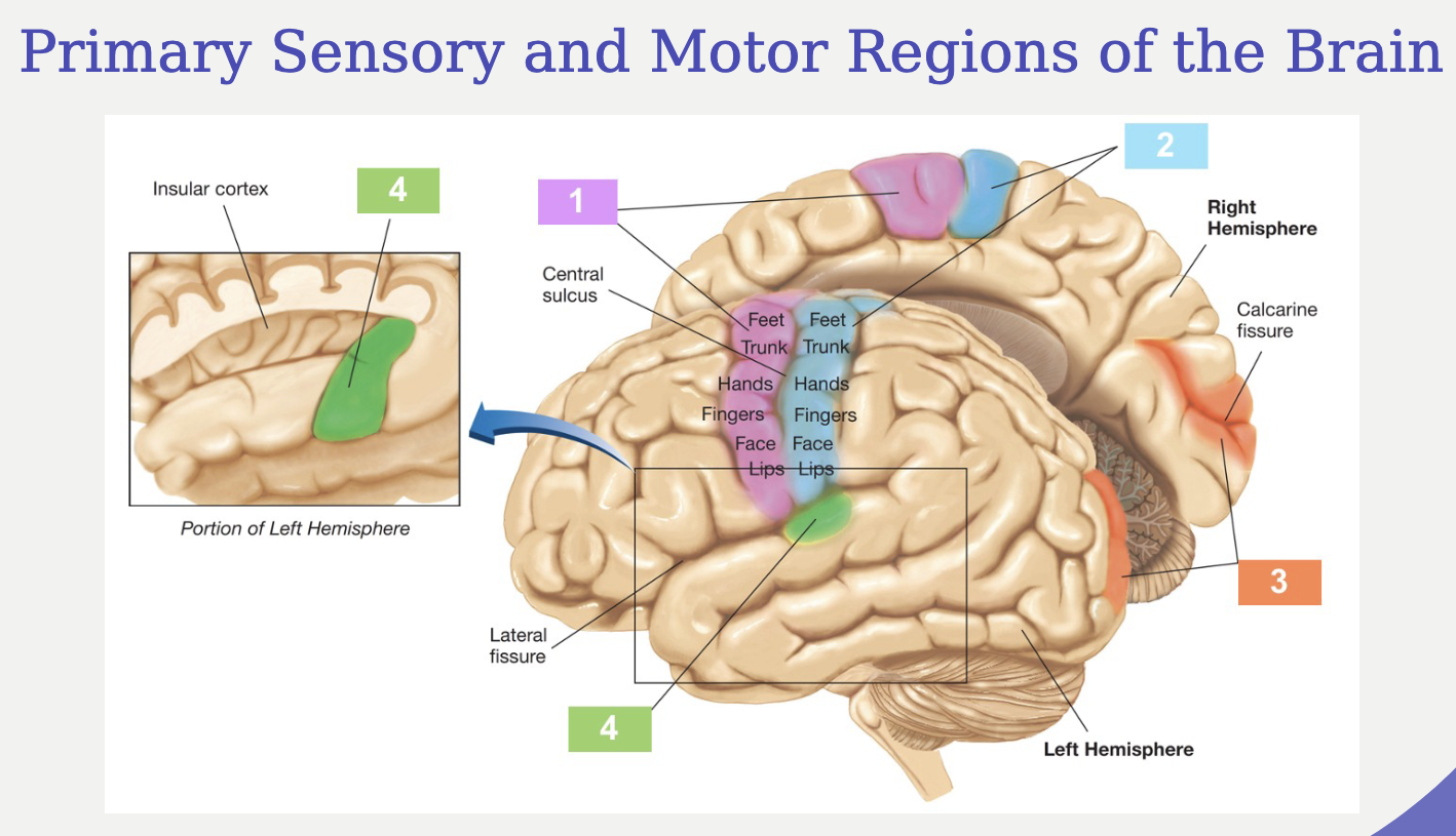
Label this diagram
Prefrontal Cortex
Involved in executive functions (i.e. formulating plans, setting goals, regulating emotions)
Limbic System
Subcortical nuclei
Involved in survival behaviors and emotional responses
Includes hippocampus, amygdala, and cingulate gyrus
Hippocampus
Learning and memory
Amygdala
Processing center for emotions
Cingulate gyrus
Processing emotions, behavior regulation, regulating autonomic motor function
Basal Ganglia
Group of subcortical nuclei
Involved in the control of movement, attention, and reward processing
Proper functioning requires dopamine
Involved in parkinson’s, SUDs, and ADHD
Thalamus
Relay for motor and sensory signals to the cerebral cortex
Hypothalamus
Controls autonomic nervous system and endocrine systems (fight, flight response)
Produces and controls secretion of hormones in conjunction with the pituitary gland
Organizes behavior related to the survival of the species
Cerebellum
Receives auditory, visual, vestibular, and somatosensory info and info about individual muscle movements
Integrates info and modifies the motor outflow; coordinates and smooths movements
Alcohol impairs functioning
Pons
Large bulb in brainstem
Sleep and arousal
Relays info from the cerebral cortex to the cerebellum
Medulla
Controls vital functions such as regulation of the cardiovascular system, respiration, and skeletal muscle tone
Somatic Nervous System
Involved in conscious activities
Spreads outward from brain and spinal cord
Receives sensory info from sensory organs and controls movements of skeletal muscles through cranial and spinal nerves
Spinal Nerves
31 pairs that begin at the junction of the nerve roots of the spinal cord
Nerves leave the vertebral column and travel to the muscles or sensory receptors branching repeatedly as they go
Efferent (motor) and afferent (sensory)
Cranial Nerves
12 pairs are attached to the ventral surface of the brain
Efferent (motor) and afferent (sensory)