US physics II midterm questions
1/99
There's no tags or description
Looks like no tags are added yet.
Name | Mastery | Learn | Test | Matching | Spaced | Call with Kai | Chat |
|---|
No analytics yet
Send a link to your students to track their progress
100 Terms
A sonographer changes from a 3MHz transducer to a 7 MHz transducer while imaging a deep abdominal structure. Which is most likely?
Improved detail resolution and decreased penetration
A sonographer increases imaging depth during a scan while maintaining the same sector width. Which change is most likely to occur?
decreased temporal resolution
Two pulses are transmitted from the same transducer.
Pulse A contains 2 cycles
Pulse B contains 6 cycles
Compared with Pulse A, Pulse B will demonstraight
Longer pulse duration and longer spatial pulse length
A sonographer wants to improve penetration while imaging a technically difficult abdomen. Which adjustment would best achieve this goal?
decrease transducer frequency
A transducer produces a narrow focused beam
compared with a wider beam, the focused beam will most likely demonstrate:
greater intensity
A sonographer increases imaging depth.
Which relationship is correct?
PRF decreases and frame rate decreases
Which imaging situation would most benefit from the use of a high frequency transducer?
evaluation of a superficial thyroid nodule
A sonographer changes from a low frequency transducer to a high frequency transducer.
Which combination is expected?
increased attenuation and shorter wavelength
A pulse with fewer cycles will generally produce:
shorter pulse duration
A sonographer wants to improve temporal resolution during real time imaging.
Which adjustment would most likely help?
reduce imaging depth
Frequency and wavelength are best described as:
Inversely related
A sonographer increases sector width during real time imaging.
Which change is most likely?
decreased frame rate
A sonographer notices reduced penetration while using a very high frequency transducer.
What is the most likely explanation?
Increased attenuation at high frequencies
Compared with a pulse containing many cycles, a pulse containing fewer cycles will generally demonstrates:
Improved ability to distinguish closely spaced reflectors
A sonographer increases imaging depth during a scan.
Which combination of changes is most likely?
Increased PRP and decreased frame rate
Which parameter most directly controls axial resolution?
Spatial pulse length
Increasing imaging depth will cause ALL of the following EXCEPT
improved temporal resolution
Which parameter best determines lateral resolution?
beam width
Low level echos appearing within a simple cyst are most likely caused by poor:
elevational resolution
A sonographer changes from a 3MHz transducer to a 10 MHz transducer.
Which change is most likely?
Improved axial resolution with decreased penetration
If an ultrasound image appears uniformly too dark, the first adjustment should be:
increased overall gain
The fair field of an image appears too dark while the near field brightness remains appropriate.
Which adjustment would best correct this problem?
Increase far field TGC
Decreasing dynamic range will result in ALL of the following except
improved temporal resolution
Adding multiple focal zones will most likely improve:
Lateral resolution while decreasing frame rate
Overall gain affects which parameter best?
returning echo amplification
A sonographer freezes an image and wants to magnify a suspicious thyroid nodule while preserving the original acquired data.
Which adjustment is most appropriate?
read zoom
A sonographer wants to improve visualization of a small superficial lesion and increase image detail within the selected region.
Which adjustment would most directly improve displayed spatial detail?
Write zoom
A sonographer activates tissue harmonic imaging during an abdominal examination.
The harmonic frequencies used to form the image are:
generated through nonlinear propagation in tissue
A technically difficult abdominal scan demonstrates near field clutter and poor tissue contrast.
Which adjustment would most likely improve image quality?
Activate harmonic imaging
A liver image demonstrates speckle and poorly defined borders. The sonographer activates a feature that acquire multiple images from different insonation directions and combines them into one final image.
Which processing technique was achieved?
Spatial compounding
Increasing persistence will most likely result in:
motion blur
A sonographer increases persistence while imaging a moving structure.
Which effect is most likely to occur?
Improved image smoothness
A carotid image demonstrates poor vessel border definition but acceptable overall image quality.
Which adjustment would most likely improve border appearance without improving true spatial resolution?
edge enhancement
During abdominal imaging, harmonic imaging is activated to improve image quality. The sonographer notices that deeper anatomy becomes more difficult to visualize.
Which factor most likely explains this change?
harmonic frequencies attenuate more rapidly
During a liver ultrasound exam, the image demonstrates excessive speckle noise but temporal resolution is already adequate.
Which adjustment is most appropriate?
activate frequency compounding
A transducer is modified to increase damping. Which change is mostly likely to occur?
Wider bandwidth and lower Q-factor
what is the primary function of the matching layer?
improve transmission of sound into tissue
A sonographer wants to improve image detail but cannot sacrifice penetration. Which adjustment is most appropriate?
add focal zones
Which feature is an advantage of electronic focusing compared with mechanical focusing?
adjustable focal location
Which transducer design most directly improves slice thickness contrast?
1.5D array
which combination would produce the deepest focus and least beam divergence?
high frequency → large diameter
Harmonic imaging most directly improves:
contrast resolution
which adjustment is most likely to improve both axial and lateral resolution?
increase transducer frequency
which transducer category provides the greatest ability to electronically steer and focus the beam?
array transducer
A sonographer is imaging a small moving cardiac structure through a narrow acoustic window and wants adjustable focus and beam steering.
which combination is most appropriate?
phased array + electronic focusing
Blood is flowing directly toward the transducer.
Compared with transmitted frequency, reflected frequency will be:
Higher
which factor would decrease doppler shift
larger doppler angle (toward 90 degrees)

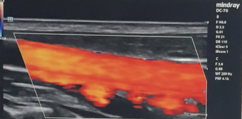
Using the displayed color map, which vessel demonstrates flow moving TOWARDS the transducer?
Vessel B and C
A sonographer needs to evaluate blood velocity at one specific location within a vessel.
Which mode should be selected?
PW doppler
Which doppler mode measures high velocities without aliasing?
CW doppler
PRF increases.
What happens?
Nyquist increases
A doppler system has a PRF of 12 kHz.
What is the Nyquist limit?
6kHz
Increasing sample volume size most likely causes:
More spectral broadening
A sonographer changes from a 5MHz transducer to an 8 MHz transducer while all other variables remain unchanged.
What happens to doppler shift?
Increases
You decrease imaging depth while evaluating flow.
What happens first?
PRF increases

A sonographer identifies Vessel A and Vessel B.
Which statement is most accurate?
flow direction must be interpreted using the color map
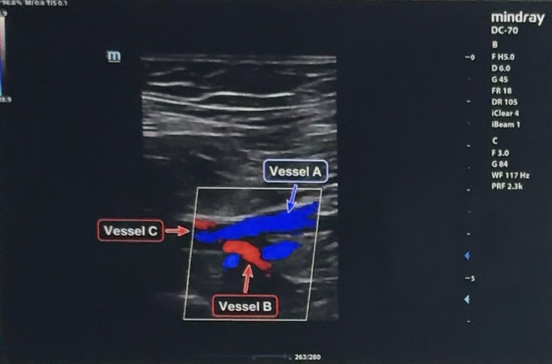
What vessel demonstrates a positive doppler shift?
Vessels B and C
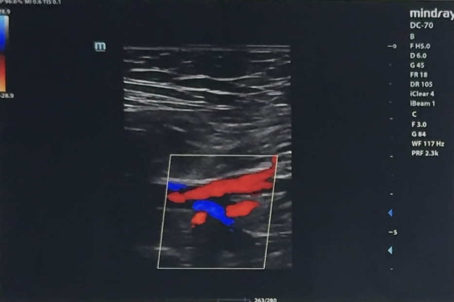
Without changing transducer position, the color map is inverted.
What happens?
display colors change only
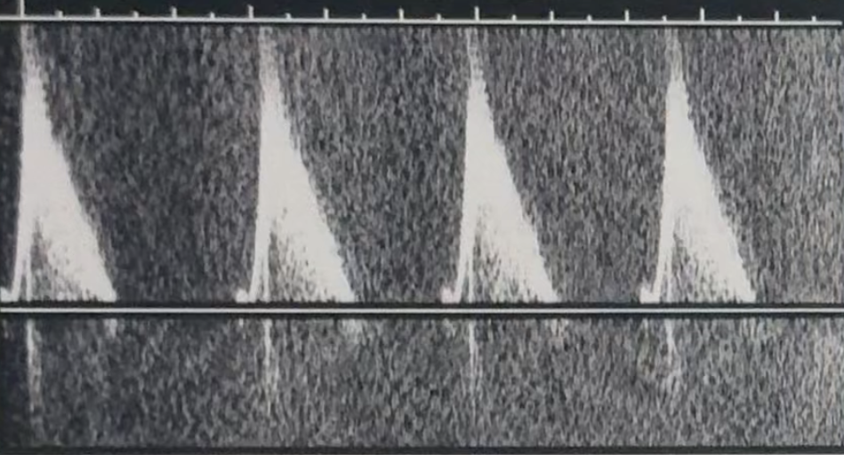
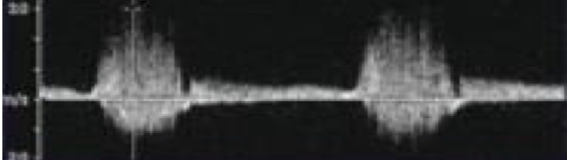
The appearance shown above is most likely caused by:
increased spectral gain
A sonographer is evaluating venous flow and notices that low velocity signals disappear from the doppler display. Which adjustment most likely caused this?
increased wall filter
What is the best first adjustment?
increase scale (PRF)
A sonographer lowers scale (PRF) while evaluating arterial flow. What is the most likely result?
improved sensitivity to slower flow
A sonographer enlarges the sample volume while evaluating a normal artery. What most likely occurs?
increased spectral broadening
Increasing color box size most directly decreases:
temporal resolution
Color suddenly disappears from a vesssel. Which adjustment should be attempted first?
increase color gain
A sonographer increase color threshold (priority) while evaluating a small vessel. What is the most likely result?
more pixels display as color
Increasing packet size most directly results in:
Improved slow flow detection
Which doppler mode is most sensitive for demonstrating very slow flow in a tiny vessel?
Power doppler
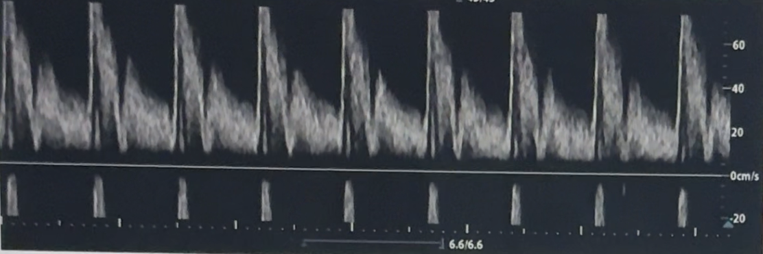
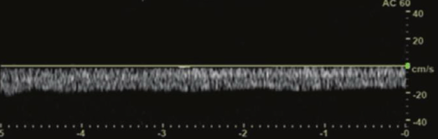
The appearance show above is most likely corrected by:
reduce color gain
A sonographer wants cleaner color display while perserving true venous flow. Which adjustment is most appropriate?
Slightly reduce color gain
A vessel narrows but the same amount of blood continues moving through it. Which changes first?
Velocity
A vessel becomes progressively narrower. What happens first?
resistance increases
A doppler waveform demonstrates filling in of the spectral window despite proper doppler optimization settings. The most likely explanation is:
turbulent flow
spectral broadening may occur because of all of the following except:
increased wall filter
which doppler finding is most consistent with increasing distal resistance?
decreased EDV
A vessel demonstrates continuous forward diastolic flow on spectral doppler.
This most likely represents:
low resistance circulation
which doppler measurement is most useful for evaluating distal resistance?
EDV
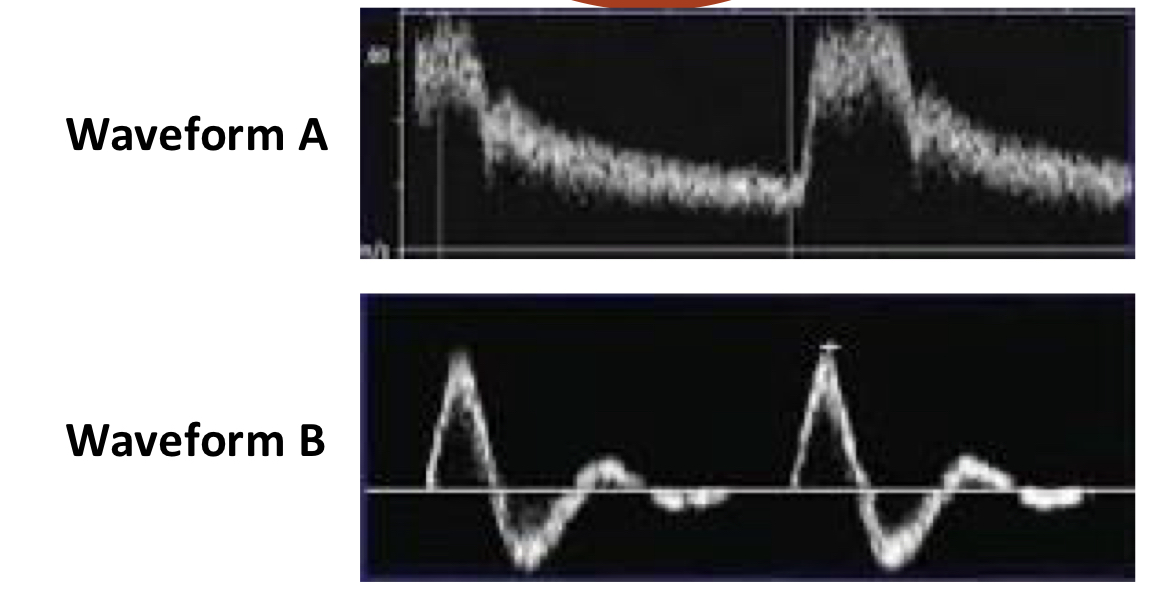
Which spectral doppler waveform most likely represents high resistance vascular bed?
waveform B
A vessel demonstrates increased forward flow during diastole. What happens to RI?
decreases
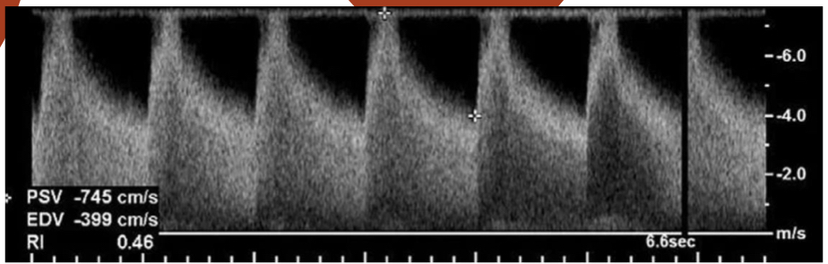
A doppler waveform demonstrates decreasing diastolic flow while PSV remains stable.
Which measurement would most likely increase?
Resistive Index (RI)
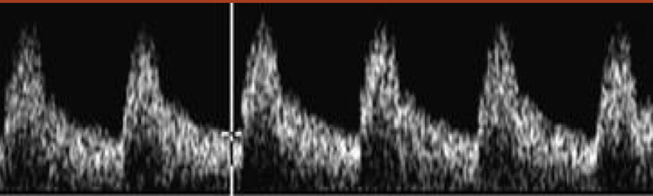
Which doppler finding is most suggested by this waveform?
Stenosis
Which resistance pattern is most consistent with this waveform?
low resistance
Despite proper doppler optimization, what is the most likely explanation?
turbulent
A venous doppler waveform demonstrates minimal respiratory variation. This most likely represents:
loss of phasicity
Mirror image artifact is most likely corrected by:
change insonation angle
crosstalk artifact is most likely corrected by:
reduce doppler gain
Spectral broadening appears during evaluation of a normal vessel. Which technical factor could explain this?
large sample volume
Which change would not reduce aliasing?
increase doppler gain
Which doppler mode is most appropriate for demonstrating blood flow in a tiny vessel with a very slow flow?
power doppler
A sonographer increases packet size while evaluating portal venous flow.
What is most likely to occur?
Improved slow flow detection
A sonographer increases color threshold during evaluation of a small vessel.
What is the most likely result?
more pixels display color
A spectral doppler waveform shows a duplicate signal appearing symmetrically on the opposite side of the baseline even though flow direction has not changed.
What is the most likely cause?
Crosstalk
A sonographer wants to improve visualization of weak slow flow. Which adjustment is most appropriate?
Lower threshold
Color appears randomly throughout tissue during breathing. Most likely cause?
clutter/ghosting
Which doppler setting should generally be lower for TDI?
wall filter
What controls velocity display range?
scale (PRF)
Increasing color box size most directly decreases:
temporal resolution
Which artifact occurs when doppler shift exceeds the system’s display limit and may falsely appear as flow reversal?
Aliasing
What most improves spectral window cleanliness?
reduce gate size
A sonographer wants cleaner color display but wants to preserve venous flow.
Which adjustment is most approriate?
slightly reduce gain