Cardiovascular System: Blood Vessels and Circulation - Notes
 Call Kai
 Learn
Learn Practice Test
 Spaced Repetition
 Match
Match Flashcards
 Knowt Play
1/95

There's no tags or description
Looks like no tags are added yet.
Name | Mastery | Learn | Test | Matching | Spaced | Call with Kai | Chat |
|---|
No analytics yet
Send a link to your students to track their progress
96 Terms
Blood Vessels
Blood travels in a closed system of vessels that begins and ends at the heart.
The three major types of vessels are arteries, capillaries, and veins.
Arteries
Carry blood away from the heart to the capillaries
There are three types of arteries:
elastic
muscular
arterioles

Capillaries
The smallest blood vessel
Its thin walls allow movement of substances between blood and interstitial fluid; endothelium
Walls consisting of a thin tunica interna, one cell thick, no muscle or connective tissue
Porous blood vessels for the exchange of substances between blood and tissues
Allow only a single RBC to pass at a time
Involved in gas exchange, nutrient-waste exchange, and delivery of various substances
Veins
Drain blood from the capillaries, transporting it back toward the heart
Structure of Blood Vessels
Arteries and veins are composed of three tunics
Tunica interna
Tunica media
Tunica externa
Capillaries are composed of endothelium with sparse basal lamina.
Lumen is the central blood-containing space surrounded by tunics.
Tunica Interna
Composed of a simple squamous epithelium layer that lines the lumen of all vessels
Provides a smooth surface for substances (nitric oxide) to regulate contraction and relaxation within the tunica media
In vessels larger than 1 mm, a subendothelial connective tissue basement membrane is present
Tunica Media
Smooth muscle and elastic fiber layer, regulated by the sympathetic nervous system
Controls vasoconstriction/vasodilation of vessels
Tunica Externa
Also known as tunica adventitia
Outermost layer of the blood vessel
Collagen fibers that protect and reinforce vessels
Larger vessels contain vasa vasorum
Companion Vessels
Arteries and veins that supply the same body region and tend to lie next to one another
Elastic (Conducting) Arteries
Largest arteries near the heart; the aorta and its major branches
Vessel Diameters from 2.5 to 1 cm
Large lumen allows low-resistance conduction of blood
Contain elastin in all three tunics, little smooth muscle
Withstand and smooth out large blood pressure fluctuations
Transport blood to the muscular arteries
Allow blood to flow fairly continuously through the body
Muscular (Distributing) Arteries and Arterioles
Medium-sized arteries
Diameters from 1 cm to 0.3 mm
Circumscribed sheets: Internal elastic lamina and external elastic lamina
Distal to elastic arteries; deliver blood to body organs
Have thick tunica media with more smooth muscle and less elastic tissue
Active in vasoconstriction
Carries blood to specific organs
Arterioles
Smallest arteries; lead to capillary beds
Diameter from 0.3 mm to 10 µm
6 layers of smooth muscles
Control flow into capillary beds & hence tissues via vasodilation and constriction
Contracted state called muscle tone
Vasomotor tone
Contracted state in blood vessels
Regulated by vasomotor center in the medulla oblongata
Three structural types of capillaries:
Continuous
Fenestrated
Sinusoids
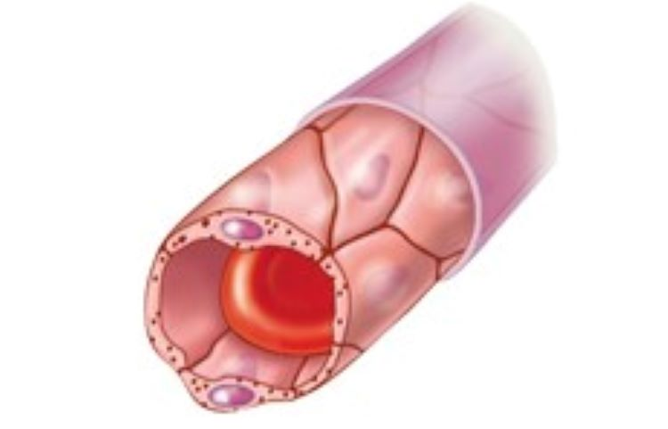
Continuous Capillaries
Abundant in skin and muscles, and have:
Endothelial cells that provide an uninterrupted lining
Adjacent cells that are held together with tight junctions
Intercellular clefts of unjoined membranes that allow the passage of fluids
Form the blood-brain barrier
Simple diffusion and Pinocytosis for materials passing through walls
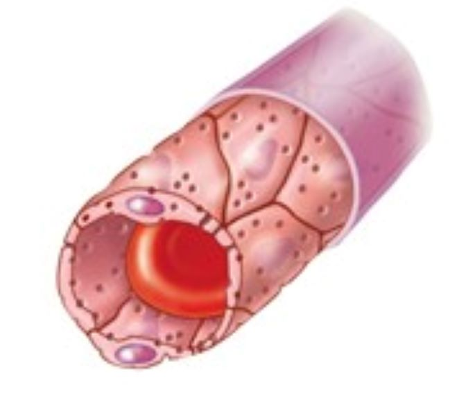
Fenestrated Capillaries
Found wherever active capillary absorption or filtrate formation occurs (e.g., small intestines, endocrine glands, and kidneys)
Characterized by:
Endothelium riddled with pores (fenestrations), gap junctions
Greater permeability to solutes and fluids than other capillaries, forms interstitial fluid

Sinusoids
Highly modified, leaky, fenestrated capillaries with large, irregular-shaped lumens
Found in the liver, bone marrow, lymphoid tissue, and in some endocrine organs
Allow large molecules (proteins & blood cells) to pass between the blood & surrounding tissues (more permeable)
Blood flows sluggishly, allowing for modification in various ways
In the liver, lined with macrophages for phagocytosis
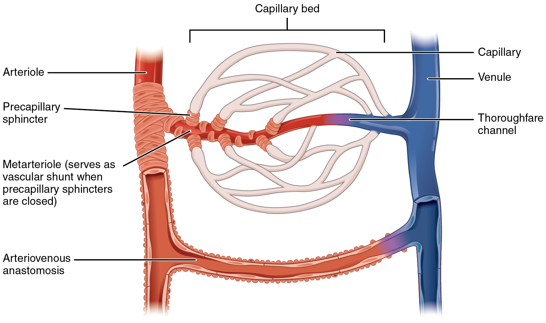
Capillary Beds
A microcirculation of interwoven networks of capillaries (10-100), consisting of:
Vascular shunts: metarteriole thoroughfare channel connecting an arteriole directly with a postcapillary venule
True capillaries: branch off the metarteriole and return to the thoroughfare channel at the distal end of the bed
Atherosclerosis
Disease in which an artery wall thickens, leaving a smaller lumen for blood flow
Result in coronary artery disease (CAD)/ peripheral artery disease (PAD)

Aneurysm
Ballooning of an artery due to a weakened vessel wall; it is susceptible to rupture, leading to severed bleeding
Venous System: Venules
The smallest type of veins, from 8 to 100 micrometers in diameter
Companion vessels with arterioles
Allow fluids and WBCs to pass from the bloodstream to tissues
Postcapillary Venules: drain blood from capillaries
Smallest venules, composed of endothelium
Large venules have one or two layers of smooth muscle (tunica media) and thin tunica externa
Venous System: Veins
Formed when venules converge
Composed of three tunics, with a thin tunica media and a thick tunica externa consisting of collagen fibers and elastic networks
Capacitance vessels (blood reservoirs) that contain 65% of the blood supply
Veins
Veins have much lower blood pressure and thinner walls than arteries
To return blood to the heart, veins have special adaptations:
Large-diameter lumens, which offer little resistance to flow
Valves (resembling semilunar heart valves), which prevent backflow of blood
Venous Sinuses
Specialized, flattened veins with extremely thin walls (e.g., coronary sinus of the heart and dural sinuses of the brain)
Blood Flow through Capillary Beds
Precapillary sphincter
Ring of smooth muscle that surrounds each true capillary (at metarteriole junction)
Regulates blood flow into the capillary
Blood flow is regulated by vasomotor nerves (ANS) and local chemical conditions, so it can either bypass or flood the capillary bed
Simple Pathway
Artery delivers blood to organ and then branches into even smaller arteries to become arterioles
Each arteriole feeds into a single capillary bed
A venule then drains blood from the capillaries and merges with other venules to form ONE MAJOR VEIN that drains from the organ or body region
Arteries can reach an organ is referred as end arteries
Example of simple pathway
Blood is transported to and from the spleen
Vascular Anastomoses
Merging blood vessels, more common in veins than arteries
Arterial anastomoses provide alternate pathways (collateral channels) for blood to reach a given body region
If one branch is blocked, the collateral channel can supply the area with adequate blood supply
Thoroughfare channels are examples of arteriovenous anastomoses
Four alternative blood vessel pathways
Simple pathway: involves blood flowing through an arteriole to a capillary bed, and out of the capillaries through a venule.
Arterial anastomosis: includes two or more arteries converging to supply the same body region
Venous anastomosis: includes two or more veins draining the same body region
Arteriovenous anastomosis: is a shunt, bypassing a capillary bed by connecting an arteriole directly to a vein
Portal system: blood flows through two capillary beds, with the two capillary beds separated by a portal vein.
Blood Flow Velocity
Rate of blood transported per unit and typically measured in cm per second
Relationship of Total Cross-Sectional Area and Velocity of Blood Flow
The greater the total cross-sectional area, the slower the blood flow
In which type of vessel is blood flow the slowest? The Fastest?
Slowest: Capillaries
Allows sufficient time for capillary to exchange nutrients and gas
Fastest: Arteries
Diffusion
Passive transport is when very small solutes (O2, CO2, glucose, ions) move from areas of higher concentration to areas of lower concentration
Vesicular transport
When endothelial cells use pinocytosis (like phagocytosis but with fluids instead of solids) to form vesicles, used for larger solutes like insulin
Bulk flow
refers to the movement of large amounts of fluids and their dissolved substances in one direction down a pressure gradient
Filtration and Reabsorption
Filtration:
arterial end of the capillary
small solutes flow from blood to tisue
Reabsorption:
arterial end of capillary
small solutes flow from tissue to blood

Hydrostatic pressure (HPb)
Pressure of blood against the capillary walls
In capillaries, it promotes filtration
HPb on the arterial end is 40 mm Hg (drops quickly) to below 20 mm Hg on venous end
Tends to force fluids through capillary walls
Is greater at the arterial end of a bed than at the venule end
Interstitial fluid
Interstitial fluid hydrostatic pressure (HPif)
Very small and for simplicity’s sake is assumed to be close to zero
Colloids Osmotic Pressure (COP)
Pressure exerted by the pull of water back into a tissue by tissue’s protein concentration
Promotes reabsorption
Net Filtration Pressure (NFP)
Difference between net hydrostatic pressure and net colloid osmotic pressure & determines a new gain or a new lot of fluids from the fluid
NFP= (HPb - HPif) - (COPb - COPif)
Positive Value = flitration
Negative Value = reabsorption
Net Filtration Pressure (NFP) continue
At the arterial end of a bed, hydrostatic forces dominate (fluids flow out)
At the venous end of a bed, osmotic forces dominate (fluids flow in)
More fluids enter the tissue beds than return to blood, and the excess fluid is returned to the blood via the lymphatic system
Lymphatic System
Responsible for picking up excess fluid and returning it to the blood
Reabsorb excess fluid → filter it → return to venous circulation
Explain the lymphatic system's role at the capillary bed
Capillary only reabsorbs 75-85% of the fluid that has passed into the interstitial fluid.
The other 15-20% is picked up by the lymphatic system, filtered, and returned to the blood.
Degree of Vasculariation
It is the extent of blood vessel distribution within a tissue
High Vascularization - brain, skeletal muscle, heart, liver
Little Vascularization - tendons and ligaments
No Vascularization - epithelial tissue, cartilage, cornea and lens of ey
Myogenic Response
Contraction and relaxation of smooth muscle within blood vessels in response to changes in stretch of the blood vessels
Example: In response to higher blood pressure, the smooth muscle vasoconstricts to slow down blood flow, and vice versa for low blood pressure.

Vasodilator
Causes smooth muscle relaxation by dilating arterioles and opens precapillary sphincters
Decrease O2 & nutrient lvls
Increased Co2, H+, K+. & lactate lvls
Histamine, bradykinin, nitric oxide, prostaglandins
Chemicals the Decrease Blood Pressure: Vasodilator
Atrial natriuretic peptide (ANP): Causes blood volume and pressure to decline
Nitric oxide (NO): Has brief but potent vasodilator effects
Inflammatory chemicals: Histamine, prostacyclin, and kinins are potent vasodilators
Alcohol: Causes BP to drop by inhibiting ADH
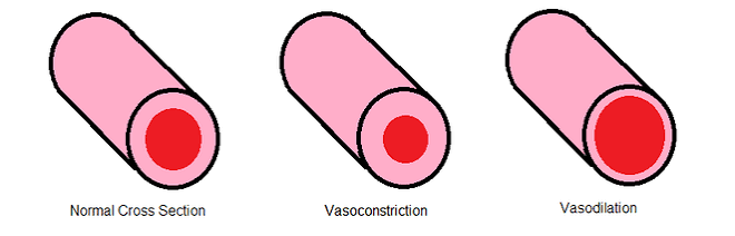
Vasoconstrictors
Causes smooth muscle contraction, which constrict arterioles and close precapillary sphincters
Increase O2 and nutrient lvls
Decrease CO2, H+, K+, & lactate lvls
Leukotrienes, Thromboxanes, endothelins
Chemicals that Increase Blood Pressure: Vasoconstrictors
Adrenal medulla hormones: Norepinephrine and epinephrine increase blood pressure
Antidiuretic hormone (ADH): Causes intense vasoconstriction in cases of extremely low BP
Angiotensin II: Kidney release of renin generates angiotensin II, which causes intense vasoconstriction and stimulates aldosterone secretion which enhances renal reabsorption and stimulates ADH release
Endothelium-derived factors: Endothelin and prostaglandin-derived growth factor (PDGF) are both vasoconstrictors
General relationship of total blood flow to local blood flow
Total blood flow is same as cardiac output → rest average of 5.25 liters per minute
Total blood flow has direct relationship and casual relationship with local blood flow
How is local blood flow dependent on total blood flow?
Cardiac output decreases, total blood flow decreases, and less blood is available to tissue
Vice versa
Blood Pressure (BP)
Force per unit area exerted on the wall of a blood vessel by its contained blood
Measured in millimeters of mercury (mm Hg)
Measured in reference to systemic arterial BP in large arteries near the heart
The differences in BP within the vascular system provide the driving force that keeps blood moving from higher to lower pressure areas
Arterial Blood Pressure
Reflects two factors of the arteries close to the heart:
Their elasticity (compliance or distensibility)
The amount of blood forced into them at any given time
Blood pressure is elastic arteries near the heart is pulsatile (BP rise and falls); in response to systolic and diastolic pressure changes
Systolic Pressure
Pressure exerted on arterial walls during ventricular contraction
Diastolic Pressure
Lowest level of arterial pressure during a ventricular cycle
Pulse Pressure
the difference between systolic and diastolic pressure
greatest in aorta
declines in muscular arteries and non-existent in arterioles
measure of the elasticity and recoil of arteries
Arterial Blood Pressure (Clinical Monitoring)
Vital signs: pulse and blood pressure, along w/ respiratory rate and body temp
Taking a pulse:
Radial pulse (taken at the wrist): most routinely used, but there are other clinically important pulse points
Pressure points: areas where arteries are close to the body surface
Can be compressed to stop blood flow in event of hemorrhaging
Capillary Blood Pressure
Ranges from 35 mm Hg at the beginning of the capillary bed to 17 mm Hg at the end of the bed
Low capillary pressure is desirable because:
High BP would rupture fragile, thin-walled capillaries
Most capillaries are very permeable, so low pressure forces filtrate into interstitial spaces
Compare and contrast blood pressure and blood pressure gradients in the arteries, capillaries, and veins
Blood pressure is highest in arteries
Lowest in Veins
In between in capillaries
Explain the mechanisms that help overcome the small pressure gradient in veins to return blood to the heart
Skeletal muscle pump moves blood through limbs
Respiratory pump moves blood through the thoracic cavity
Venous Blood Pressure
Steady and changes little during the cardiac cycle
The pressure gradient in the venous system is only about 15-20 mm Hg; falls to almost 0mm Hg in vena cava
A cut vein has even blood flow; a lacerated artery flows in spurts
Maintaining Blood Pressure
The main factors influencing blood pressure are:
Cardiac output (CO)
Peripheral resistance (PR)
Blood volume
BP = CO*PR
Blood Flow
Actual volume of blood flowing through a vessel, an organ, or the entire circulation in a given period:
Measured in ml per min.
In the entire vascular system it is equivalent to cardiac output (CO)
Relatively constant when at rest
Varies widely through individual organs, according to function
Determined by blood pressure & resistance
Resistance
Any opposition to blood flow; increases pressure
Measure of the amount of friction blood encounters as it passes through vessels
Generally encountered in the systemic circulation
Referred to as peripheral resistance (PR)
Sources of Resistance
Blood viscosity
Total blood vessel length
Blood vessel diameter
Blood Viscosity
Thickness or “stickiness” of the blood
Determined by the number of cells and amount of protein
Blood Vessel Length
The longer the vessel, the greater the resistance encountered
Blood Vessel Radius
Most common way resistance is altered is because of vasoconstriction/vasodilation
Greater width of vessels (arteries) increases laminar flow
The difference between the rate of flow at the center vs the rate of flow at the periphery where friction occurs
Relationship between blood pressure gradient and resistance to total blood flow
Direct relationship between the blood pressure gradient and total blood flow
Inverse relationship between resistance and total blood flow
Why does blood pressure increases with increased resistance in the systemic circulation
More resistance = greater pressure gradient to overcome
ensure adequate perfusion = blood pressure increases
Relationship between Blood Flow, Blood Pressure, and Resistance Formula
F=Δ P/R
Blood flow = F
Difference in blood pressure = Δ P
Resistance = R
Relationship between Blood Flow, Blood Pressure, and Resistance
Blood flow (F ) is directly proportional to blood pressure gradient (ΔP).
If ΔP increases, blood flow speeds up.
Blood flow is inversely proportional to peripheral resistance (R).
If R increases, blood flow decreases, so
R is more important in influencing local blood flow because it is easily changed by altering blood vessel diameter
Systemic Blood Pressure
Pumping action = heart generating blood flow through vessels along a pressure gradient
high to lower pressure
Pressure results when flow is opposed by resistance
Systemic Pressure
Highest in the aorta
Declines throughout the length of the pathway
Is 0 mm Hg in the right atrium
The steepest change in blood pressure occurs n the arterioes
Factors Aiding Venous Return
Venous BP alone is low to promote adequate blood return and is aided by the Respiratory pump, Muscular pump, & the sympathetic venoconstriction
Respiratory Pump
Pressure changes created during breathing suck blood toward the heart by squeezing local veins
Muscular Pump
Contractions of skeletal muscles “milk” blood toward the heart
Sympathetic Venoconstriction
Smooth muscles constrict, pushing blood back toward the heart
Valves prevent backflow during venous return
Formed from tunica intima
Cardiac Output (CO)
Determined by venous return and neural and hormonal controls
Resting heart rate is controlled by the cardioinhibitory center via the vagus nerves
Stroke volume is controlled by venous return (end diastolic volume, or EDV)
Under stress, the cardioacceleratory center increases heart rate and stroke volume
The end systolic volume (ESV) decreases and MAP increases
Controls of Blood Pressure: Short-term
Are mediated by the nervous system and bloodborne chemicals
Counteract moment-to-moment fluctuations in blood pressure by altering peripheral resistance and cardiac output
Controls of Blood Pressure: Long-term
Regulate blood voluem
Short-Term Mechanisms: Neural Controls
Neural controls of peripheral resistance:
Alter blood distribution to respond to specific demands
Maintain MAP by altering blood vessel diameter
Neural controls operate via reflex arcs involving:
Baroreceptors
Vasomotor centers of the medulla and vasomotor fibers
Vascular smooth muscle
Baroreceptors
Significant types of sensory receptors in regulating blood pressure
Specialized sensory nerve endings in blood vessel walls that respond to stretch
Location: Carotid Sinuses and Aortic Arch in the tunica externa
Carotid Sinus Reflect
Prevents major changes in blood supply to the brain; rapid response to short-term changes
Mediated by baroreceptors in carotid sinus
Regulates the activity of the CIC and the CAC
Located within internal carotid artery
Detects changes in blood pressure
Monitors BP changes in head and neck
Aortic Sinus Reflex
Regulates blood flow to the systemic blood vessels
Action is similar to the carotid sinus reflex
Short-Term Mechanisms: Vasomotor Center
Vasomotor center
A cluster of sympathetic neurons in the medulla that oversees changes in blood vessel diameter
Maintains blood vessel tone by innervating smooth muscles of blood vessels, especially arterioles
Short-Term Mechanisms: Vasomotor Activity
Sympathetic activity causes:
Vasoconstriction and a rise in blood pressure if increased
Blood pressure to decline to basal levels if decreased
Vasomotor activity is modified by:
Baroreceptors (pressure-sensitive), chemoreceptors (O2, CO2, and H+ sensitive), higher brain centers, bloodborne chemicals, and hormones
Vasoactive
Chemicals classified as either vasodilators or vasoconstrictors
Alter local blood flow collectively
Angiogenesis
Is the formation of new blood vessels
Takes place:
As the number of vessels to a region increases
When existing vessels enlarge
When a heart vessel becomes partly occluded
Routinely in people in high altitudes, where oxygen content of the air is low
In response to exercise
Short-Term Mechanisms: Chemical & Neural Controls
Blood pressure is regulated by chemoreceptor reflexes sensitive to oxygen, H+ and carbon dioxide
Prominent chemoreceptors are in the aortic arch, & the carotid and aortic bodies
Reflexes that regulate blood pressure are integrated in the medulla
Higher brain centers (cortex and hypothalamus) can modify BP via relays to medullary centers
Mean Arterial Pressure (MAP)
Working pressure; pressure that propels the blood to the tissues
MAP = diastolic pressure + (1/3*pulse pressure)
70-110 mm Hg typically indicates good perfusion
Cardiovascular center
Vasomotor center plus the cardiac centers that integrate blood pressure control by altering cardiac output and blood vessel diameter
Angiotensinogen
Liver produces the plasma protein and continuously releases into the blood
The kidney releases renin and converts it into angiotensin I
Angiotensin I is converted to Angiotensin II by angiotensin-converting enzyme (ACE)
Associated with the capillary endothelium
ACE found in high concentrations in pulmonary capillaries
Conversion occurs in blood vessels of lungs
Long-Term Mechanisms: Renal Regulation
Baroreceptors adapt to chronic high or low blood pressure
Long-term mechanisms control BP by altering blood volume via kidney action
Direct renal mechanism:
Increased BP stimulates the kidneys to eliminate water, thus reducing BP
Decreased BP stimulates the kidneys to increase blood volume and BP
Indirect renal mechanism
Renin release produces angiotensin II; leads to aldosterone and ADH release which leads to water reabsorption; also leads to vasoconstriction; also thirst sensation