Thorax 1 - Views, Vessels, Nodules
1/42
There's no tags or description
Looks like no tags are added yet.
Name | Mastery | Learn | Test | Matching | Spaced | Call with Kai |
|---|
No analytics yet
Send a link to your students to track their progress
43 Terms
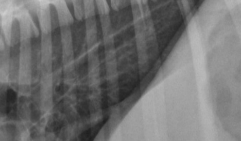
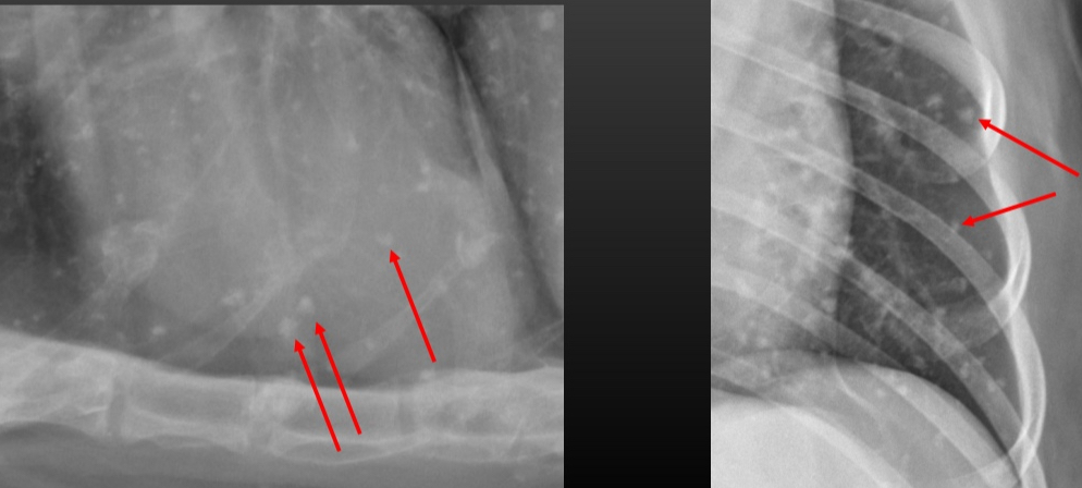
What structure is highlighted?
Cranioventral mediastinal reflection
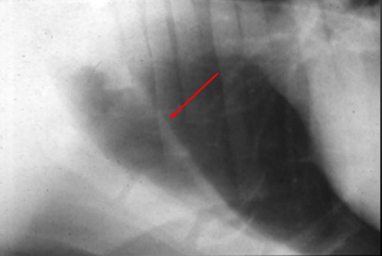
What structure is shown in this image?
Cranioventral mediastinal reflection
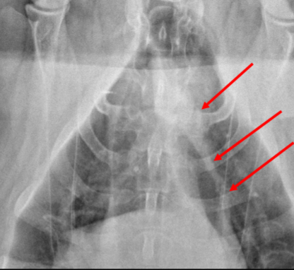
What structure is highlighted?
Caudal mediastinal reflection
What view is this?
VD
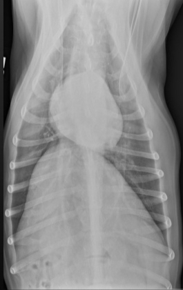
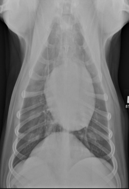
What view is this?
DV
What structures can be see on a thorax VD?
Diaphragm cupula
Left hemidiaphragm
Right hemidiaphragm
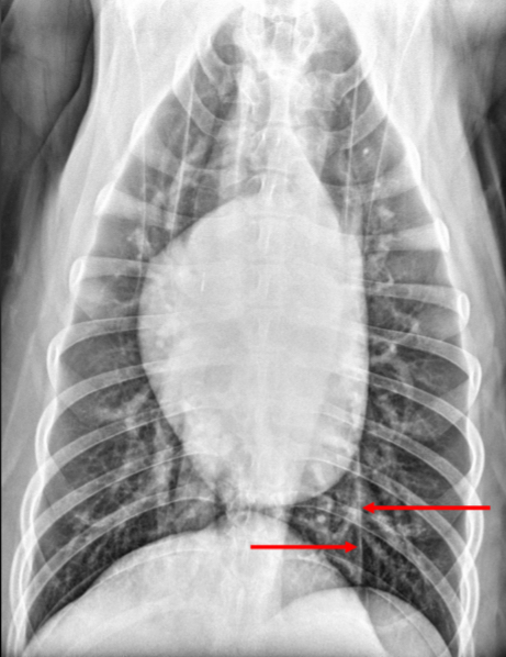
What structures can be seen on a thorax DV?
Cupula
Caudodorsal vessels
Positioning VD/DV thorax
Roughly equal thorax space each side of the spine
Sternum over spine
Spinous process straight (end-on tear drop shape)
Positioning laterals Thorax
Rib heads superimposed
Rib curvature should match
Pull limbs out of image - minimize bone & soft tissue on top of lungs
Veins are ______ and _____ to bronchus
Ventral and Central
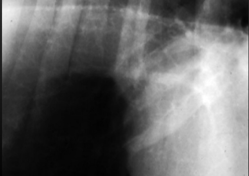
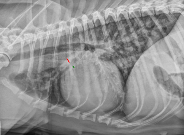
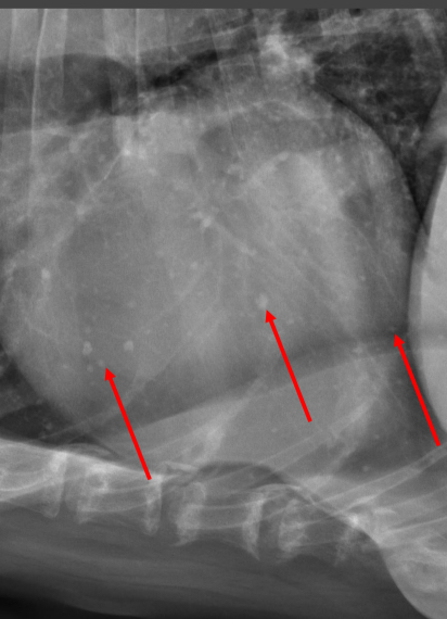
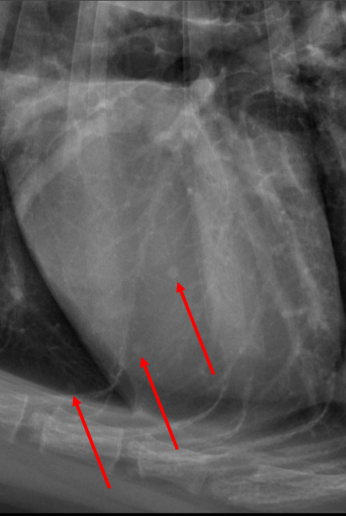
What is being shown on this image?
Artery, bronchus, & vein - lateral projection
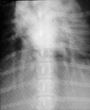
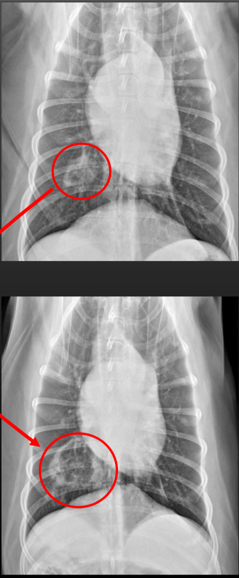
What structures are shown in this image?
Artery, bronchus & veins - VD/DV projection
Normal vasculature - lateral projection
Arteries & veins equal in size
Normal vasculature - DV or VD
Arteries & veins equal in size
No larger than 9th ribs where they cross
____ arteries and ____ veins
Big, Big (“vascular pattern”)
____ arteries and ____ veins
Small, small
____ arteries, _____ veins
Big, normal
_____ veins, _____ arteries
Big, normal
Causes of Big arteries and veins
L —> R cardiac shunt
Patent Ductus Arteriosus (PDA)
Ventricular Septal Defect (VSD)
Atrial Septal Defect (ASD)
Iatrogenic IV fluid overload
Causes of small arteries and veins
Hypovolemia
Shock
Addison’s disease
Severe pulmonic stenosis
R —> L cardiac shunt
Tetralogy of Fallot
Any L —> R shunt that reversed direction due to high R heart pressure (aka Eisenmenger syndrome)
Causes of Big arteries, normal veins
Pulmonary Hypertension
Heartworm disease
Thromboembolism (rare)
Causes of Big veins, normal arteries
Venous congestion due to:
L-sided heart congestion
Mitral valve degeneration
Cardiomyopathy (Dilated, hypertrophic)
Rare
Mitral valve dysplasia (congenital)
L atrial obstruction
Mass at heart base
Thrombosis
Cor triatriatum sinister (congenital)
Radiographic appearances of pulmonary Nodules
Soft tissue opaque
Round
Radiographic appearance of pulmonary Masses
Soft tissue opaque
Round or irregularly shaped
Can have ill-defined margins
Can have air bronchograms
Causes of Multiple pulmonary nodules/masses
Metastatic neoplasia
Granuloma
Fungal disease
Feline asthma
Certain inflammatory disease
Hematogenous pneumonia (rare)
Causes of Solitary pulmonary nodule/masses
Primary lung tumor
Early metastatic neoplasia (1 small nodule)
Granuloma (uncommon)
Causes of weird stuff pulmonary nodules/masses
Bulla (uncommon)
Cavitation nodule (rare)
Hematocele (rare)
Abscess (rare)
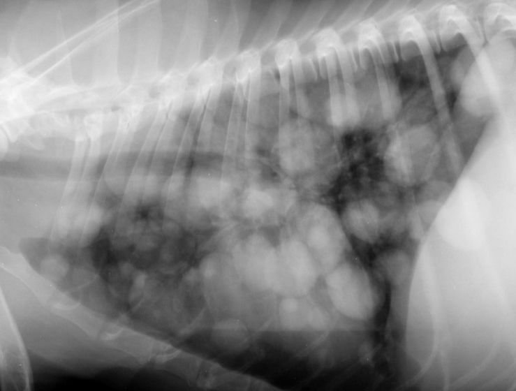
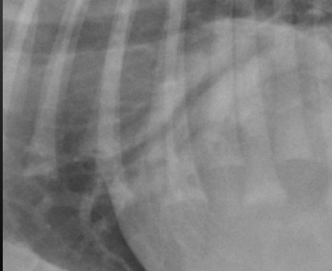
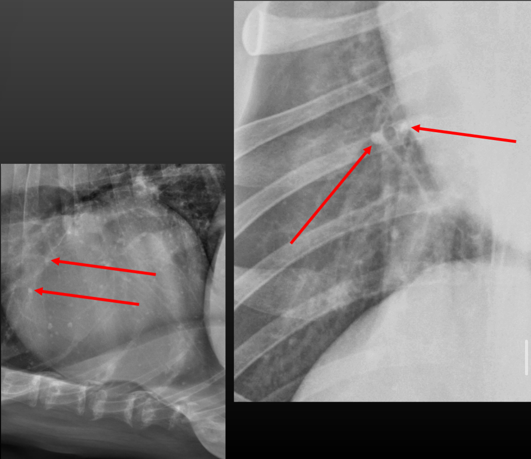
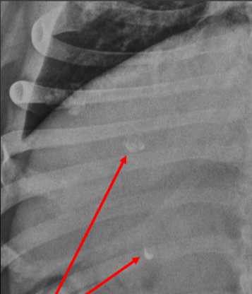
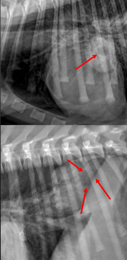
What cause of pulmonary nodules/masses is this image showing?
Metastatic neoplasia
Things that mimic pulmonary nodules
Pulmonary osteomas
End-on vessels
Dermal structures
Dermal nodules/masses
Nipples
Costochondral junction remodeling
What is being highlighted?
Pulmonary osteomas
Pulmonary osteomas
mineral opaque
Visible at smaller size
Sharp edges or weird shapes
Appearance of Pulmonary nodules on rads
round, smooth
Similar opacity as similar size vessel
End-on vessels
in vessel’s path - same size as vessel
More opaque than a same-size soft tissue nodule
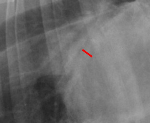
What structure is highlighted?
End-on vessel
What structure is highlighted?
Cutaneous nodules/nipples
Appearance of Costochondral junction remodeling on radiographs
between bone & cartilage w/in a rib
Mineral opaque
Usually irregular, can be smooth/round
Humanoid projection
move scapulae & soft tissues away from cranial lung lobes
Thoracic limbs pulled caudally
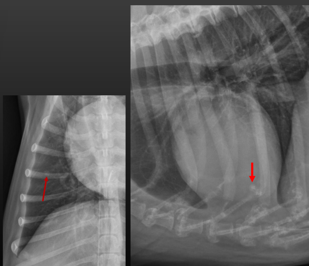
What structure is this image showing?
Bulla
Bulla
gas-filled round structure w/ very thin soft tissue opaque rim
What structure is highlighted?
Cavitation lesions
What do Cavitary lesions look like on radiographs?
thicker more irregular soft tissue opaque outer tissue (compared to bulla)
Gas-filled center
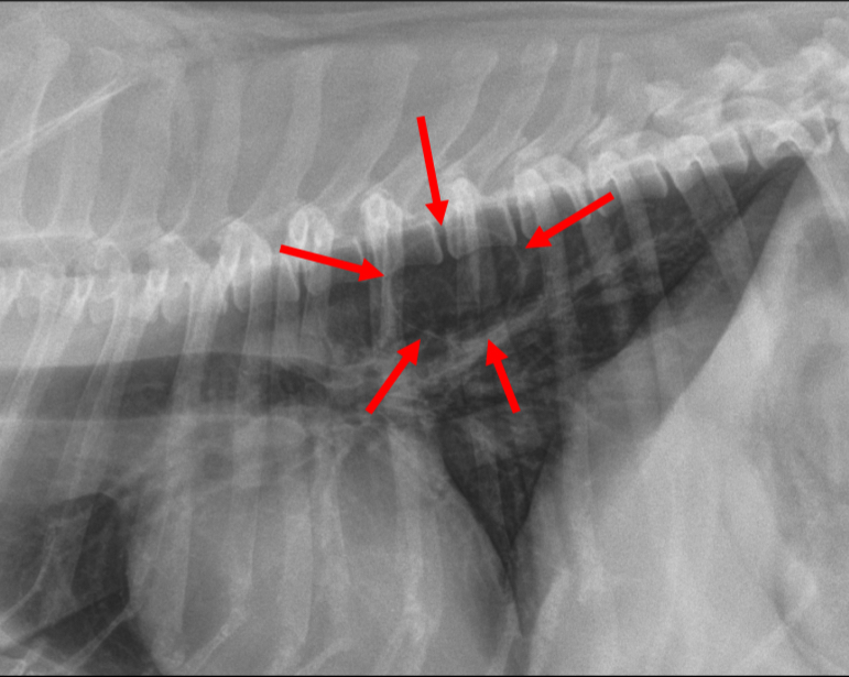
What structure is highlighted?
Abscess
What do Abscess look like on radiographs?
Gas bubbles in soft tissue mass