[AP Bio] Unit 2
1/116
Earn XP
Description and Tags
Chapters 4.2-4.7 & 5.1-5.5
Name | Mastery | Learn | Test | Matching | Spaced |
|---|
No study sessions yet.
117 Terms
Eukaryotic Cells
a type of cell with a membrane-enclosed nucleus and membrane-enclosed organelles
Prokaryotic Cells
a type of cell lacking a membrane-enclosed nucleus and membrane-enclosed organelles
Cytosol
a semifluid, jellylike substance in which subcellular components are suspended
Nucleoid
a non-membrane enclosed region in a prokaryotic cell where its chromosome is located
Cytoplasm
the contents of the cell enclosed by the plasma membrane
Cell organelles are located in the _______ of a eukaryotic cell, whereas organelles are largely absent in prokaryotic cells
Plasma Membrane
a selective barrier that allows passage of enough oxygen, nutrients, and wastes to service the entire cell
For each square micrometer of membrane, only a limited amount of a particular substance can cross per second, making the ratio of surface area to volume important
As a cell (or any other object) increases in size, its surface area grows proportionately less than its volume
Nucleus (Cell)
the organelle of a eukaryotic cell that contains the genetic material in the form of chromosomes, made up of chromatin
Nuclear Envelope
the double membrane that surrounds the nucleus, perforated with pores that regulate traffic with the cytoplasm
The double membrane is made of a phospholipid bilayer with associated proteins
Nuclear Pore Complex
an intricate protein structure that lines each pore, regulating the entry and exit of proteins and RNAs, as well as large complexes of macromolecules
Nuclear Lamina
a netlike array of protein filaments that lines the inner surface of the nuclear envelope and helps maintain the shape of the nucleus
Chromosomes
a cellular structure consisting of one DNA molecule and associated protein molecules
Eukaryotic cells typically carry multiple, whereas prokaryotic cells often only have 1 singular, circular chromosome
Chromatin
the complex of DNA and proteins that makes up eukaryotic chromosomes
When the cell is not dividing, chromatin exists in its dispersed form, as a mass of very long, thin fibers not visible with a light microscope
Nucleolus
a specialized structure in the nucleus which serves as the site of rRNA (ribosomal RNA) synthesis and ribosomal subunit assembly
These subunits then exit the nucleus through the nuclear pores to the cytoplasm, where a large and a small subunit can assemble into a ribosome
Ribosomes
the complex of DNA and proteins that makes up eukaryotic chromosomes
When the cell is not dividing, chromatin exists in its dispersed form, as a mass of very long, thin fibers not visible with a light microscope
What are the 2 types of ribosomes and where are they found?
Free Ribosomes: found in a cell’s cytoplasm
Bound Ribosomes: attached to the outside of the rough ER or nuclear envelope
Endomembrane System
the collection of membranes inside and surrounding a eukaryotic cell, related either through direct physical contact or by the transfer of membranous vesicles
Includes the plasma membrane, the nuclear envelope, the smooth and rough endoplasmic reticulum, the Golgi apparatus, lysosomes, vesicles, and vacuoles
What are the tasks the endomembrane system carries out?
Protein Synthesis + Transport
Metabolism + Movement of Lipids
Detoxification of Poisons
Endoplasmic Reticulum (ER)
an extensive membranous network in eukaryotic cells, continuous with the outer nuclear membrane and composed of ribosome-studded (rough) and ribosome-free (smooth) regions
Consists of a network of membranous tubules and sacs called cisternae
ER membrane separates the internal compartment of the ER, called the ER lumen (cavity) or cisternal space, from the cytosol
Smooth ER
the portion of the endoplasmic reticulum that is free of ribosomes
What are the functions of the Smooth ER?
Deals with variety of metabolic processes
lipid synthesis
metabolism of carbohydrates
detoxification of drugs and poisons
storage of calcium ions
Rough ER
the portion of the endoplasmic reticulum with ribosomes attached
What are the functions of the Rough ER?
Proteins are secreted through the ribosomes attached to the Rough ER, and are threaded into the ER lumen to fold into their functional shapes
keeps secretory proteins separate from proteins made in free ribosomes—these proteins get packaged into vescicles
acts like a “membrane factory” for the cell, adding membrane proteins and phospholipids to its own membrane
Glycoproteins
a secretory protein with one or more covalently attached carbohydrates
Transport Vescicle
small membranous sac in a eukaryotic cell’s cytoplasm carrying molecules produced by the cell
Membrane Phospholipids
phospholipids that are assembled when enzymes built into the ER use precursors in the cytosol, with the phospholipids contributing to the ER membrane’s growth
Golgi Apparatus
an organelle in eukaryotic cells that consists of stacks of flat membranous sacs that modify, store, and route products of the endoplasmic reticulum (ER)
Cisternae
flattened membranous sacs found in both the ER and Golgi apparatus
What are the two sides of a Golgi stack referred to as?
The “cis face” and the “trans face”
The cis face is usually located near the ER, as cis means “on the same side”, and it receives material from the ER
As products from the ER move from the cis to the trans face, they get modified
Cisternae Maturation Model
Scientific model that depicts cisternae moving from the cis to the trans face, carrying and modifying their “cargo” are they journey to the trans face
Lysosome
a membranous sac of hydrolytic enzymes that many eukaryotic cells use to digest (hydrolyze) macromolecules, and are found in the cytoplasm of animal cells and some protists
Rough ER make the hydrolytic enzymes in lysosomes, and they have 3-dimensional shapes to protect vulnerable bonds from attacks by enzymes
Phagocytosis
when large particulate substances or small organisms are taken up by a cell
This is a type of endocytosis—where cells absorb molecules/particles from their surroundings by engulfing them with their plasma membrane
Autophagy
where a lysosome recycles a cell’s own components
Vacuole
a membrane-bounded vesicle derived from the ER and Golgi apparatus whose specialized function varies in different kinds of cells
Food Vacuole
a membranous sac formed by phagocytosis of microorganisms or particles to be used as food by the cell
Contractile Vacuole
a membranous sac that helps move excess water out of certain freshwater protists, maintaining a suitable concentration of ions and molecules inside the cell
Central Vacuole
a large membranous sac with diverse roles in growth, storage, and sequestration of toxic substances, found in a mature plant cell
As this organelle in a plant cell absorbs water, the plant cell grows
Mitochondria
an organelle in eukaryotic cells that serves as the site of cellular respiration, and uses oxygen to break down organic molecules and synthesize ATP
Chloroplasts
an organelle found in plants and photosynthetic protists that absorbs sunlight and uses it to drive the synthesis of organic compounds from carbon dioxide and water
Endosymbiont Theory
theory that mitochondria and plastids, including chloroplasts, originated as prokaryotic cells engulfed by host cells. The engulfed cell and its host cell then evolved into a single organism
Mitochondria and chloroplasts are autonomous (somewhat independent) organelles that grow and reproduce within the cell
Cristae
an infolding of the inner membrane of a mitochondrion that houses electron transport chains and molecules of the enzyme catalyzing ATP synthesis
Mitochondria have a phospholipid bilayer, with the outside membrane being smooth, but the inner membrane being convoluted with cristae infoldings
Mitochondrial Matrix
the compartment of the mitochondrion enclosed by the inner membrane and containing enzymes and substrates for the citric acid cycle, as well as ribosomes and DNA
The cristae give the inner mitochondrial membrane a large surface area, thus enhancing the productivity of cellular respiration
Chlorophyll
the green pigment crucial for photosynthesis, located in the thylakoid membranes
Thylakoids
a flattened, membranous sac inside a chloroplast
Granum (plural, grana)
a stack of membrane-bounded thylakoids in the chloroplast that function in the light reactions of photosynthesis
Stroma
the dense fluid within the chloroplast surrounding the thylakoid membrane containing ribosomes and DNA, and is involved in the synthesis of organic molecules
Plastids
one of a family of closely related organelles that includes chloroplasts, chromoplasts, and amyloplasts
Amyloplast: a colorless organelle that stores starch (amylose), mainly in roots and tubers
Chromoplast: contains pigments that give fruits and flowers their orange and yellow hues
Peroxisomes
an organelle containing enzymes that transfer hydrogen atoms from various substrates to oxygen (O2), producing and then degrading hydrogen peroxide (H2O2)
Peroxisomes in the liver detoxify alcohol by transferring hydrogen from poisonous compounds to oxygen molecules
Hydrogen peroxide is toxic by itself, but it eventually gets converted into water (H2O)
Cytoskeleton
a network of microtubules, microfilaments, and intermediate filaments that extends throughout the cytoplasm and serves a variety of mechanical, transport, and signaling functions
Cell Motility
the movement of cells and their components
Motor Proteins
a protein that interacts with cytoskeletal elements and other cell components, producing movement of the whole cell or parts of the cell
Microtubules
hollow rods composed of tubulin proteins making up part of the cytoskeleton in all eukaryotic cells—found in cilia and flagella
Centrosome
a structure present in the cytoplasm of animal cells that functions as a microtubule-organizing center, typically located near the nucleus
Contains 2 centrioles and is important for cell division
Centriole
a structure in the centrosome of an animal cell composed of a cylinder of microtubule triplets arranged in a “9 plus 0” pattern
What’s the difference between 9+0 and 9+2 arrangements of microtubules in cilia and flagella?
9+2 Pattern: consists of nine doublets of microtubules arranged in a ring, with two single microtubules in the center
Found in motile cilia and flagella
9+0 Pattern: lacks the central pair of microtubules, having only the nine outer doublets
Found in nonmotile primary cilia
Cilia
short, hair-like structures that contain microtubules that are found on the surface of eukaryotic cells
Two Main Types of Cilia: Motile Cilia & Primary Cilia
Motile Cilia: typically occurs in large numbers on a cell’s surface (since they’re shorter), and flagella are usually limited to 1 or a couple per cell (since they’re longer)
Primary Cilia: acts as a signal-receiving antenna for the cell that transmits molecular signals from the cell’s environment to its interior, and are generally nonmotile, with only 1 per cell
Flagella
long, whip-like structures that contain microtubules that help cells move
Basal Body
a eukaryotic cell structure consisting of a “9+0” arrangement of microtubule triplets that anchors the microtubule assembly of a cilium or flagellum in the cell
Structurally similar to a centriole
Dyneins
a large motor protein extending from one microtubule doublet to the adjacent doublet, leading to the bending of cilia and flagella
A typical dynein protein has two “feet” that “walk” along the microtubule of the adjacent doublet, using ATP for energy. One foot maintains contact, while the other releases and reattaches one step farther along the microtubule.
Microfilament
a cable composed of actin proteins in the cytoplasm of almost every eukaryotic cell, making up part of the cytoskeleton—also called actin filaments
The structural role of microfilaments in the cytoskeleton is to bear tension (pulling forces) to help support the cell’s shape
Actin
a globular protein that links into chains, forming microfilaments (actin filaments) in muscle and other kinds of cells
Myosin
a type of motor protein that associates into filaments that interact with actin filaments, causing cell contraction
Intermediate Filaments
a component of the cytoskeleton that includes filaments between the size of microtubules and microfilaments
These are only found in the cells of some animals, including vertebrates
Cell Wall
a protective layer external to the plasma membrane in the cells of plants, prokaryotes, fungi, and some protists
Protects the plant cell, maintains its shape, and prevents excessive uptake of water
Primary Cell Wall
a relatively thin and flexible layer that surrounds the plasma membrane of a young plant cell
When a cell matures and stops growing, it strengthens its wall. Some plant cells do this simply by secreting hardening substances into the primary wall, and others add a secondary cell wall
Middle Lamella
a thin layer of adhesive extracellular material, primarily pectins, found between the primary walls of adjacent young plant cells
Secondary Cell Wall
a strong and durable matrix that is often deposited in several laminated layers around the plasma membrane and that provides protection and support in plant cells
When a cell matures and stops growing, it strengthens its wall. Some plant cells do this simply by secreting hardening substances into the primary wall, and others add a secondary cell wall
Extracellular Matrix (ECM)
the meshwork surrounding animal cells, consisting of glycoproteins, polysaccharides, and proteoglycans synthesized and secreted by the cells
Collagen
a glycoprotein in the extracellular matrix of animal cells that forms strong fibers, found extensively in connective tissue and bone
This is the most abundant protein in the animal kingdom
Proteoglycans
a large molecule consisting of a small core protein with many carbohydrate chains attached, found in the extracellular matrix of animal cells
Fibronectin
an extracellular glycoprotein secreted by animal cells that helps them attach to the extracellular matrix
Integrins
a transmembrane receptor protein with two subunits that interconnects the extracellular matrix and the cytoskeleton in animal cells
By communicating with a cell through integrins, the ECM can regulate a cell’s behavior
Plasmodesmata
an open channel through the cell wall that connects the cytoplasm of adjacent plant cells, allowing water, small solutes, and some larger molecules to pass between the cells
Plasmodesmata unify most of a plant into one living thing by joining adjacent cells
What are the 3 main types of cell junctions?
Tight Junctions
Desmosomes
Gap Junctions
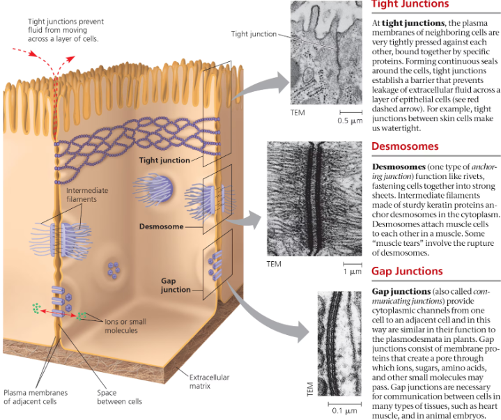
Tight Junctions
a web-like structure where the plasma membranes of neighboring cells are tightly pressed against each other, facilitated by proteins binding the membranes together
In humans, these structures between skin cells make our skin watertight
Desmosomes
a type of intercellular junction found in animal cells, functioning like rivets to fasten cells together into strong sheets, made by intermediate filaments
Helps maintain a tissue’s structural strength by preventing cells from being pulled apart
Gap Junctions
specialized intercellular connections in animal cells that facilitate communication between adjacent cells, small molecules to pass directly from one cell to another through pores
Helps important cells communicate, like heart muscle cells & embryos
Amphipathic
something that has both a hydrophilic region and a hydrophobic region
In a phospholipid bilayer, the tails on the inside are hydrophobic, and the heads on each side are hydrophilic
What steroid is sandwiched between phospholipid molecules in a cell membrane, and helps resist changes in fluidity?
Cholesterol
Reduces phospholipid movement at high temperatures
Reduces the temperature phospholipids need to solidify
A membrane’s fluidity is extremely important in its ability to function
Integral Proteins
a transmembrane protein with hydrophobic regions that either extend into or completely span the hydrophobic interior of the membrane
The hydrophobic regions of an integral protein consist of one or more stretches of nonpolar amino acids
The hydrophilic parts of the molecule are exposed to the aqueous solutions on either side of the membrane
Peripheral Proteins
a protein loosely bound to the surface of a membrane or to part of an integral protein and not embedded in the lipid bilayer
What are the 6 functions of a Membrane Protein?
Transport
Enzymatic Activity
Signal Transduction
Cell-Cell Recognition
Intercellular Joining
Attachment to the Cytoskeleton and ECM
Cell-Cell Recognition
a cell’s ability to distinguish one type of neighboring cell from another through recognizing other cells by binding to molecules on a cell’s extracellular surface
Important in the sorting of cells into tissues and organs in an animal embryo
Serves as basis for the rejection of foreign cells by the immune system
The four human blood types designated A, B, AB, and O reflect variation in the carbohydrate part of glycoproteins on the surface of red blood cells
Glycolipids
a lipid with one or more covalently attached carbohydrates
Glycoproteins
a protein with one or more covalently attached carbohydrates
Transport Proteins
transmembrane protein that helps a certain substance or class of closely related substances to cross the membrane
Two types of Transport Proteins: Channel Proteins & Carrier Proteins
Channel Proteins
provides hydrophilic corridors that allow water molecules or small ions to diffuse very quickly from one side of the membrane to the other
Carrier Proteins
specialized transport proteins that assist in moving solutes across cell membranes without the need for energy
Changes shape in a way that shuttles them across the membrane
Aquaporins
a channel protein in the plasma membrane of a plant, animal, or microorganism cell that specifically facilitates osmosis, the diffusion of free water across the membrane
Diffusion
the net movement of a substance from a region where it is more concentrated to a region where it is less concentrated
Passive Transport
diffusion of a substance across a biological membrane with no expenditure of energy
Osmosis
the diffusion of free water molecules across a selectively permeable membrane
Tonicity
the ability of a solution surrounding a cell to cause that cell to gain or lose water
Depends in part on its concentration of solutes that cannot cross the membrane relative to that inside the cell
Isotonic
referring to a solution that, when surrounding a cell, causes no net movement of water into or out of the cell
Dynamic Equilibrium (water is still moving in/out of the cell, but at an equal pace)
Hypertonic
referring to a solution that, when surrounding a cell, will cause the cell to lose water
The cell will lose water, shrivel, and probably die—explaining why an increase in the salinity (saltiness) of a lake can kill the animals there
Hypotonic
a solution that, when surrounding a cell, will cause the cell to take up water
Water will enter the cell faster than it leaves, and the cell will swell and lyse (burst) like an overfilled water balloon
Osmoregulation
regulation of solute concentrations and water balance by a cell or organism
Organisms that must live in hypertonic/hypotonic environments must develop adaptations for osmoregulation, such as possessing a contractile vacuole
Turgor Pressure
the outward force exerted by the fluid inside a plant cell against its rigid cell wall, resulting when a plant cell takes in the maximum amount of water it can handle
Turgid
swollen or distended, as in plant cells
Plant cells become turgid when in a hypotonic solution
Flaccid
limp; lacking turgor, as in plant cells in surroundings where there is a tendency for water to leave the cell
A walled cell becomes like this if it has a higher water potential than its surroundings (aka an isotonic solution), resulting in the loss of water
Plasmolysis
a phenomenon in walled cells in which the cytoplasm shrivels and the plasma membrane pulls away from the cell wall
Occurs when the cell loses water to a hypertonic environment