Synaptic Transmission
1/40
There's no tags or description
Looks like no tags are added yet.
Name | Mastery | Learn | Test | Matching | Spaced |
|---|
No study sessions yet.
41 Terms
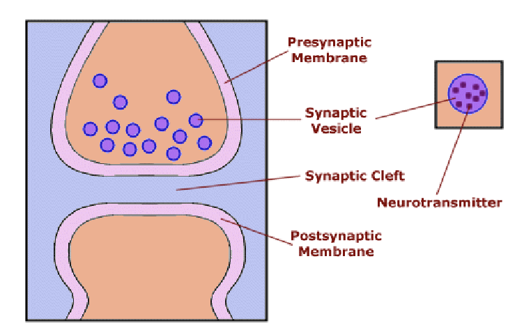
what is a synpase?
junction between a presynaptic and a postsynaptic neuron
presynaptic and postsynaptic neurons do not actually make physical contact. therefore, there is no direct electrical transmission of the signal from one neruon to another. what happens instead?
presynaptic neuron(s) release a chemical neurotransmitter that binds to receptors on the postsynaptic neuron to initiate a new action potential.
the process of presynaptic neuron(s) releasing a chemical neurotransmitter that binds to receptors on the postsynaptic neuron to initiate a new action potential is called…?
transduction
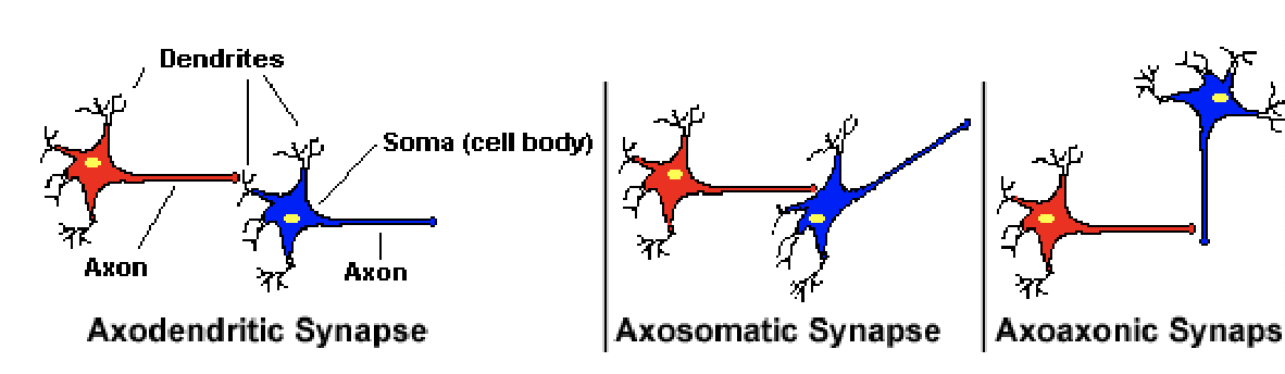
what are the different anatomic variations of synapses?
Presynaptic Events in transmission occur in
axon terminals
axon terminals contain many _______, each containing a _________
axon terminals contains many synaptic vesicles, each containing a quantum (~3000-5000 molecules) of neurotransmitte
what are some examples of neurotransmitters?
acetylcholine, norepinephrine, glutamine, serotonin, glycine, etc.
axon terminals expand into ________ that contain numerous ________ that contain the chemical ________- that will excite the postsynaptic cell
knobs (terminal boutons)
small membrane-bound synaptic vesicles (~ 50 nm diameter)
neurotransmitters
many synpatic vesicles found in knobs lie close to what zone?
specialized zone in the terminal called active zone
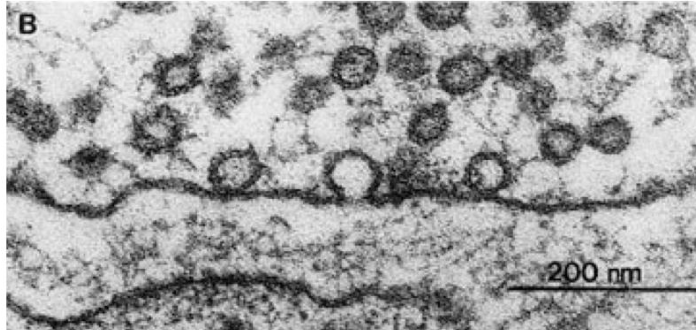
what happens in response to an action potential arriving at the terminal?
vesicles fuse to plasma membrane in active zone realasing their content
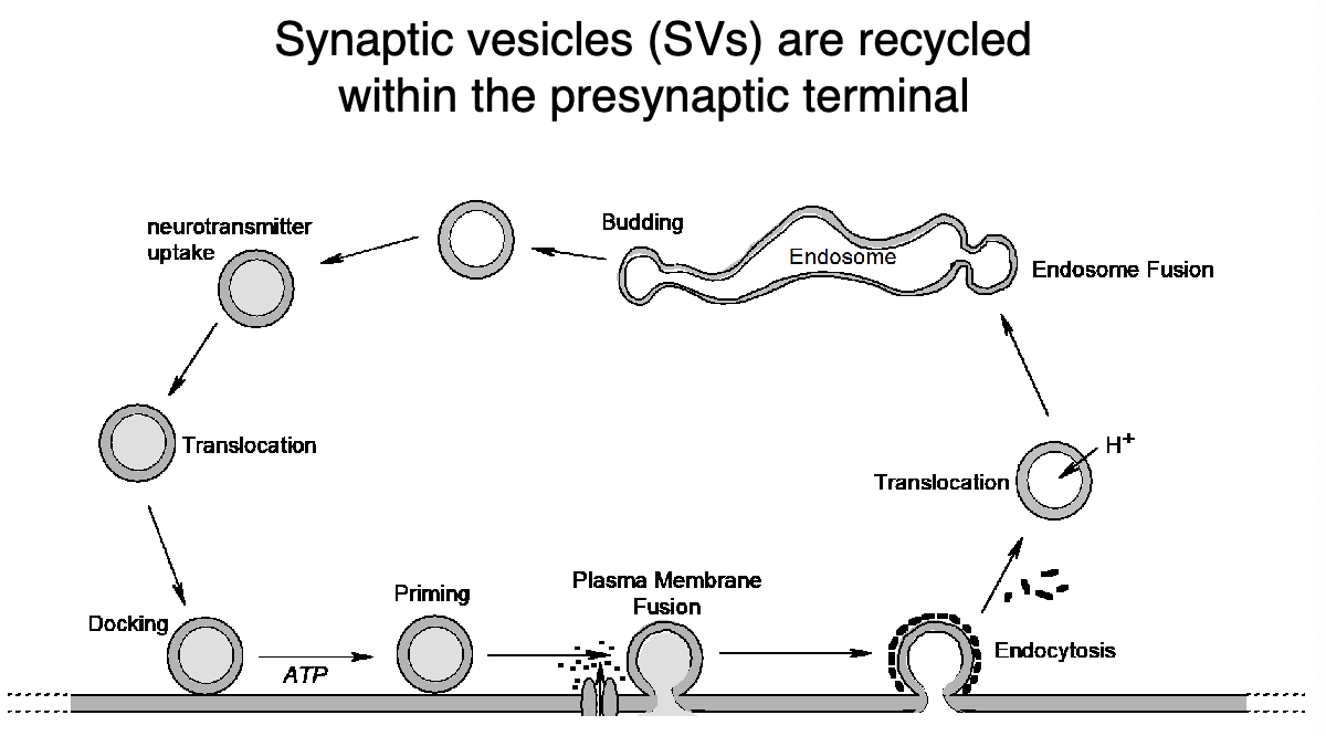
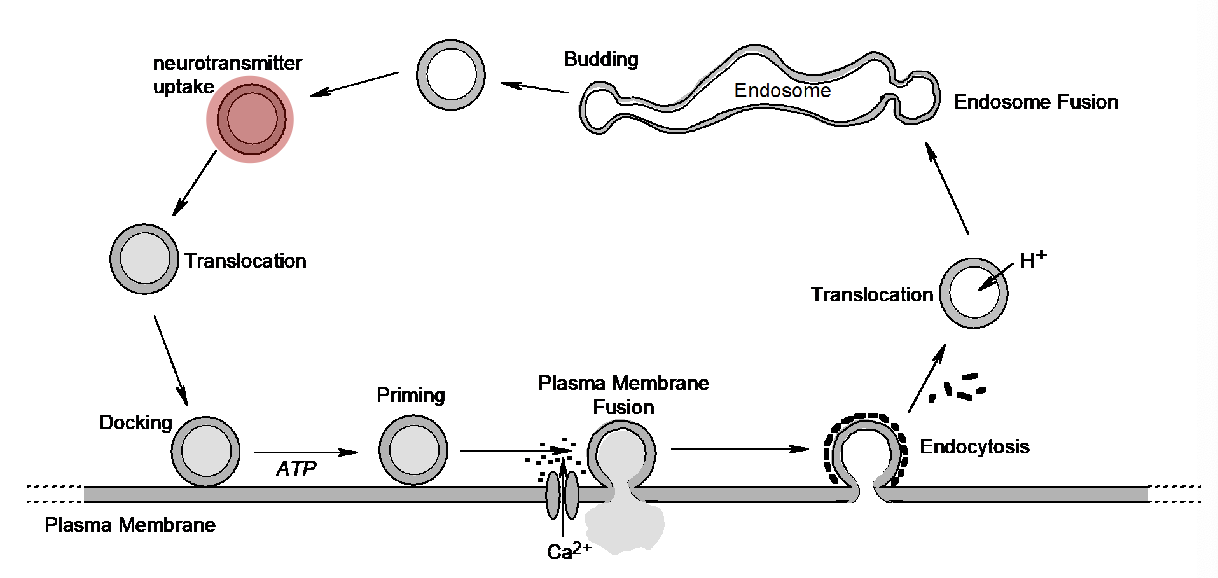
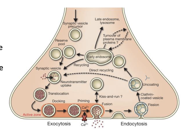
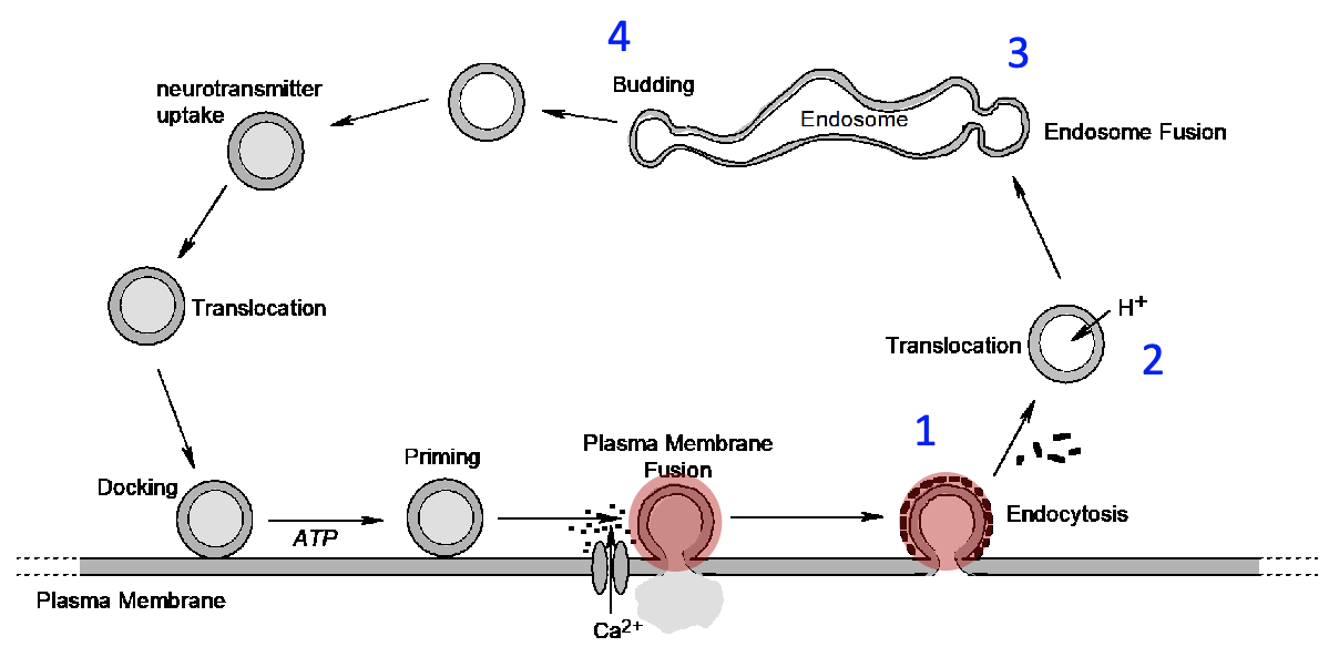
walk through the cycle of neurotransmitter release from presynaptic neurons
Each synaptic vesicle is filled with a quantum of neurotransmitter ( about 3000-5000 molecules).
Vesicles then move toward the plasma membrane in the active zone
Where they are “docked” and “primed” and ready for fusion with the plasma membrane
each synaptic vesicle is filled with
a quantum of neurotransmitter (3000-5000 molecules)
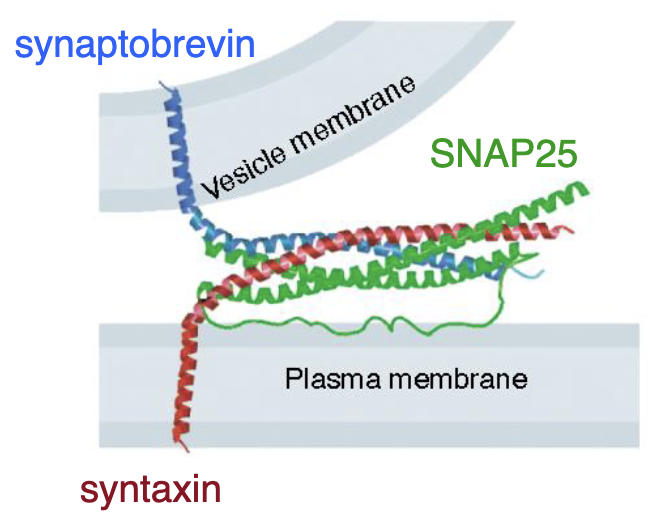
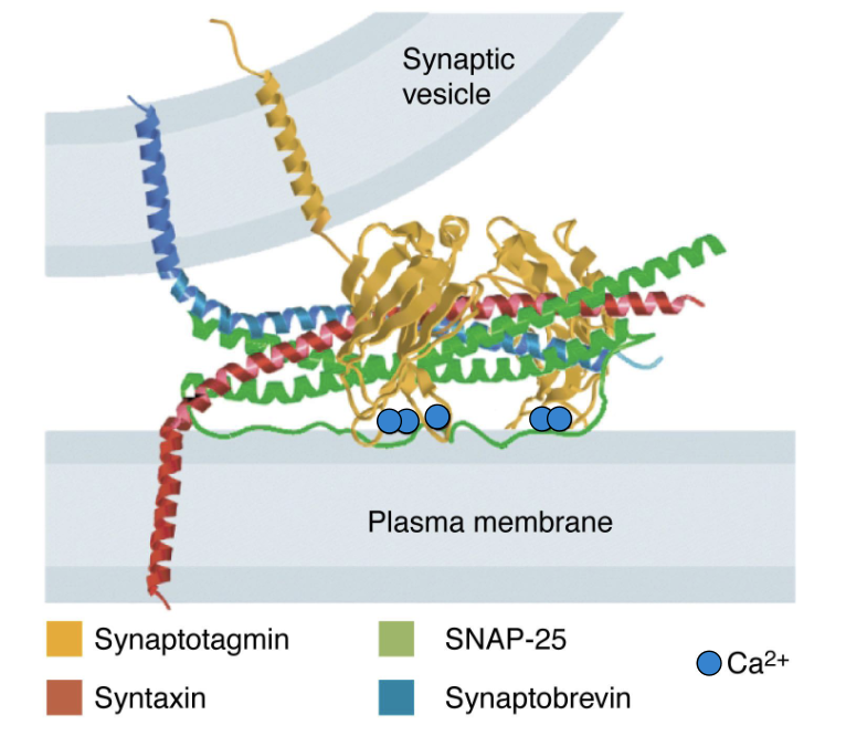
docking and priming involve 3 snare proteins that do what
a coiled-coil that holds the vesicle close to the plasma membrane and prepares it for fusion
what are the 3 snare proteins invovled in docking and priming?
synaptobrevin
SNAP25
syntaxin
(Many of these proteins are the same as those involved in non-synaptic vesicle release such as the exocytosis of peptide hormones)
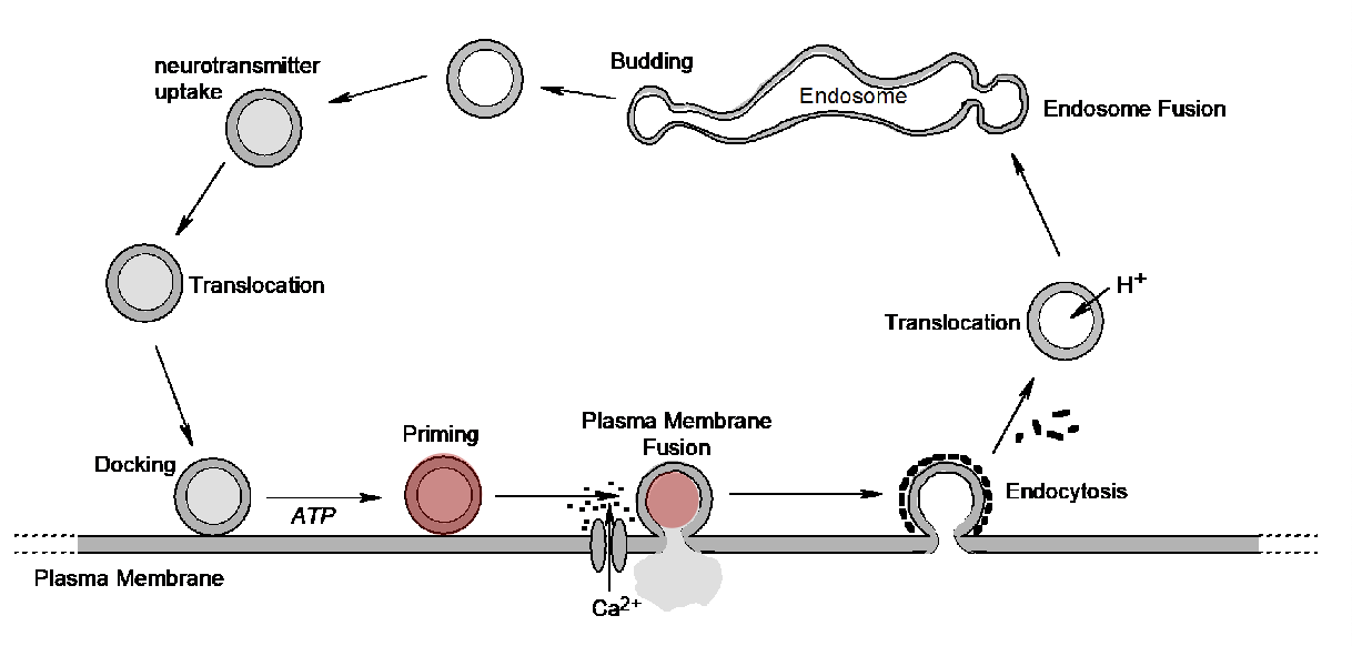
what is fusion triggered by?
presynaptic action potential that also opens voltage-gated Ca++ channels located in the active zone of the plasma membrane.
what is a 4th protein that binds to the vesicle membrane and prevents spontaneous fusion of the vesicle with the plasma membrane under resting conditions?
synaptotagmin
what happens during fusion?
synaptotagmin binds to vesicle memrane and prevent spontaneous fusion of vesicle with plasma membrane under resting conditions
upon activation by an action potential, Ca++ entering the cell binds to synaptotagmin, causing it to change its conformation and interactions with the snare proteins to promote vesicle fusion and the release of neurotransmitter
describe the steps in the release of neurotransmitter from presynaptic terminal
arrival of the action potential at the axon terminal also causes the opening of local voltage-gated Ca++ channels
Ca++ enters the cell and binds to a fourth protein called synaptotagmin
this triggers fusion of the vesicle with the axon membrane and the release of the neurotransmitter into the synaptic cleft: = calcium-dependent exocytosis
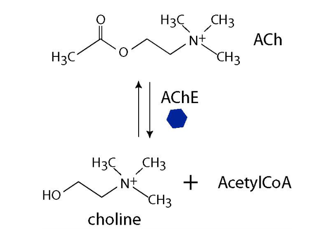
ACh is rapidly degraded by the enzyme AChesterase present in the cleft
empty vesicles are recovered by cell and recycled
what happens to synaptic vesciles after release of neurotransmitter into the synaptic cleft?
SVs are endocytosed (1, 2), fuse with the endosome (3) , and ultimately pinched off to form a new vesicle (4).
the presynaptic cell stores transmitter in
membrane-bound synaptic vesicles (SVs)
A rise in intracellular Ca2+ concentration promotes
SV fusion with the plasma membrane
release of a quantum of transmitter into the synaptic cleft
the essential nature of the three SNARE proteins is underscored by the fact that each can be hydrolyzed by one of several deadly neurotoxins produced by the anaerobic bacterium, _______________.
Any one of these toxins blocks exocytosis, paralyzing muscle and leading to respiratory failure and death if untreated. Humans are most commonly exposed through ingestion of improperly canned foods
Clostridium botulinum
Released ACh is rapidly destroyed by the enzyme _______ which cleaves it into two inactive components.
ACH-esterase
what are some of the numerous neurotransmitters expressed in peripheral and central nervous system neruons. what are some common ones?
• acetylcholine (A neurotransmitter used by neurons in the PNS and CNS in the control of functions ranging from muscle contraction and heart rate to digestion and memory)
• norepinephrine, (epinephrine)
• serotonin.
• dopamine.
• GABA. (-aminobutyric acid)
• glutamate..
• endorphin
(postsynaptic events) Once neurotransmitter is released into the synaptic cleft, it is available to bind receptors on the postsynaptic nerve or ______ to elicit a post-synaptic or _______. These cells have many receptors in the vicinity of the presynaptic active zone
neuromuscular junction (NMJ)
end-plate potential
what are 2 possiblee fates of neurotransmitters in the cleft during postsynaptic events?
hydrolyzed by extracellular enzymes present in cleft (acetylcholine esterase)
transported back into terminal via high-affinity transporters (repackaged into SVs for reuse)
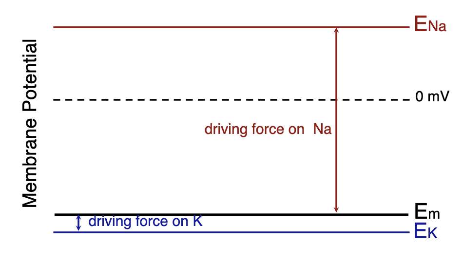
why does the cell depolarize even when permeability of Na and K+ is equal during postsynaptic events?
since the resting potential (Em) of the postsynaptic cell is far from the ENa and close to EK, the electrical driving force on Na+ is greater than that for K+. This causes the inward Na+ current to be much larger than the outward K+ current, and the cell depolarizes even when the permeability is equal.
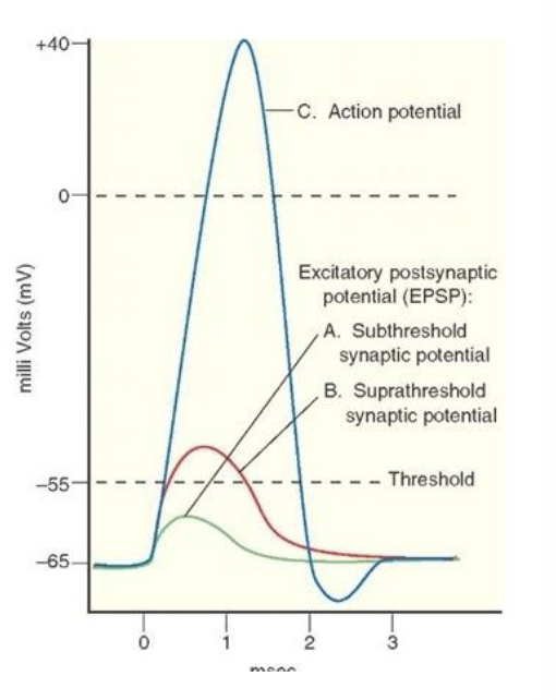
the response of the postsynaptic neuron is a depolarization called an
excitatory post-synaptic potential or EPSP
what does it mean that excitatory post-synaptic potential or EPSP are graded?
their amplitude depends upon:
how many quanta of ACh were released by the presynaptic cell
how much was destroyed in the cleft by the esterase
how many ACh receptors are occupied by 2 ACh molecules and for how long
what are possible responses to excitatory post-synaptic potential or EPSP?
subthreshold EPSP
suprathreshold EPSP
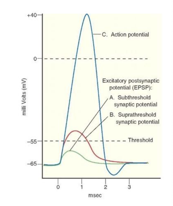
role of EPSP
trigger opening of voltage gated channel (to allow for action potential in the postsynaptic neuron, EPSP itself is NOT an action potential)
chemically induced by neurotransmitters
depolarize the postsynaptic cell enough to reach the threshold of the voltage-gated channels and produce an action potential
what are examples of neuroinhibitors?
lambda-aminobutyric acid or GABA
what does lambda-aminobutyric acid or GABA (neuroinhibitor) do?
binds to GABA receptors on the postsynaptic membrane and opens Cl- channels
Cl- diffuses into postsynaptic cell and makes Em more polar (hyperpolarized)
moves further from threshold causing inhibition
what provide a mechanisms to control the excitability of a postsynaptic neuron?
inhibitory postsynaptic potentials
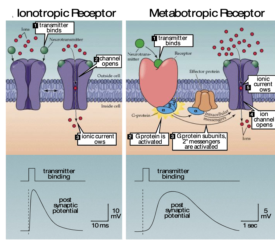
what is a ionotropic receptor?
transmitter binds → channel opens → ionic current flows
very rapid
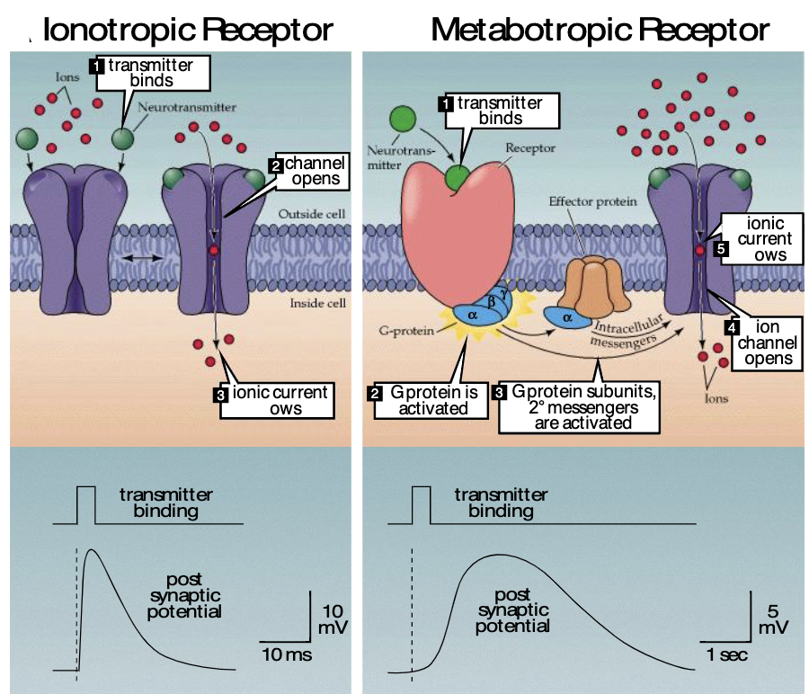
what are metabotropic receptors?
transmitter binds → G-protein activated → G-protein subunits/2nd messengers are activated → ion channel opens → ionic current flows
slower
what factors determine post-synaptic neurons being either excited or inhibited?
type of neurotransmitter
type of change in permeability of post-synaptic membrane
How do we increase the probability of a postsynaptic action potential?
summation
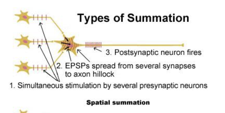
what are types of summation?
spatial summation
temporal summation
what is spatial summation?
simultaneous stimulation by several presynaptic neurons
EPSPs spread from several synapses to axon hillock
postsynaptic neuron fires
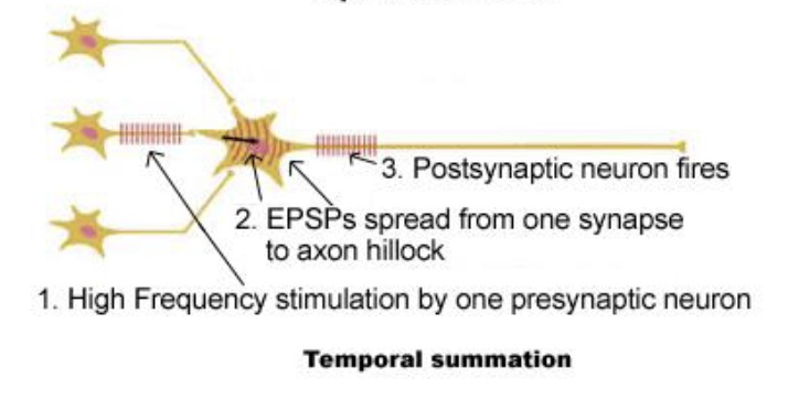
what is temporal summation?
high frequency stimulation by 1 presynaptic neuron
EPSps spread from 1 synapse to axon hillock
postsynaptic neuron fires