Anatomy wk3 tables and exam questions
 Call Kai
 Learn
Learn Practice Test
 Spaced Repetition
 Match
Match Flashcards
 Knowt Play
1/19

There's no tags or description
Looks like no tags are added yet.
Name | Mastery | Learn | Test | Matching | Spaced | Call with Kai | Chat |
|---|
No analytics yet
Send a link to your students to track their progress
20 Terms
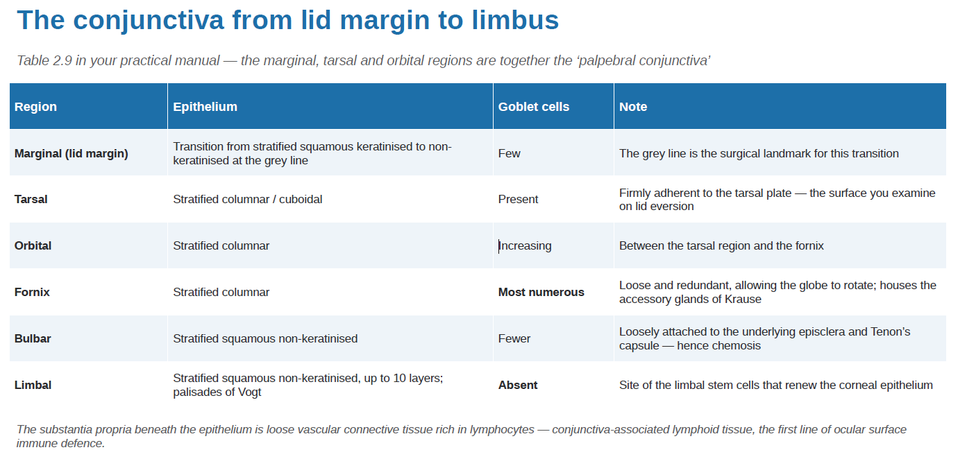
The conjunctiva from lid margin to limbus - slide 10
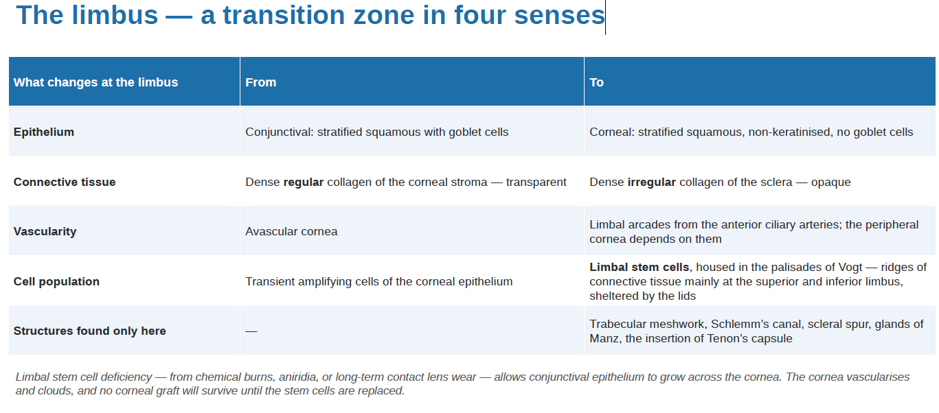
The limbus — a transition zone in four senses - slide 12
Q1 Bulbar conjunctiva joins the keratinised epidermis of the eyelid skin - true or false? **Palpebral conjunctiva joins the skin slide 13
False
Q2 Goblet cells produce --------through unicellular ----------------------- that contain clusters of ------------------------. slide 14
mucin
apocrine
secretory vesicles
The tear film — three layers, four functions - slide 14
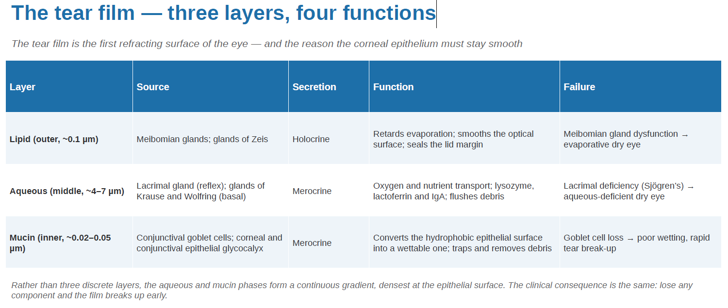
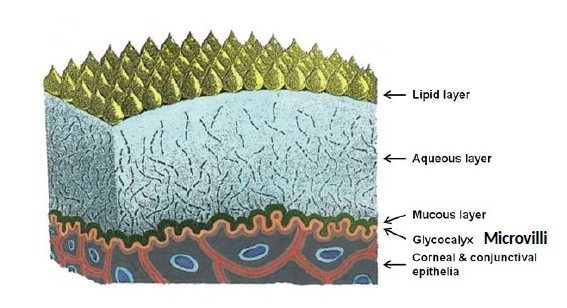
Q4 What is the innermost layer of the tear film? - S17
Mucin layer
Q5 Basal secretion of the aqueous layer is done by - S18
Gland of Krause (fornix)
Gland of Wolfring (palpebral conjunctiva)
The five layers of the cornea - s23
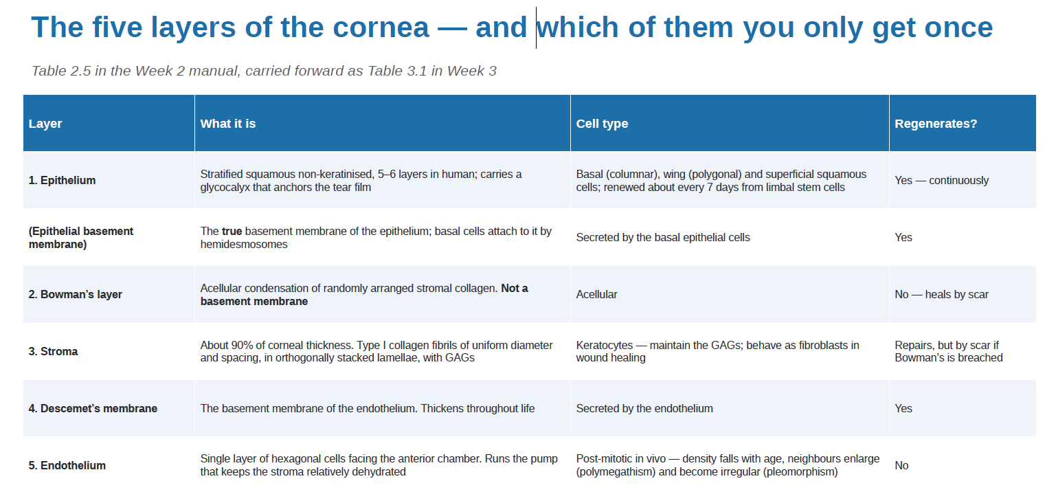
Q9 Corneal epithelium contains -------------- for better adherence with tears, and --------------------------------------to prevent passive uptake of excess tear film fluid. s24
Microvilli
tight junctions and desmosomes
** (a) The glycocalyx is a carbohydrate-rich coating present on the external surface of plasma membranes
(b) The major component is glycans composed of monosaccharides linked to each other
(c) Maximises the surface area with the mucinous layer of the tear film
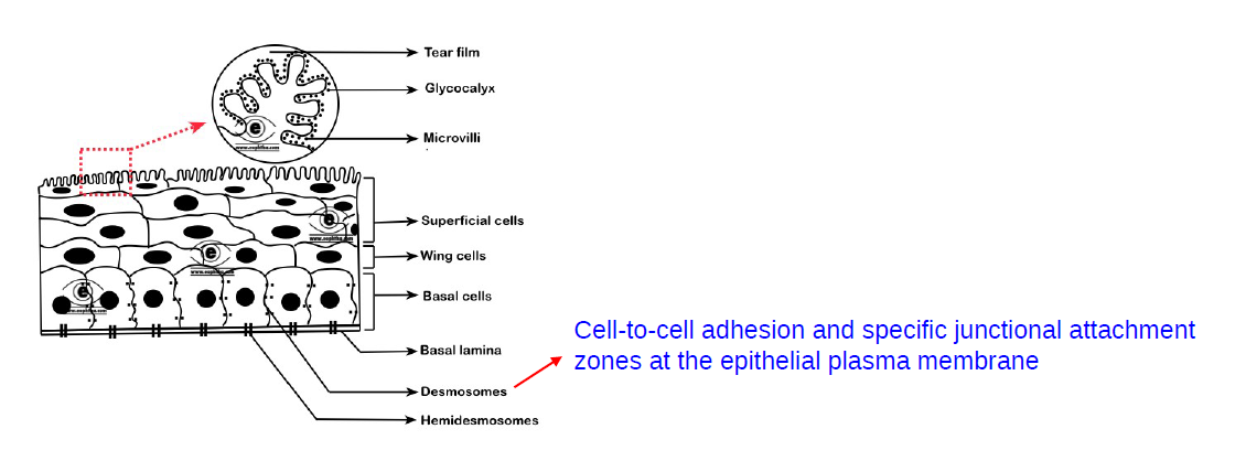
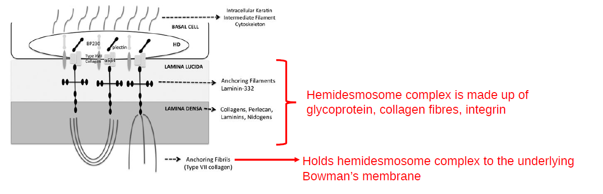
Q10 Basal corneal layer is attached to the acellular basement membrane (or basal lamina) via ----------------------------. s27
hemidesmosome complex
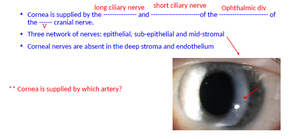
Q12 Cornea is supplied by the --------------- and ----------------------of the ---------------------- of the ------ cranial nerve.
What are the 3x networks of nerves?
Which 2x areas are the corneal nerves absent? s32
long ciliary nerve, short ciliary nerve, Ophthalmic div, V
Three network of nerves: epithelial, sub-epithelial and mid-stromal
Corneal nerves are absent in the deep stroma and endothelium
How an avascular tissue stays alive requirements, sources and consequences (all x5) - s33
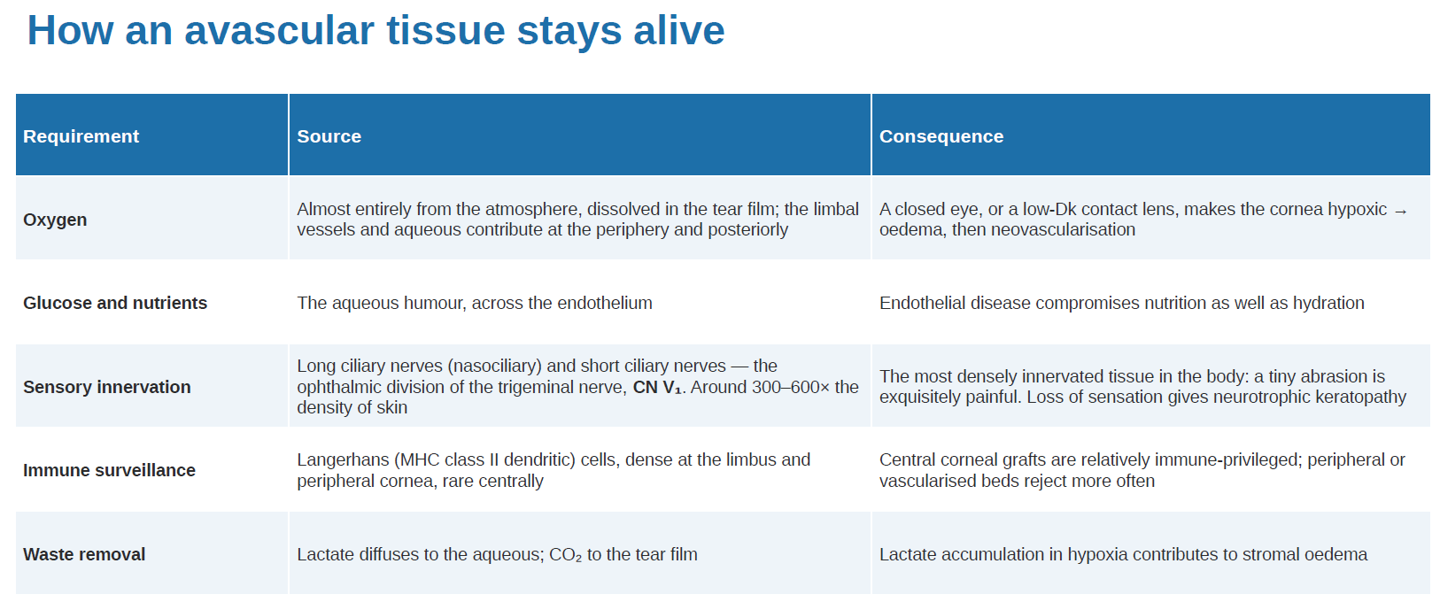
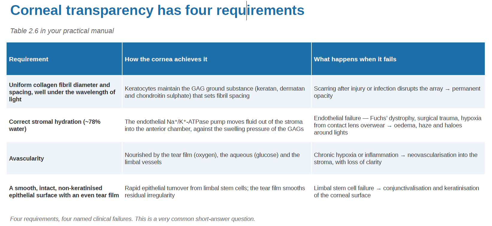
Q13 Name 4 important anatomical factors important for corneal transparency? s34 ***WILL BE IN EXAM
Regular collagen fibril diameter and spacing (read Lattice and Goldman theories)
Regular collagen lamellae separation
Sparsely cellular
Avascular
Smooth tear film
Non-pigmented
Regulated thickness due to stable water content (approx. 78%) and regulated IOP
Corneal transparency requirements, how and clinical failures (all x4) - s35
Q14 ----------------------- ion-pump present in the corneal ------------is the most important moderator of corneal hydration. s36
Na+/K-/ATP-ase, endothelium
**For every ATP molecule that the pump uses, three sodium ions are exported and two potassium ions are imported
Sclera layers x3 openings x2 - s39
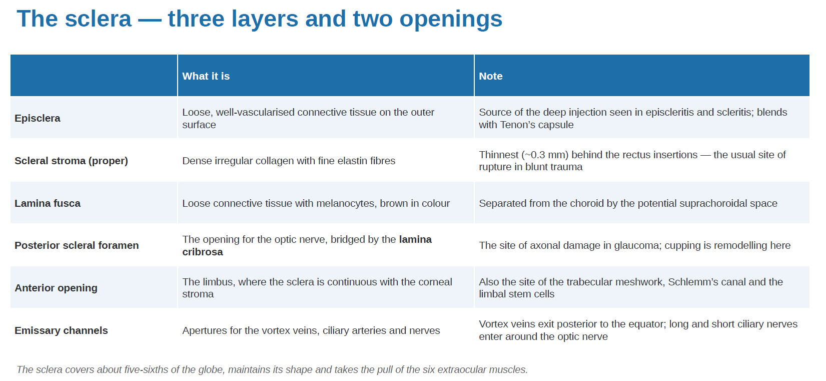
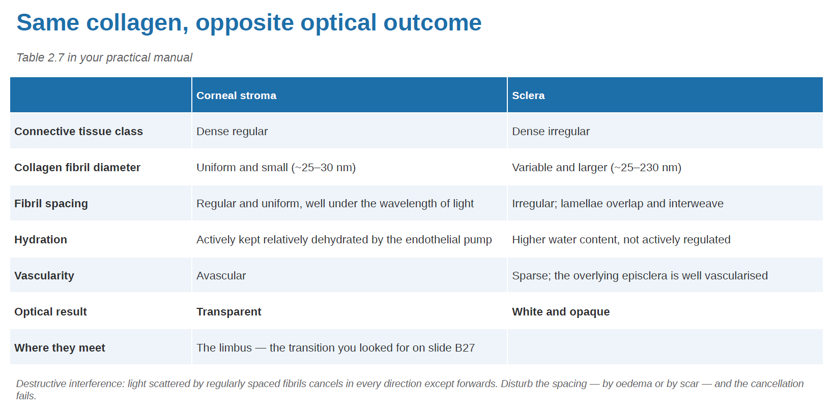
Q15 Why is the sclera opaque and cornea transparent? s40 → leading to table next flashcard
Because of the variable collagen fibril diameter and overlapping collagen lamellae in the sclera
Cornea vs Sclera - Same collagen, opposite optical outcome (vs x5, result, and where does the corneal stroma and sclera meet?) - s41
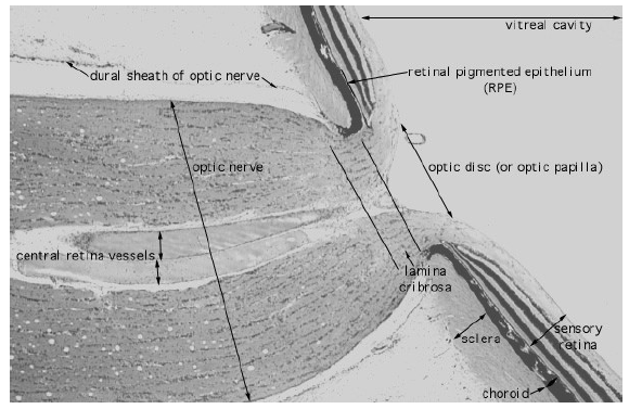
Q16 Optic nerve exits the sclera through -------------------------------------- that is occupied by a collagenous sieve-like structure called the --------------------. s42
posterior scleral foramen
lamina cribrosa
Q17 Outermost layer of the sclera ------------- is covered by the ------------------- posteriorly and the ----------------- anteriorly.
-------------------- separates episclera from the Tenon’s capsule. S43
episclera, Tenon’s capsule, conjunctiva, episcleral space