Blood Vessels and Circulation APR - Lab
 Call Kai
 Learn
Learn Practice Test
 Spaced Repetition
 Match
Match Flashcards
 Knowt Play
1/74

There's no tags or description
Looks like no tags are added yet.
Name | Mastery | Learn | Test | Matching | Spaced | Call with Kai | Chat |
|---|
No analytics yet
Send a link to your students to track their progress
75 Terms
Elastic lamellae
Location: Walls of medium and large arteries
Function: Provide elastic properties of some blood vessel walls
Description: Wavy, fenestrated (perforated) sheets composed of the protein elastin
Comment: Human aorta contains >40-50 elastic lamellae

Tunica externa of elastic artery
Location: Outermost layer of vessel wall
Function: Helps anchor blood vessel to surrounding structures
Description:
Elastic and collagen fibers
Contains lymphatic capillaries, nutritive arteries (vasa vasorum), and small nerves
Also known as:
Tunica adventitia of elastic artery

Tunica intima of elastic artery
Location: Innermost layer of vessel wall
Function:
Selectively permeable barrier to blood solutes
Secretes vasoconstrictors and vasodilators
Provides smooth inner lining that repels blood cells and platelets
Description:
Endothelium, its basement membrane, elastic laminae and elastic fibers
External limit formed by internal elastic lamina (not clearly defined)
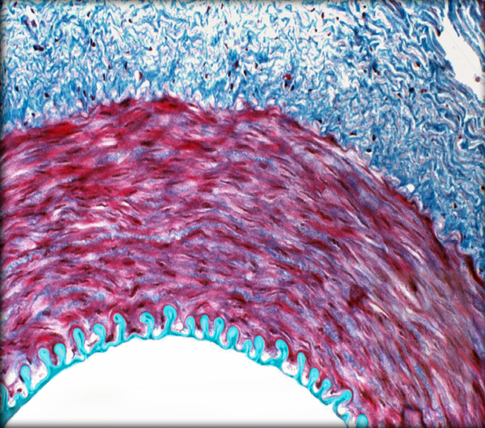
Tunica media of elastic artery
Location: Middle layer of vessel wall
Function: Vasoconstriction and vasodilation
Description:
Circularly arranged smooth muscle layers and connective tissue
Fenestrated (perforated) elastic laminae between muscle cells
External limit formed by external elastic lamina
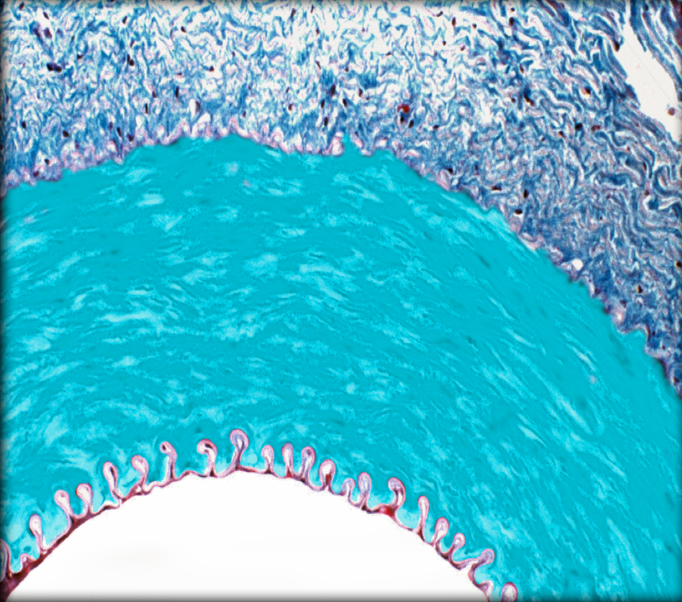
Vasa vasorum
Location: Tunica externa
Function: Supply outer 1/2-2/3 of tunica media
Description: Small blood vessels

Internal elastic lamella
Location: Layer of elastic fibers
Description:
Tunica intima of medium and large arteries
External to endothelium and subendothelium (loose connective tissue)

Smooth muscle bundles in large vein
Location: Tunica externa of large veins
Function: Vasoconstriction and vasodilation
Description: Large, wide, longitudinal bundles of smooth muscle
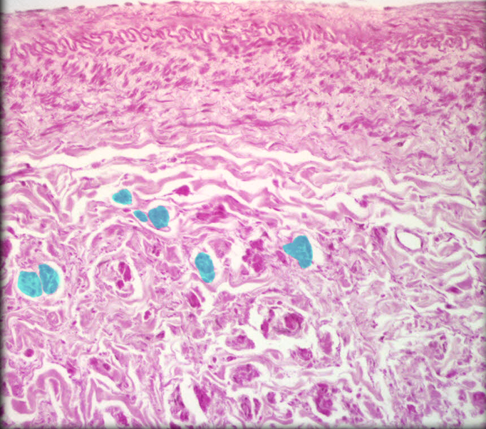
Tunica externa of large vein
Location: Outermost layer of vessel wall
Function: Anchor blood vessel to surrounding structures
Description:
Thickest of three tunics
Prominent bundles of longitudinal smooth muscle
Longitudinal bundles of collagen fibers with some elastic fibers
Also known as:
Tunica adventitia of large vein

Tunica intima of large vein
Location: ocation:
Innermost layer of vessel wall
Function:
Barrier to blood solutes
Provides smooth inner lining that repels blood cells and platelets
Description:
Endothelium, its basement membrane, and connective tissue
Incomplete internal elastic lamina
Comment:
Valves (folds of tunica intima) common in veins of all sizes
Tunica intima also known as tunica interna
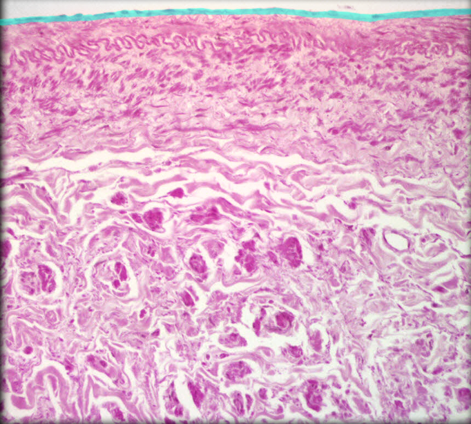
Tunica media of large vein
Location: Middle layer of vessel wall
Function: Vasoconstriction and vasodilation (when muscular tunica media present)
Description:
Poorly developed, with a few layers of smooth muscle
Collagen and elastic fibers present

Lumen of medium-sized vein
Location: Medium vein (e.g., radial vein)
Function: Conducts blood toward larger veins
Description:
Central opening in medium-sized vein
Up to 10 mm in diameter
Collapsed when empty
Comment:
Many medium-sized veins, especially in the limbs, exhibit infoldings of the tunica interna that meet in the middle of the lumen, forming venous valves directed toward the heart.

Lumen of muscular artery
Location: Muscular artery (e.g., brachial)
Function: Conducts blood to specific organs
Description:
Central opening in muscular artery
Has up to 40 layers of smooth muscle
Smooth muscle constitutes approximately 3/4 of the wall’s thickness
Comment:
The smooth muscle in these arteries is more conspicuous than the elastic tissue. Muscular arteries are also known as distributing arteries or medium-sized arteries.

Medium size vein
Location: Connects small veins or large veins
Function: Returns blood toward heart
Description:
Less smooth muscle than in comparable size artery
Collagen and/or elastic fibers predominate in tunica media
Diameter of 2-9 mm
Comment:
Companion vein for muscular artery
Examples include radial and small saphenous veins

Muscular artery
Location: Between elastic artery and arteriole
Function: Distributes blood to organs and regions of body
Description:
Predominance of smooth muscle layers in tunica media
Wide range in diameter
Also known as: Distributing artery
Comment:
Companion to medium-sized vein
Examples include brachial, coronary, and mesenteric arteries

Tunica externa of medium-sized vein
Location: Outermost layer of vessel wall
Function:
Vasoconstriction and vasodilation
Helps anchor blood vessel to surrounding structures
Description: Thickest of three tunics
Also known as: Tunica adventitia of medium sized vein
Comment:
In arteries, tunica media is thickest layer; in veins, tunica externa is thickest layer
Inner 1/3 of tunica media and all tunica intima supplied by diffusion of oxygen and nutrients from lumen of blood vessel
In large vessels, outer 1/2-2/3 of tunica media supplied by nutritive arteries (vasa vasorum) located in tunica externa
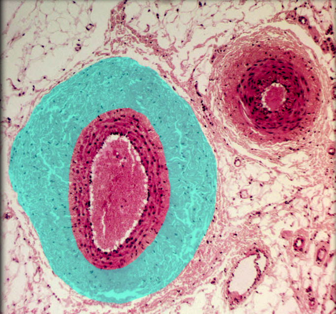
Tunica externa muscular artery
Location: Outermost layer of vessel wall
Function: Helps anchor blood vessel to surrounding structures
Description:
Variable thickness, sometimes comparable to tunica intima
Elastic fibers, collagen, and smooth muscle
Contains lymphatic capillaries, nutritive arteries (vasa vasorum), and small nerves
Also known as:
Tunica adventitia of muscular artery

Tunica intima of medium-sized vein
Location: Innermost layer of vessel wall
Function:
Barrier to blood solutes
Provides smooth inner lining that repels blood cells and platelets
Description:
Endothelium, its basement membrane and minimal connective tissue
Incomplete internal elastic lamina

Tunica intima of musclar artery
Location: Innermost layer of vessel wall
Function:
Selectively permeable barrier to blood solutes
Secretes vasoconstrictors and vasodilators
Provides smooth inner lining that repels blood cells and platelets
Description:
Endothelium, its basement membrane and collagen
Thick internal elastic lamina
Comment:
Tunica intima also known as tunica interna

Tunica media of medium-sized vein
Location: Middle layer of vessel wall
Function: Vasoconstriction and vasodilation (when muscular tunica media present)
Description: Few layers of smooth muscle, with interspersed collagen and elastic fibers
Comment:
Some veins of limbs (e.g., great saphenous vein) have prominent muscular tunica media
Cerebral and dural veins lack smooth muscle cells in tunica media

Tunica media of muscular artery
Location: Middle layer of vessel wall
Function: Vasoconstriction and vasodilation
Description:
5-30 circular or spiral layers of smooth muscle
External elastic lamina

Arteriole
Location: Connect muscular (distributing) arteries with capillaries
Function: Supplies blood to capillaries
Description:
Arteries with overall diameter 40-200 µm
Tunica media has one to three layers of smooth muscle
Comment:
Companion vessels to venules
Primary point for control of blood flow to organs
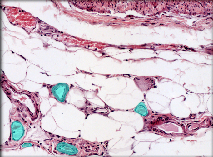
Tunica externa of arteriole
Location: Outermost layer of vessel wall
Function: Anchor blood vessel to surrounding structures
Description: Composed of elastic and collagen fibers
Also known as: Tunica adventitia of arteriole
Comment: In arteries, tunica media is thickest layer; in veins, thickest layer is tunica externa

Tunica externa of venule
Location: Outermost layer of vessel wall
Function: Anchor blood vessel to surrounding structures
Description: Minimal tunica externa with a few collagen fibers and some fibroblasts
Also known as: Tunica adventitia of venule
Comment: In arteries, tunica media is thickest layer; in veins, tunica externa is thickest layer

Tunica intima of arteriole
Location: innermost layer of vessel wall
Function:
Selectively permeable barrier to blood solutes
Secretes vasoconstrictors and vasodilators
Provides smooth inner lining that repels blood cells and platelets
Description:
Composed of endothelium and its basement membrane
External limit formed by internal elastic lamina
Comment: Tunica intima also known as tunica interna

Tunica intima of venule
Location: Innermost layer of vessel wall
Function:
Selectively permeable barrier to blood solutes
Secretes vasoconstrictors and vasodilators
Provides smooth inner lining that repels blood cells and platelets
Description: Composed of endothelium and its basement membrane
Comment: Tunica intima also known as tunica interna

Tunica media of arteriole
Location: Middle layer of vessel wall
Function: Vasoconstriction and vasodilation
Description:
Composed of one to three layers of circularly arranged smooth muscle cells and connective tissue
External elastic lamina inconspicuous
Comment: In arteries, tunica media is thickest layer; in veins, tunica externa is thickest layer

Tunica media of venule
Location: Middle layer of vessel wall
Function: Vasoconstriction and vasodilation (when muscular tunica media present)
Description:
Small venules lack tunica media
Larger venules acquire incomplete tunica media of smooth muscle
Comment: In arteries, tunica media is thickest layer; in veins, tunica externa is thickest layer
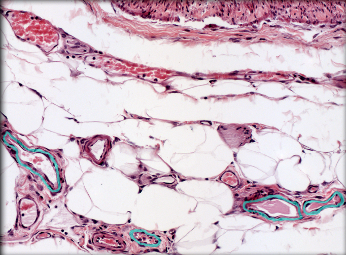
Vasa vasorum
Location: Tunica externa
Function: Supply outer 1/2-2/3 of tunica media
Description: Small blood vessels
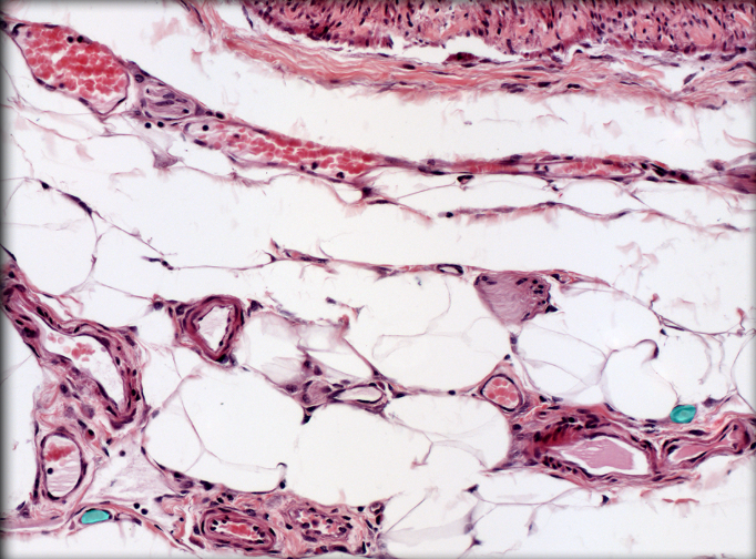
Venule
Location: Connects capillaries with small and medium-sized veins
Function:
Exchange fluid with surrounding tissues
Diapedesis (migration of leukocytes from blood vessel into interstitial fluid) occurs in postcapillary venules
Description:
Veins with overall diameter 15-100 µm
Acquires tunica media of smooth muscle in larger venules
Comment:
Companion vessels to arterioles
Smallest venules called postcapillary venules

Anterior cerebral artery
Origin: Internal carotid
Course: Passes anteriorly along medial aspect of frontal lobe of cerebral cortex
Distribution: Medial aspect of frontal lobe of cerebral cortex

Anterior communication artery
Origin: Anterior cerebral
Course:
Short, unpaired vessel
Connects paired anterior cerebral arteries

Anterior spinal artery
Origin: Vertbral
Course:
Descends on anterior surface medulla oblongata
Arteries from each side unite to form single anterior median vessel along anterior spinal cord
Distribution: Central branches supply anterior 2/3 of spinal cord

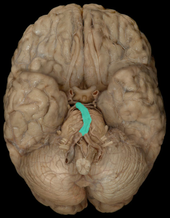
Basilar artery
Origin: Junction of paired vertebral arteries
Course: Unpaired (midline) vessel ascends on anterior (ventral) surface of pons
Distribution: Pons and Cerebellum
Branches: Pontine, Anterior inferior cerebellar, Superior cerebellar, and Posterior cerebral
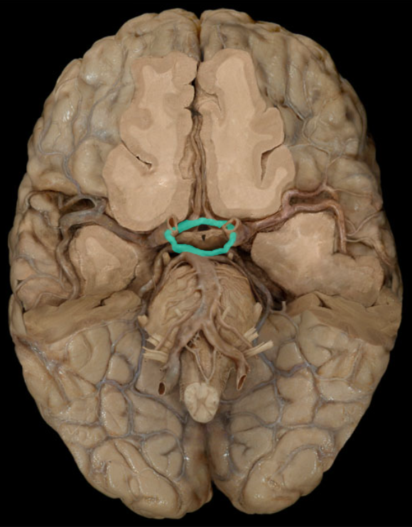
Cerebral arterial circle
Origin: Internal carotid and basilar arteries
Course: Circular anastomosis on ventral surface of brain
Also known as: Circle of Willis
Comment:
Branches include paired anterior and posterior cerebral arteries, posterior communicating arteries, and unpaired anterior communicating artery
Anastomosis refers to the end-to-end union of vessels
Internal carotid artery
Origin: Common carotid
Course:
Ascends in neck to base of cranium
Enters cranial cavity via carotid canal
Distribution: Eye, Cerebrum and Cerebral arterial circle (of Willis)
Branches: Ophthalmic, Anterior cerebral, and Middle cerebral
Comment:
Enclosed in carotid sheath
Carotid sheath is a fascial sleeve which contains common and internal carotid arteries, internal jugular vein, vagus nerve (CN X), and lymphatic channels

Posterior communicating artery
Origin: Internal carotid and basilar arteries
Course: Paired vessel connects basilar artery with internal carotid artery on each side

Vertebral artery
Origin: Subclavian
Course:
Paired vessel ascends through neck, via transverse foramina in cervical vertebrae
Enters cranial cavity via foramen magnum
Distribution: Brain, Spinal cord, and Vertebral column
Branches:
Posterior inferior cerebellar
Spinal arteries
Continues as basilar artery (formed by union of right and left vertebral arteries)
Comment: Large arteries do not by themselves supply structures, but do so through their branches

Anterior jugular vein
Drainage: Anterior Neck
Course: Descends in neck from level of hyoid bone as paired or single vessel
Termination: External jugular or subclavian
Comment: Size inversely proportional to external jugular

Carotid sinus
Location: Distal common carotid artery or proximal internal carotid artery
Function: Reacts to changes in blood pressure to effect heart rate and artery diameter
Description: At this location, tunica externa (tunica adventitia) contains collection of baroreceptors
Comment: Innervated primarily by glossopharyngeal (CN IX) nerve
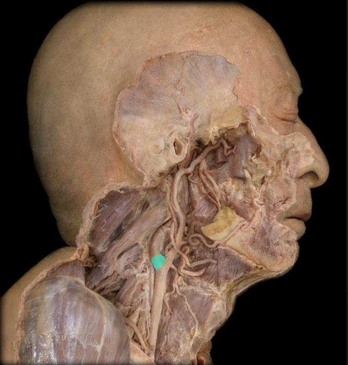
Common carotid artery
Origin:
Right: brachiocephalic trunk from arch of aorta
Left: arch of aorta
Course: Exits thorax and ascends in neck
Distribution: Structures of head and neck
Branches: Internal and external carotid arteries (at level of C4 vertebra)
Comment:
Has no branches before terminal branches
Enclosed in carotid sheath
Carotid sheath is a fascial sleeve which contains common and internal carotid arteries, internal jugular vein, vagus nerve (CN X), and lymphatic channels
Large arteries do not by themselves supply structures, but do so through their branches

External carotid artery
Origin: Common carotid (at bifurcation)
Course: Ascends in neck
Distribution: Exterior of head (except orbit), Face, Meninges, and Neck structures
Branches: Superior thyroid, Ascending pharyngeal, Lingual, Facial, Occipital, Posterior auricular, Maxillary, and Superficial temporal
Comment: Not contained in carotid sheath
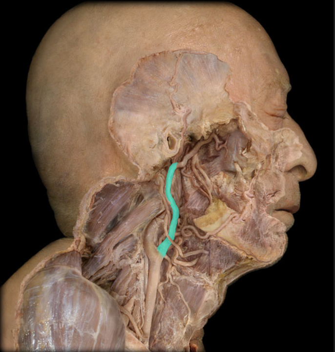
External jugular vein
Drainage: Scalp and Face
Course:
Subcutaneous vessel formed by union of retromandibular and posterior auricular veins near angle of mandible
Descends toward middle of clavicle
Crosses anterior surface of sternocleidomastoid muscle
Tributaries: Retromandibular, Posterior auricular, Transverse cervical, Suprascapular, and Anterior jugula
Termination: Subclavian
Comment:
Often communicates with internal jugular vein (near parotid gland)
Valves present at inferior end
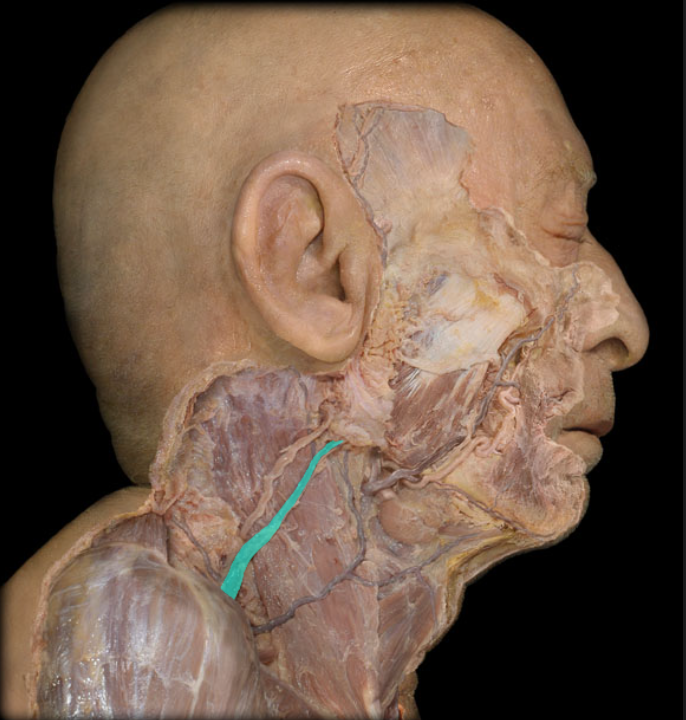
Facial artery
Origin: External carotid
Course:
Arches over submandibular gland medial to body of mandible
Curves around inferior mandibular border to ascend anteriorly from across face towards medial corner of eye
Ends as angular artery at side of nose
Distribution: Face, Palatine tonsil, Pharyngotympanic (Eustachian) tube, Soft palate, and Submandibular gland
Branches: Submental, Tonsillar, Labial, and Continues as angular
Comment:
Anastomosis with ophthalmic artery via dorsal nasal artery
Anastomosis refers to the end-to-end union of vessels

Facial vein
Drainage: Orbit, Superficial and deep face, Palatine tonsil, and Anterior neck
Tributaries: Angular, Tonsillar, Labial, and Submental
Course:
Descends, lateral to angle of mouth, from ala of nose
Crosses body of mandible anterior to masseter
Arches over submandibular gland medial to body of mandible
Enters anterior neck
Termination: Internal jugular

Lingual artery
Origin: External carotid
Course: Ascends toward oral cavity
Distribution: Tongue and Floor of oral cavity
Branches: Dorsal lingual, sublingual, and continues as deep lingual

Maxilary artery
Origin: External carotid
Course: Passes anteriorly and medially to deep face
Distribution: Mandible and teeth, maxilla and teeth, muscles of mastication, meninges, soft and hard palate, nasal cavity, face, and middle and inner ears
Comment: Largest branch of external carotid artery

Maxillary vein
Drainage: Mandible and teeth, maxilla and teeth, muscles of mastication, meninges, soft and hard palate, nasal cavity, face, and middle and inner ears
Tributaries: Pterygoid venous plexus
Course: Passes posteriorly from pterygoid venous plexus, medial to neck of mandible
Termination: Unites with superficial temporal vein to form retromandibular vein

Occipital artery
Origin: External carotid
Course: Ascends toward the posterior head
Distribution:
Posterior scalp
Sternocleidomastoid, digastric, stylohyoid, splenius, and longissimus muscles
Dura mater and bone of posterior cranial fossa
Comment: Crossed near origin by hypoglossal nerve (CN XII)

Anterior circumflex humeral artery
Origin: Axillary
Course: Passes anteriorly around surgical neck of humerous
Distribution: Deltoid muscle, Anterior proximal arm muscles, and Shoulder joint
Comment:
Smaller than posterior circumflex humeral
Anastomosis with posterior circumflex humeral
Anastomosis refers to the end-to-end union of vessels
Surgical neck of humerus is proximal portion of shaft (common area of fracture)
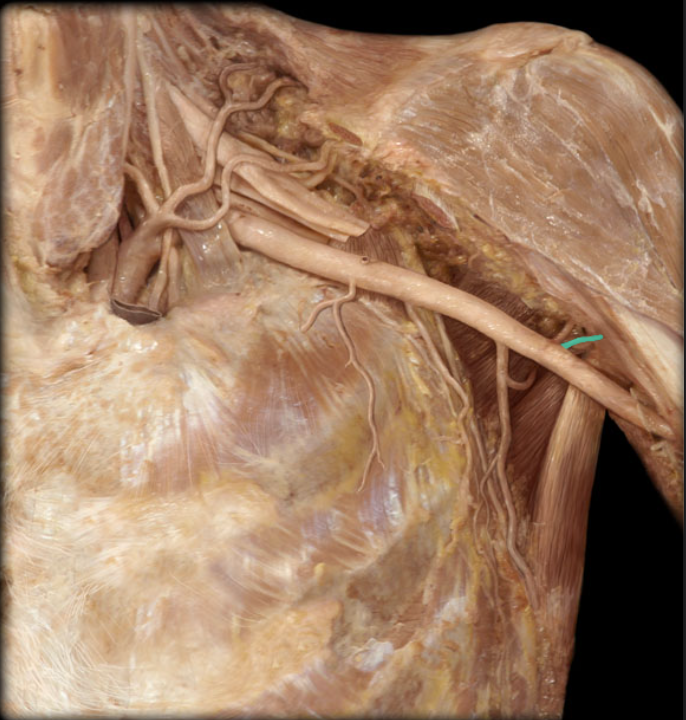
Axillary artery
Origin: Subclavian (at lateral border of rib 1)
Course:
Passes through axilla surrounded by cords and branches of brachial plexus
Divided into 3 parts by pectoralis minor m. First part is proximal to pectoralis minor m., second part is posterior to muscle, and third part is distal to muscle
Distribution:
Pectoral, shoulder, and scapular regions
Superior and lateral aspects of thoracic wall
Branches:
Superior thoracic (from first part of axillary a.)
Lateral thoracic (from second part of axillary a.)
Thoraco-acromial (from second part of axillary a.)
Subscapular (from third part of axillary a.)
Anterior and posterior circumflex humeral (from third part of axillary a.)
Continues as brachial artery (at inferior border of teres major m.)
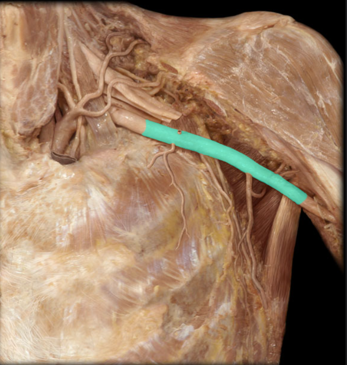
Brachial artery
Origin: Axillary (at inferior border of teres major m.)
Course: Passes inferiorly along medial aspect of arm
Distribution: Arm muscles, Humerus, and Elbow joint
Branches:
Deep brachial (deep artery of the arm, or profound brachii a.)
Superior ulnar collateral
Inferior ulnar collateral
Radial
Ulnar
Comment:
Large arteries do not by themselves supply structures, but do so through their branches
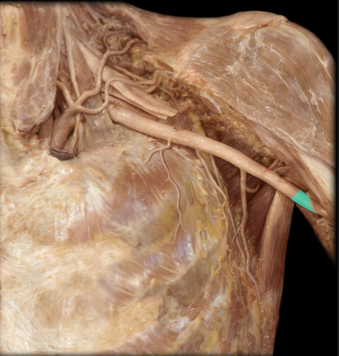
Left subclavian artery
Origin: Arch of aorta
Course: Arches over rib 1
Branches:
Vertebral
Internal thoracic
Costocervical trunk
Thyrocervical trunk
Suprascapular (sometimes)
Continues as axillary artery at lateral border of rib 1

Suprascapular artery
Origin: Thyrocervical trunk or subclavian artery
Course:
Passes across inferior neck towards scapula
Passes onto posterior aspect of scapula
Distribution:
Supra- and infraspinatus muscles
Acromioclavicular and glenohumeral joints
Comment:
Contributes to anastomoses around scapula
Anastomosis refers to the end-to-end union of vessels
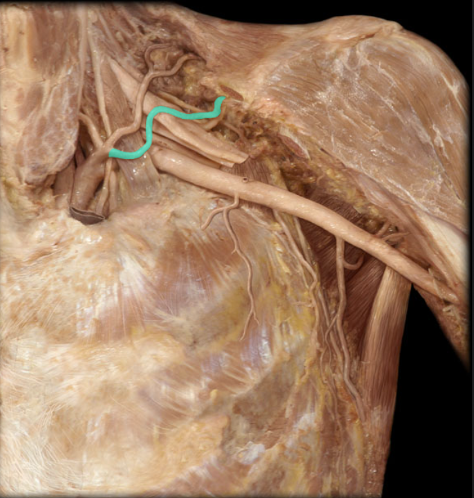
Transverse cervical artery
Origin: Thyrocervical trunk
Course: Passes posteriorly across neck to scapular muscles
Distribution: Trapezius muscle, Levator scapulae muscle, and Rhomboid muscles
Branches: Superficial and deep
Comment:
Contributes to anastomoses around scapula
Anastomosis refers to the end-to-end union of vessels

Radial artery
Origin: Brachial
Course: Passes distally along lateral aspect of forearm
Distribution:
Superficial posterior forearm muscles
Deep hand muscles
Thumb and lateral side of index finger
Joints of elbow and hand
Branches:
Radial recurrent
Superficial palmar branch (to superficial palmar arterial arch)
Princeps pollicis
Continues as deep palmar arterial arch
Comment:
Commonly used for taking pulse at wrist
Anastomosis with ulnar artery through superficial and deep palmar arterial arches
Anastomosis refers to the end-to-end union of vessels

Ulnar
Origin: Brachial
Course: Passes distally along medial aspect of forearm
Distribution: Medial forearm muscles, Hand muscles, and Joints of elbow and hand
Branches:
Common interosseous
Ulnar recurrent
Deep palmar branch (to deep palmar arterial arch)
Continues as superficial palmar arterial arch
Comment:
Larger than radial artery
Anastomosis with radial artery through superficial and deep palmar arterial arches
Anastomosis refers to the end-to-end union of vessels
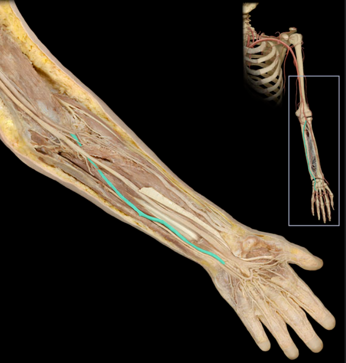
Abdominal aorta
Origin: Thoracic aorta
Course:
Parallels vertebral column, slightly left of midline
Divides into common iliac arteries on body of L4 vertebra
Distribution:
Abdominal and pelvic viscera
Walls of abdomen and pelvis
Ovaries and testis
Branches: Celiac, Superior mesenteric, Inferior mesenteric, Middle suprarenal, Renal, Gonadal, Inferior phrenic, Lumbar, Median sacral, and continues as right and left common iliac

Common hepatic artery
Origin: Celiac artery
Course:
Passes anterior and to the right to enter lesser omentum
Ends at superior border of first (superior) part of duodenum
Distribution: Liver, Pancreas, Stomach. Gallbladder, and Duodenum
Branches: Hepatic artery proper and Gastroduodenal
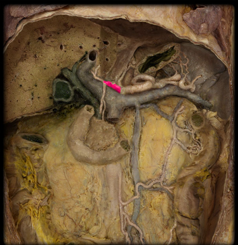
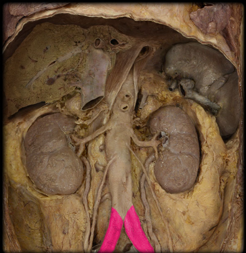
Common iliac artery
Origin: Abdominal aorta
Course:
Begins anterior to L4 vertebral body
Passes obliquely across L4-5 vertebral bodies
Ends at the level of the sacro-iliac joint (L5-S1)
Distribution: Pelvis, Posterior abdominal wall, Perineum, and Lower limb
Branches: Internal iliac and External iliac
Gastroduodenal artery
Origin: Common hepatic
Course: Descends between superior (first) part of duodenum and neck of pancreas
Distribution: Stomach, Pancreas,and Duodenum
Branches: Right gastro-omental and Superior pancreaticoduodenal

Gonadal artery
Origin: Abdominal aorta
Course:
Descends on posterior abdominal wall
Male (testicular artery): enters inguinal canal to join contents of spermatic cord
Female (ovarian artery): in pelvis, enters suspensory ligament of ovary
Distribution:
Ureter
Male: testis and spermatic cord
Female: ovary and uterine tube

Left renal artery
Origin: Abdominal aorta
Course: Passes laterally to enter kidney
Distribution: Kidney, ureter (upper part), and Suprarenal gland
Branches:
Inferior suprarenal
Ureteric
Four or five branches near hilum of kidney
Comment: Left renal artery shorter than right renal artery

Lumbar artery
Origin: Abdominal aorta
Course: Four, paired branches course laterally on posterior abdominal wall
Distribution: Posterior abdominal wall, back, and spinal cord
Comment: Segmental pattern similar to posterior intercostal arteries

Right renal artery
Origin: Abdominal aorta
Course: Passes laterally to enter kidney
Distribution: Kidney, Ureter (upper part), and Suprarenal gland
Branches: Four or five branches near hilum of kidneyy
Comment: Right renal artery longer than left renal artery

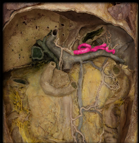
Splenic artery
Origin: Celic artery
Course: Serpenmtine (snake-like) path of left along superior border of pancreas
Distribution: Spleen, pacnreas, and stomach (fundus
Branches:
Comment:
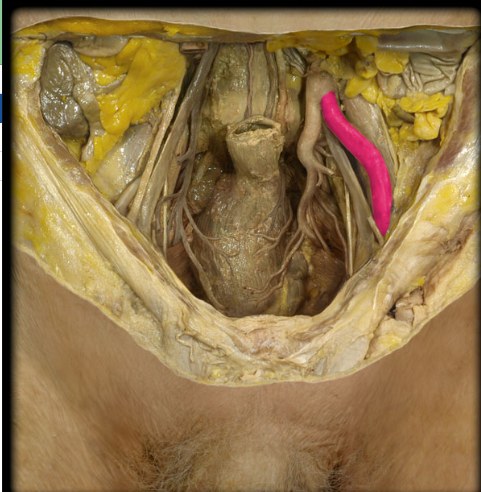
Left external illac artery (female pelvis)
Origin: Bifurcation of common iliac artery
Course: Descends laterally along medial border of psoas major muscle from level of sacro-iliac joint to inguinal ligament
Distribution: Abdominal wall (inferior) and Lower limb
Branches: Inferior epigastric, Deep circumflex iliac, and Continues as femoral artery
Comment: At origin usually crossed by ureter and gonadal vessels

Left internal iliac artery (female pelvis)
Origin: Common iliac
Course:
Begins at the level of the sacro-iliac joint (L5-S1)
Descends into pelvis and passes posteriorly toward superior margin of greater sciatic notch (ilium)
Distribution: Posterior abdominal wall, Pelvic wall, Pelvic organs, Gluteal region, and Perineum
Branches: Iliolumbar, Lateral sacral, Superior gluteal, Umbilical, Uterine (female), Inferior vesical (male), Vaginal (female), Obturator, Middle rectal, Inferior gluteal, and Internal pudendal
Comment:
Commonly divides into anterior and posterior divisions
Branching pattern has considerable variation

Uterine artery (female pelvis)
Origin: Internal iliac (anterior division)
Course:
Passes medially to reach uterus at junction of uterine body and cervix
Reflects along side of uterus
Distribution: Uterus and Vagina
Branches: Vaginal
Comment:
Commonly a branch of umbilical artery
Forms anastomosis with ovarian and vaginal arteries
Anastomosis refers to the end-to-end union of vessels

External iliac artery (male pelvis)
Origin: Common iliac
Course: Descends laterally along medial border of psoas muscle from level of sacro-iliac joint to inguinal ligament
Distribution: Abdominal wall (inferior) and Lower limb
Branches: Inferior epigastric, Deep circumflex iliac, and Continues as femoral artery
Comment:
At origin usually crossed by ureter and gonadal vessels
Formed by bifurcation of common iliac
Internal iliac artery (male pelvis)
Origin: Common illiac
Course:
Begins at the level of the sacro-iliac joint (L5-S1)
Descends into pelvis and passes posteriorly toward superior margin of greater sciatic notch (ilium)
Distribution: Posterior abdominal wall, Pelvic wall, Pelvic organs, Gluteal region, and Perineum
Branches: Iliolumbar, Lateral sacral, Superior gluteal, Umbilical, Uterine (female), Inferior vesical (male), Vaginal (female), Obturator, Middle rectal, Inferior gluteal, and Internal pudendal
Comment:
Commonly divides into anterior and posterior divisions
Branching pattern has considerable variation

Obturator artery (male pelvis)
Origin: Internal iliac
Course:
Passes along lateral pelvic wall to superior part of obturator foramen
Passes through obturator canal to reach medial thigh
Distribution:
Muscles and skin of medial thigh
Head and neck of femur

Deep femoralartery
Origin: Femoral
Course: Passes distally through medial thigh
Distribution: Thigh muscles and Hip and knee joints
Branches: Nutrient artery to humerus, Medial circumflex femoral, Lateral circumflex femoral, and Three to four perforating arteries
Also known as:
Deep artery of thigh
Profunda femoris a.
Comment: Largest branch of femoral

Femoral artery
Origin: External iliac
Course:
Descends in anterior and medial thigh
Extends from inguinal ligament to adductor hiatus
Distribution: Abdominal wall (anterior inferior), External genitalia, Thigh muscles, Femur, and Hip and knee joints
Branches:
Superficial epigastric
External pudendal
Superficial circumflex iliac
Muscular branches
Deep artery of thigh (profunda femoris a.)
Continues as popliteal (at adductor hiatus)
Comment:
Major artery of lower limb
Superficial in proximal thigh (femoral pulse)

Anterior tibial artery
Origin: Popliteal
Course:
Passes through proximal part of interosseous membrane
Descends on anterior surface of interosseous membrane (proximal 2/3) and tibia (distal 1/3)
Crosses ankle midway between malleoli
Distribution: Anterior (extensor) compartment of leg
Branches: Continues onto dorsum of foot as dorsal artery of foot
Comment: Terminal branch of popliteal a.

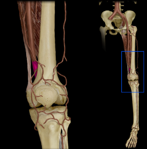
Popliteal artery
Origin: Femoral
Course: Descends posterior to knee joint in popliteal fossa
Distribution: Structures around knee and Knee joint
Branches: Posterior and anterior tibial and Genicular arteries
Comment:
Continuation of femoral artery at adductor hiatus
Terminal branches (posterior and anterior tibial) supply everything below knee