A&P Lab 2: Integumentary System
 Call Kai
 Learn
Learn Practice Test
 Spaced Repetition
 Match
Match Flashcards
 Knowt Play
1/83

There's no tags or description
Looks like no tags are added yet.
Name | Mastery | Learn | Test | Matching | Spaced | Call with Kai | Chat |
|---|
No analytics yet
Send a link to your students to track their progress
84 Terms
Simple Epithelial Tissues
squamous, cuboidal, columnar, pseudostratified columnar
Stratified Epithelial Tissues
squamous, columnar, transitional
Tissue (definition)
collections of cells and the extracellular substances surrounding them that work together to perform a specific function
Characteristics of Epithelium (cellular arrangment)
tightly packed sheets
Characteristics of Epithelium (superficial surfaces)
referred to as apical surface, exposed to either external environment or an internal body cavity, may have microvilli or cilia
Characteristics of Epithelium (lateral surfaces)
may have intercellular junctions
Characteristics of Epithelium (deep surface)
called basal surface, attaches to a basement membrane by connective tissue
Characteristics of Epithelium (regeneration capacity)
high level of regeneration since damage is caused by exposure to external environments
Characteristics of Epithelium (avascular)
receives blood supply from connective tissue it is attached to, nutrient exchange at the apical surface or diffused across basal surface from connective tissue blood vessels
Characteristics of Epithelium (innervation)
lots of nerves to detect changes in the environment that the apical surface is exposed to

Simple Squamous Epithelium
number of layers: one
cell shape: flat cells
nucleus: flattened or ovular
locations in the body: vessels, serous membranes, lungs, kidneys
functions: filtration, diffusion, and secretion (thin for gas exchange, directly attached to basement membrane)
honors information: Endothelium - vessels and heart
Mesothelium - serous membranes

Simple Cuboidal Epithelium
number of layers: one
cell shape: cube-like cells
nucleus: centralized
locations in the body: lens of eye, ovaries, kidney tubules, duct of glands
functions: secretion and absorption
honors information: none

Simple Columnar Epithelium
number of layers: one
cell shape: column
nucleus: extend even heights
locations in the body: non-ciliated - gastrointestinal tract, ducts of glands, gallbladder
ciliated - fallopian tube, bronchioles
functions: secretion and absorption
ciliated - “beating” of cilia cause movement
honors information: contains goblet cells that secrete mucin which mixes with water to form mucus
ciliated: mucus on apical surface
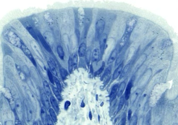
Pseudostratified Columnar Epithelium
number of layers: one
cell shape: column
nucleus: cells at different levels and appear to be multiple layers
locations in the body: upper respiratory tract, trachea
functions: secretion and absorption, trap particles in mucous and sweep them away with the use of cilia
honors information: non-ciliated - rare form found in vas deferens
stereociliated- found in epididymis
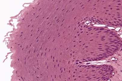
Stratified Squamous Epithelium
number of layers: multiple
cell shape: flat
locations in the body: keratinized - superficial layer of the skin (apical layer filled with keratin, keratin-though, fibrous protein)
non-keratinized - oral cavity, esophagus, vagina, tongue
functions: keratinized - protein
non-keratinized - keep areas moistened by mucous

Stratified Cuboidal Epithelium
number of layers: multiple
cell shape: cube-like with round edges
locations in the body: ducts of sweat glands, male urethra, esophageal glands
functions: protection and some secretion and absorption

Stratified Columnar Epithelium
number of layers: multiple
cell shape: column
locations in the body: conjunctiva or the eye, part of urethra, ducts
functions: protection and secretion

Transitional Epithelium
number of layers: multiple
cell shape: relaxed - cube-like
stretched - squamous
locations in the body: urinary bladder, portions of ureters and urethra
functions: allows urinary organs to stretch without rupturing
Characteristics of Connective Tissues (cellular arrangement)
there is usually a fair amount of space between each connective tissue cell, there are exceptions such as adipose tissue
Characteristics of Connective Tissues (protein fibers)
collagen fibers, elastic fibers, and reticular fibers
Characteristics of Connective Tissues (ground substance)
materials between the cells and the protein fibers, the ground substance varies widely by tissue type
Characteristics of Connective Tissues (extra cellular matrix)
non-cell portion, protein fibers and ground substance
Characteristics of Connective Tissues (vascular)
most connective tissue is very vascular
Loose Connective Tissue (definition)
fibers are loosely arranged between cells, cells are much more abundant than in dense connective tissues

Areolar Connective Tissue
function: strength, elasticity, support
location: in and around almost every body structure as a packaging material
notable things: one of the most widely distributed connective tissue, contains fibers and numerous cells with random arrangement in a semifluid ground substance
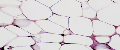
Adipose Tissue
function: reduces heat loss, energy reserve, supports and protects organs
location: wherever areolar connective tissue is
notable things: stores fat, cells derived from fibroblasts called adipocytes
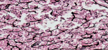
Reticular Connective Tissue
function: stroma of organs, connects to smooth muscle, filters and removes
location: stroma of liver, spleen, lymph nodes, red bone marrow
notable things: meshwork of type III collagen fibers and reticular cells
Dense Connective Tissue (definition)
consists of thicker and more densely packed fibers and much fewer cells than loose connective tissue

Dense Regular Connective Tissue
function: provides strong attachments between structures, absorbs energy
location: tendons, ligaments, aponeurosis
notable things: shiny white appearance, mainly collagen fibers, arranged in bundles with fibroblasts between them, fibers run in one direction that is parallel to the force being applied

Dense Irregular Connective Tissue
function: provides pulling strength in many directions
location: fibrous pericardium of heart, joint capsules, fasciae and dermis
notable things: often occurs in sheaths, collagen fibers irregularly arranged with a few fibroblasts, increased strength due to arrangement, compare to dense regular connective tissue

Elastic Connective Tissue
function: allows stretching of various organs and is able to recoil back to shape
location: lung tissues, elastic arteries, trachea, bronchial tubes, true vocal cords
notable things: mostly elastic fibers with fibroblasts, do not confuse with transitional epithelium
Osseous Connective Tissue (definition)
provides support and protection for the organs of the body, hard and rigid due to mineralization of the extracellular matrix, bone also serves as a reservoir for calcium and other nutrients

Trabecular Bone
cancellous or spongy bone
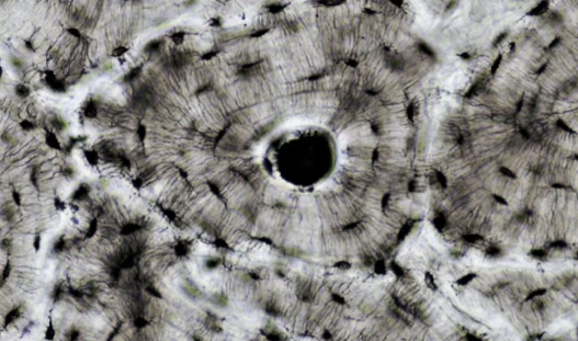
Cortical Bone
compact

Blood
structure: contains formed elements, dissolved protein within a liquid ground substance (plasma)
function: transports respiratory gases, leukocytes, platelets, plasma transports nutrients, wastes, and hormones
location: vessels and. organs of the circulatory system
Lymph
structure: derived from blood plasma, contains dissolved solutes, protein, cell debris and pathogens
function: transports antibodies, lymphocytes, metabolic waste
location: lymph vessels, lymphoid structures
Cartilage (definition)
composed of cells, fibers, and highly-hydrated (gel like) ground substance, the fibers are densely packed to provide tensile strength, while proteoglycans in the ground substance make cartilage resilient by trapping water, cartilage is avascular and its cells rely on diffusion for nutrients, cartilage is aneural, any pain felt with an injury to cartilage is stemming from surrounding tissues and structures
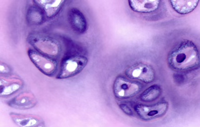
Hyaline Cartilage Tissue
function: provides smooth surfaces for movements of joints, flexibility and support
location: ends of long bones, costal cartilage, respiratory tract
notable things: weakest cartilage and can fracture easily, most abundant cartilage, blue-white color, perichondrium present
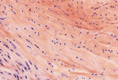
Fibrocartilage
function: support joining structure together, shock absorber
location: pubic symphysis, menisci, intervertebral discs
notable things: strongest cartilage, thick bundles of collagen fibers, no perichondrium present
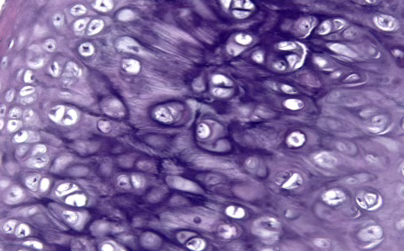
Elastic Cartilage
function: strength and elasticity
location: larynx, external ear, epiglottis and eustachian tubes
notable things: chondrocytes in threadlike network, perichondrium present
Fiber Composition
Hyaline Cartilage - type II collagen
Fibrocartilage - type I and II collagen
Elastic Cartilage
elastic fibers and type II collagen
Embryonic Connective Tissues
Mesenchyme and Mucous Connective Tissue (Wharton’s Jelly)
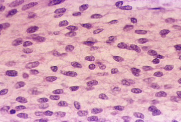
Mesenchyme
structure: stellate (star shaped) or spindle shaped, ground substance is viscous fluid and immature protein fibers
function: first connective tissue to develop, all connective tissue is formed from mesenchyme, adult connective tissue has mesenchymal cells that act as stem cells to repair tissue
location: throughout early embryo, fills space between germ layers and organs
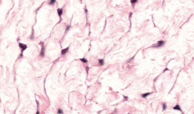
Mucous Connective Tissue (Wharton’s Jelly)
structure: same as mesenchyme except immature protein fibers are MORE abundant
function: support the structures in the umbilical cord
location: inside the umbilical cord, where it surrounds and cushions the two umbilical arteries and one umbilical vein

Skeletal Muscle Tissue
voluntarily controlled, striated, has several nuclei per cell, found attached to bones, allows you to direct your eyeballs, concerned with locomotion of the body as a whole

Cardiac Muscle Tissue
involuntarily controlled, striated, has a single nucleus in each cell, tissue of the heart

Smooth Muscle
involuntarily controlled, has a single nucleus in each cell, found in walls of stomach, uterus, and arteries, changes the internal volume of an organ as it contracts

Nervous Tissues
neurons - the cells specialized for the propagation of electrochemical signals
neuroglia - supporting cells of nervous tissue, multiple types
Functions of the Integumentary System
sensation - nerve endings, tactile cells, and corpuscles sense and distinguish touch and pain signals
water storage - stores a large amount of blood in the body; maintains homeostasis by regulating water loss and absorption
metabolic - regulates body processes by secreting hormones, cytokines, and growth factors, synthesizes vitamin D
protection - protects body from external environment
excretion - regulates electrolyte levels by secreting water, salt, and. urea through exocrine and apocrine glands
thermoregulation - constriction and dilation of dermal vessels regulates body temperature to protect vital internal organs, dermal sweat glands help decrease body temperature
Epidermis (definition)
the most superficial part of the skin, composed of epithelial tissue, protects the underlying tissues from abrasion, water loss, and provides immune support by inhibiting pathogen growth, composed of four or five layers depending on its location on the body (the stratum lucidum is only found on the palmar and plantar regions)
Stratum Corneum
thickest most superficial layer, composed of twenty - thirty layers of tightly interwoven, anucleate keratinized cells, the cells are keratinized stratified squamous epithelial cells, continuously shed and regenerated by deeper skin layers
Stratum Lucidum
the most translucent layer that only exists in thick skin (palmar and plantar regions), made of keratinized stratified squamous cells
Stratum Granulosum
the third deepest layer, contains keratinocytes that are undergoing keratinization, cells begin to die further from the nutrition source and fill with keratin protein causing the nucleus and organelles to disintegrate, contains lamellar granules which release a fatty water-repellent secretion, this layer demarcates the transition from the deeper living strata to the more superficial dead strata, can be easily identified microscopically by its dark, granulated appearance
Stratum Spinosum
deep to the stratum granulosum, cells begin to mature and produce keratinocytes, cells here are bound to each other by desmosomes, stem cells in the stratum basale divide and one cell moves to the stratum spinosum and differentiates while the other remains in the stratum basale, langerhans cells are found in this layer and serve as part of the skin’s immune system
Stratum Basale
single layer of cuboidal or low columnar keratinocyte stem cells joined by hemidesmosomes atop the basement membrane, main cells here are keratinocyte stem cells, melanocytes and tactile cells, are located here, contain epidermal ridges that interlock with dermal papillae (giving us our fingerprints)
Thick Skin
found on palmar and plantar regions, all five layers of epidermis are present and combine for a thickness of 0.4 mm - 0.6 mm, sweat glands but no hair follicles or sebaceous glands
Thin Skin
covers most of the body and does not have a stratum lucidum layer, the thickness is between 0.075 mm - 0.150 mm, hair follicles, sebaceous glands, and sweat glands
Langerhans Cells
the phagocytic immune cells in the stratum spinosum, granulosum, and less often in the stratum corneum and lucidum
Keratinocytes
the majority of cells in the epidermis that produce keratin, a protein that strengthens the epidermis by contributing to the cytoskeleton as intermediate filaments, this protein also imparts water resistance to the skin
Merkel Cells
cells in the stratum basale that help with tactile sensation, stimulate nerve endings to allow identification of touch
Melanocytes
cells that produce melanin, long projections on cells send melanin within melanosomes to more superficial layers, melanin contributes to skin color and aids in protection from UV light, originates in stratum basale
Dermis (definition)
the layer deep to the epidermis and is made of two layers, the papillary and reticular layers, the tissue type located here is connective tissue proper, contains blood capillaries, lymph vessels, nerves, sweat glands and smooth muscle
Papillary Region
superficial layer of the dermis, made of loose areolar connective tissue, contains collagen and fine elastic fibers, blood supply, and tactile nerve endings, this layer is responsible for supplying nutrients and removing waste from the epidermis
Dermal Papillae
upward projections of the papillary region of the dermis, increase connection between epidermis and dermis, contain capillaries that provide nutrients to the epidermis and tactile sensation, interlock with epidermal ridges to form fingerprints
Capillary Loops
blood vessels found in the dermal papillae
Meissner Corpuscle
nerve endings that detect light touch, located in dermal papillae
Free Nerve Endings
detect temperature, pain, tickling, and itching, found in dermal papillae
Reticular Region
deepest layer of skin, composed of dense irregular connective tissue, this layer is highly vascular, and is responsible for the skin’s strength, extensibility, and elasticity due to the presence of collagen
Hair Follicle
composed of external and internal root sheath and surrounds the root of the hair; where hair development occurs
Arrector Pilli Muscle
smooth muscle that attaches to the hair follicle, during physical or emotional stress this muscles contracts and causes goose bumps
Pacinian Corpuscles
composed of multilayered connective tissue capsules enclosed by dendrite, detects deep pressure
Glands
sebaceous glands - holocrine gland that produces sebum into hair follicle and onto hair; keeps hair and skin from drying out and inhibits growth of some bacteria, mainly located on hair follicles in the face, neck, and superior chest, not found on palmar or plantar regions, activated during puberty and stimulated by hormones
sudoriferous glands - sweat glands that empty substance onto the surface of the skin or the hair follicle
merocrine sweat glands - simple, coiled tubular glands that discharge secretion to surface of skin, function in thermoregulation, activated shortly after birth
apocrine sweat glands - simple coiled tubular glands, found in axilla, groin and areolae, and bearded regions on men, releases sweat into hair follicles, secretion is viscous cloudy and contain proteins and lipids that produce an odor during bacterial interaction, acts as a pheromone and is involved in emotional sweating, activated during puberty
Subcutaneous Layer (Hypodermis)
not part of the skin, composed of areolar and adipose tissue, pads and protects body, acts as energy reservoir and provides thermal insulation
First-Degree Burn
partial-thickness burn, affects epidermis, appears red with mild edema and no blisters, mildly painful, heals in three - five days, typically doesn’t scar, to treat immerse in cool water or apply cool, wet compress
Second-Degree Burn
partial-thickness burn, affects epidermis and part of dermis, appears red tan or white with blistering and edema, very painful, heals in two - four weeks, slight scarring, to treat immerse in cool water or apply cool, wet compress and elevate limbs to prevent swelling, avoid ointments, and don’t break blisters
Third-Degree Burn
full-thickness burn, affects epidermis dermis and subcutaneous layers, appears marble-white to mahogany to dark wounds with edema, little pain since nerves are damages, heals depending on the burn and treatment plan, problematic due to large loss of water, infection, reduced circulation of blood, decreased urine production, and diminished immune response, excessive scarring, to treat hospitalize, antibiotics, skin graft, treatment for dehydration
Cutaneous Membranes
this refers to the dermal and epidermal layers of the skin, the epidermis is the superficial, thinner portion comprised of keratinized stratified squamous epithelial tissue, and is avascular, the deeper, thicker portion is the vascular dermis composed of connective tissue which protects the body, detects external sensations, and contributes to thermoregulation
Mucous Membranes
these membranes line cavities that open directly to the external environment, such as the digestive, respiratory, and reproductive tracts, they consist of a surface epithelium (that can change based on function) that protects and lubricates, and a connective tissue called the lamina propria (loose areolar connective tissue), this membrane functions in absorption, secretion, and protection, sometimes the mucous membrane has a deep, smooth muscle layer called the muscularis mucosae
Serous Membranes
these membranes are closed to the external environment, they have a specialized layer of epithelium called mesothelium (simple squamous epithelium) on a loose areolar connective tissue component, serous membranes have a visceral layer, which covers the organ directly, and a parietal layer, that lines the body cavity, between the two layers is serous fluid that lubricates and cushions the organs, the pericardium, peritoneum, and pleura are serous membranes and reside in the thoracic and abdominal cavities
Serous Membranes (Serosa vs Adventitia)
the serosa and adventitia serve two different functions and are found on different sections of the body, the serosa is prevalent on intraperitoneal organs and aids in lubrication and cushion between surfaces, on the other hand, adventitia’s function is to attach organs to other organs or tissues and is found on retroperitoneal organ surfaces
Synovial Membranes
these membranes are found in synovial joints, and are composed of connective tissue, these membranes secrete synovial fluid, which lubricate moving bone parts in a synovial joint and provide nutrients to the cartilage associated with these bones
Decubitus Ulcers
causes: prolonged contact with moisture, immobility, impaired circulation due to pressure
common locations: heels, ankles, toes, hip, sacrum, back of head, ears, knees, elbows, shoulders, and shoulder blades
stages: stage one - redness on skin surface, blue-grey discoloration
stage two - redness along the abrasion, blisters or a shallow crater
stage three - all skin layers are broken down, subcutaneous layer is exposed
stage four - extends deeper into tissues, muscle, bones, and joints

Cleavage Lines
collagen fibers in the dermis are often overall orientated in a predictable direction, they usually run parallel to muscle fibers, skin will heal better when cut along these lines, used to plan surgical incisions