Physiology Exam #1
 Call Kai
 Learn
Learn Practice Test
 Spaced Repetition
 Match
Match Flashcards
 Knowt Play
1/157

There's no tags or description
Looks like no tags are added yet.
Name | Mastery | Learn | Test | Matching | Spaced | Call with Kai | Chat |
|---|
No analytics yet
Send a link to your students to track their progress
158 Terms
3 types of muscles
skeletal, cardiac, and smooth muscle
cardiac muscle
striated
involuntary
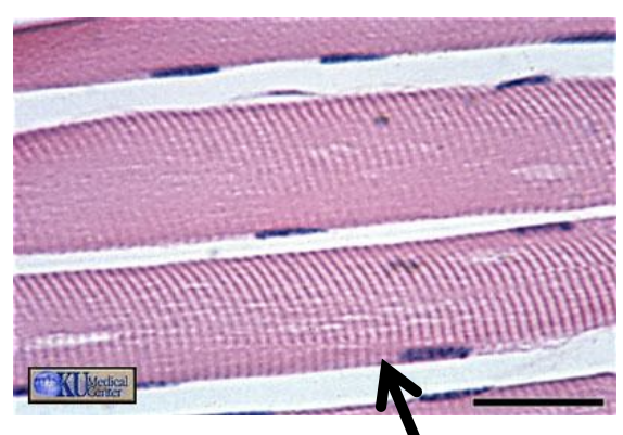
skeletal muscle
striated
voluntary
smooth muscle
non striated
involuntary
cardiac muscle

skeletal muscle
smooth muscle

Factors that contribute to skeletal muscle diversity
shape
size
number of fibers per area
length of fiber
angle of pennation
why is skeletal muscle diverse
diversity in skeletal muscle shape and structure allows for diverse function
Pennation angle
the angle between muscle fibers and tendons
A greater pennation angle can
allows more fibers per area
allows muscle to produce more force within a limited anatomical space
result in a smaller length change during contraction
Biomechanical Advantage
pennation —> more fibers in an area —> greater ability to produce force
Characteristics of skeletal muscle
Excitable & Contractile
Excitable
controlled voluntarily by the nervous system
Contractile
able to contract and produce force
Skeletal Muscle functions
movement
the only tissue capable of this function
support
protection
energy storage
heat generation
Skeletal muscle: organ —> cell
whole skeletal muscle —> fascicle —> muscle fiber —> myofibril —> sarcomere
Fascicle (2)
a group of muscle fibers packed tightly together
Muscle Fiber (3)
groups of myofibrils packed together
Myofibrils (4)
cylindrical organells that contain sarcomeres inside the muscle fiber
Sarcomere (5)
the contractile unit of a myofibril
3 layers of Connective Tissue
1) epimysium
2) perimysium
3) endomysium
Epimysium
surrounds the entire muscle organ
Perimysium
surrounds the fascicles
it contains lots of blood vessels and nerves
Endomysium
thin layer surrounds the individual muscle fibers
contains tiny capillaries individual neurons
Sarcolemma
cell/plasma membrane (lipid bilayer) surrounding each muscle fiber
NOT A CT
Connective Tissue
resists excessive passive stretching of the muscle and distributes forces to minimize damage to the muscle fibers
Function of Connective Tissues
provide a scaffolding for muscle fibers
holds muscle fibers together
help determine the gross structure of the muscle belly
provides a pathway/conduit for blood vessels and nerves = perimysium
resists excessive passive stretching
helps distribute forces to minimize damage to muscle fibers
Tendon
tough, fibrous, cord like tissue
extension of the epimysium, perimysium, and endomysium
the CT layers don’t just stop at the muscle, but continue into the tendon
Functions of the tendon
physically connects muscle to bone (or another structure)
wraps around and within the muscle to transmit force
Myotendinous Junction
muscle contracts —> force —> MTJ —> tendon —> bone

Myotendinous Junction (MTJ)
the MTJ is the site where the muscle and tendon connect
function = force transmission
the contractile proteins of the muscle fivers transmit force to the
extracellular connective tissue proteins of the tendon
Blood Vessels and Nerve Fibers
provide nutrient delivery
help regulate muscle contraction
Skeletal Muscle within the Cell
1) sarcolemma
2) sarcoplasm
3) sarcoplasmic reticulum
4) sarcomere
5) myofibril
6) myofiber
sarcolemma (1)
the lipid bilayer surrounding the muscle fiber
also called: plasma membrane, cell membrane
functions of sarcolemma
Regulates ion concentrations
Receives and transmits action potentials
signals at the neuromuscular junction
carries AP into muscle fiber through T-tubules
provides scaffolding
fiber regeneration
neighboring muscle cell attachment
Transverse (T) Tubules (2)
extensions of the sarcolemma
run deep into muscle fiver
carry action potential deep
interacts w/ end of the terminal cisternae of the sarcoplasmic reticulum
Sarcoplasmic Reticulum (SR) (3)
Function = stores calcium
runs parallel to the muscle fiber
is a membrane bound structure
functions similarly to the ER in other cells
Sarcoplasm (4)
the cytosol/cytoplasm of the muscle cell
stores: glycogen, myoglobin, proteins, minerals, fats, organelles
Myofibrils (5)
is a structure inside the muscle fiber that contains sarcomeres
sarcomere (5)
is the contractile unit of a myofibril
thick filament = Myosin
thin filament = Actin
the arrangement of the filaments creates the striated appearance
Myosin
thick filament
Head
binds to actin
contains ATPase activity
Actin
thin filament
contain myosin binding sites
Tropomyosin
part of thin filament
wraps around actin
blocks the myosin binding sites
Troponin
part of thin filament
bound to actin
holds tropomyosin in place
Nuclei/Myonuclei (6)
skeletal muscle fibers = multinucleated
myonuclei located near the edge of muscle fibers
function of nuclei/myonuclei
genetic control
control gene expression
replication of DNA during the cell cycle
Mitochondria (7)
are responsible for producing ATP
use oxygen and water to convert energy into a form the cell can use
muscle cells need ATP to generate force
Mitochondria produce ATP w/ oxidative metabolism including:
krebs cycle
electron transport chain
oxidative phosphorylation
What is membrane potential (MP)
the electrical potential difference/voltage between the intracellular and extracellular spaces
What is Resting Membrane Potential (RMP)
for a neuron, -70mV (at rest)
the inside of the cell is more negative
outside is more positive
What determines RMP
ion channels
gated channels
ion pumps
ion channels
always open
ions diffuse through them down their gradient
gated channels
closed until a stimulus acts on the channel
once open, ions diffuse through the channel down their gradient
ion pumps
actively transport ions across the membrane
requires ATP
Potassium (K+) channels
allow K+ to leak out of the cell
Voltage gated Sodium (Na+) channels
open/close depending on membrane voltage
important during the action potential
Voltage gated Potassium (K+) channels
open/close depending on membrane voltage
important during action potential
Sodium/Potassium Pump (Na+/K+ pump)
pumps 3 Na+ OUT
pumps 2 K+ IN
requires ATP
what maintains RMP
primarily through the Na+/K+ pump and K+ channels
Action Potential (AP)
change in membrane potential that occurs when the cell reaches threshold
1) stimulus
2) depolarization
3) repolarization
4) hyperpolarization
5) resting membrane potential (RMP)
Threshold
MUST be reached to initiate an action potential
once threshold is reached:
both Na+ and K+ voltage gated channels open
but they open at DIFFERENT speeds
At Rest of AP
cell is -70mV
no movement
voltage gated Na+ channel = closed
voltage gated K+ channel = closed
Threshold of AP
stimulus caused MP to move toward threshold
trigger point for AP
a little Na+ move IN
voltage gated Na+ channel = stimulated to open
voltage gated K+ channel = stimulated to open
Depolarization of AP
rapid upward portion
Na+ moves into the cell A LOT
voltage gated Na+ channels = open
voltage gated K+ channels = closed
the inside of cell become more positive = graph goes up
Peak of AP
top of waveform
a little Na+ moves IN
a little K+ moves OUT
VG Na+ channels = closing
VG K+ channels = opening
transition point
Repolarization of AP
downward portion of AP
a lot of K+ moves OUT
VG Na+ channels = closed
VG K+ channels = open
inside becomes more negative
Hyperpolarization of AP
membrane potential becomes more negative than normal RMP
a little K+ move OUT
VG Na+ channels = closed
VG K+ channels = closing
then the membrane return to normal RMP
EPSP & IPSPs
summation of all incoming signals determines whether threshold is reached and an AP is generated
Excitatory Postsynaptic Potential (EPSP)
depolarizes the membrane
makes it more positive
brings the cell closer to threshold
Ex: opening of a ligand gated Na+ channel
Inhibitory Postsynaptic Potential (IPSP)
makes membrane more negative
moves cell further away from threshold
Ex: opening of ligand gated K+ channel
How does action potential travel
Na+ moves more negative areas inside the cell
causes next section to reach threshold
a new AP begins in next section
doesn’t travel backwards b/c of VG Na+ channels
Orthodromic (Electrical Nerve Stimulation)
AP travels in the same direction as physiological signaling
Antidromic
AP travels in the opposite direction from physiological signaling
What determines Action Potential Conduction Velocity?
1) Myelination
more myelin = faster conduction
2) Axon Diameter
larger axon = faster conduction
Motor Unit
consists of the motor neuron and all the scattered muscle fibers which it innervates
Motor Neuron
is the nerve cell that communicates with the skeletal muscle
Motor Unit = motor neuron + all muscle fibers it controls
Parts of the motor neuron
Dendrites
Cell body
Axon
Axon Terminals
Dendrites
receive input/signals from the dendrites
Cell body
receives the signals from the dendrites
decides what to do with the summed inputs
Axon
transmits the signal along the length of the neuron
Axon Terminals
transmit signal to the adjacent neurons/cells
Synapse
connection between axon terminal from neuron 1 and dendrites of neuron 2
Neuromuscular Junction (NMJ)
the synapse that allows communication between the motor neuron and ALL the muscle fibers it innervates
Neurotransmitter
Acetylcholine (ACh)
Receptor
Nicotinic Acetylcholine receptor
Motor End Plate
specialized area of the sarcolemma at the NMJ
Motor Endplates Contain
Post Junctional Folds
within the sarcolemma
increase the surface area of the motor endplate by 8-10x
Nicotinic Acetylcholine Receptors
motor endplate is covered w/ Nicotinic ACh receptors
binding of ACh opens ligand gated Na+ channels
Na+ enter cell and MP changes
EXCITATION
getting the signal from the motor neuron —> along the muscle fiber
Excitation Step 1
Action potential reaches the motor neuron axon terminal
signal travels down motor neuron toward axon terminal
axon terminal = motor neuron communicates w/ muscle
Excitation Step 2
ACh is released from synaptic vesicles
then diffuses across the synaptic cleft
Excitation Step 3
ACh binds to Nicotinic Acetylcholine Receptor on motor endplate
opens Ligand Gated Na+ channel
Excitation Step 4
Na + enters the muscle fiber
changes the membrane potential
if threshold reached, AP is generated
Excitation Step 5
action potential propagates along the sarcolemma
neuron RMP = -70 mV
skeletal muscle RMP = -85 mV
skeletal muscle threshold = -55 mV
Characteristic of Action Potential
resting membrane potential = - 85 mV (neuron -70 mV)
threshold potential of skeletal muscle = - 55mV
all or nothing
propagated wave of sarcolemma depolarization at a constant velocity
Triad
is the interface between: T tubule + sarcoplasmic reticulum (SR) on both sides
Excitation-Contraction Coupling
rapid communication between electrical events at the sarcolemma of skeletal muscle fibers Ca2+ release from the SR
Step 1 of EC Coupling
initiation and propagation of an AP along the plasma membrane
Step 2 of EC Coupling
the electrical signal spreads radially/deep into the muscle fiber through the T=tubule
Why?
b/c the muscle fiber is large and the signal needs to reach the deeper portions of the cell