fluid/electrolyte imbalances
 Call Kai
 Learn
Learn Practice Test
 Spaced Repetition
 Match
Match Flashcards
 Knowt Play
1/63

Earn XP
Description and Tags
fundamentals in nursing
Name | Mastery | Learn | Test | Matching | Spaced | Call with Kai | Chat |
|---|
No analytics yet
Send a link to your students to track their progress
64 Terms
Hyponatremia (Na²+ < 135)
system affected: neuro
patho: low sodium in ECF→ water shifts into cells (ICF) → brain cells swell
common causes: excess water intake, SIADH, heart failure, thiazide diuretics
s&s: headache, confusion, seizures, decreased LOC, n/v
common treatments: hypertonic saline (3% NaCl), fluid restriction
⭐️ correct slowly!
hypernatremia (Na²+ >145)
system affected: neuro
patho: high sodium in ECF→ water shifts out of cells → cells shrink
common causes: dehydration, diabetes, excessive salt intake, fever, diarrhea
s&s: thirst, irritability, restlessness, seizures
common treatments: hypotonic solutions (0.45% NaCl, D5W), PO water
hypokalemia (k+ < 3.5)
system affected: cardiac, muscle
patho: low potassium→ decreased excitability of cardiac and skeletal muscle
common causes: GI losses (vomitting, diarrhea), diuretics, NG suction, alkalosis
s&s: weakness, cramps, paralytic ileus, arrhythmias, flat T waves
common treatments: PO or IV K+ replacement, ekg and tele monitoring
⭐️ Never give potassium IVP
hyperkalemia (K+ > 5.0)
system affected: cardiac, muscle
patho: high potassium → increased excitability but then conduction blocks
common causes: renal failure, potassium-sparing diuretics, ACE inhibitors, tissue breakdown (burns, trauma)
s&s: Tall T waves, bradycardia, arrhythmias, muscle weakness
common treatments: IV calcium gluconate, insulin and glucose, sodium polystyrene, dialysis
hypocalcemia ( Ca²+ < 9.0)
system affected: neuro, renal, bones
patho: low calcium → increased neuromuscular excitability
common causes: hypoparathyroidism, renal disease, low vitamin D, pancreatitis
s&s: tetany, numbness, Chvostek/ Trousseau signs, seizures
common treatments: PO/IV calcium, vitamin D, seizure precautions
hypercalcemia ( Ca ²+ > 10.5)
system affected: neuro, renal, bones
patho: high calcium → decreased excitability, increased bone resorption
common causes: hyperparathyroidism, bone metastases, prolonged immobility, excessice calcium/ vitamin D
s&s: lethargy, weakness, constipation, kidney stones, arrhythmias
common treatments: IV NS, loop diuretics, bisphosphonates
hypomagnesmia (Mg ²+ < 1.3)
system affected: neuro, cardiac
patho: low magnesium → increased excitability
common causes: alcoholism, diarrhea, malnutrition, diuretics
s&s: tremors, tetany, seizures, arrhythmias
common treatments: IV magnesium sulfate, PO Mg, seizure precautions
hypermagnesmia (Mg ²+ > 2.1)
system affected: neuro, cardiac
patho: high magnesium → decreased excitability
common causes: renal failure, excess antacids/ laxatives containinf Mg, overcorrection
s&s: flushing, hypotension, decreased reflexes, respiratory depression
common treatments: IV calcium gluconate (antagonist), dialysis, stop Mg intake
isotonic fluid deficit (hypovolemia)
system affected: vascualar/perfusion
patho: equal loss of water and sodium → decrease in ECF volume
common causes: hemorrhage, GI losses (vomitting, diarrhea), third-spacing, diuretics
s&s: dry mucous membranes, hypotension, tachycardia, decreased urine output
common treatments: isotonic fluids (0.9% NS, LR)
isotonic fluid volume excess (hypervolemia)
system affected: vascular/pulmonary
patho: excess water / sodium → increase ECF volume
common causes: heart failure, renal failure, liver cirrhosis, excess IV fluids
s&s: edema, crackles, increased BP, bounding pulses
common treatments: diuretics, fluid / salt restriction
hypotonic fluid excess (water intoxication)
system affected: neuro
patho: too much free water → ECF is hypotonic → water into cells
common causes: excessive water intake, SIADH, psychogenic polydipsia
s&s: confusion, seizures, cerebral edema
common treatments: hypertonic saline (3% NaCl), fluid restriction
hypertonic dehyration
system affected: neuro
patho: more water loss than sodium loss → ECF hypertonic → cells shrink
common causes: fever, sweating, osmotic diuresis (uncontrolled diabetes), diarrhea
s&s: thirst, confusion, seizures
common treatments: hypotonic fluids (0.45% NS, D5W)
hyperphosphatemia ( PO4- > 4.8)
system affected: neuromuscular, musculoskeletal
patho: increased phosphate→ low calcium ⭐️twitchy
common causes: Chronic kidney disease, hypoparathyroidism, cell/tissue breakdown, excess intake
s&s: arrhythmias, muscle spasms, numbness/tingling, seizures, bone/joint pain, rash/itchy skin
common treatments: treat underlying cause, phosphate binders, restrict intake, dialysis (severe)
hypophosphatemia ( PO4- < 2.5)
system affected: neuromuscular, musculoskeletal
patho: low phosphate → insufficient ATP → low energy ⭐️ weak
common causes: hypoparathyroidism, DKA, malabsorption, refeeding syndrome
s&s: confusion, muscle weakness, fatigue, bone/muscle pain, seizures, numbness, confusion/ irritability
common treatments: replacement, check calcium
Importance of fluids and electrolytes
maintain homeostasis
transport nutrients, electrolytes, and oxygen to cells
remove waste
illness or injury can disrupt homeostasis leading to an imbalance of fluids and electrolyes (often secondary to diagnosis)
body water content
human body is made of mostly water
accounts for 50% - 60% body weight in the adult
water content varies with age, gender, and fat content
preterm fetus has highest content, decreasing with age
older adults more likely to have imbalances and dehydration
fluid comparments
intracellular fluid (ICF)
found within cells
approx 2/3 body water
extracellular fluid (ECF)
found outside the cells
approx 1/3 body water
calculation of fluid gain/loss
1 Liter of water weighs 2.2 pounds (1 Kg)
hemodialysis: weigh before and after
daily weights are best way to recognize fluid volume status
electrolyte composition

ICF: cation potassium, anion phosphate
ECF: cation sodium, anion chloride
simple diffusion
- movement of molecules across a permeable membrane from an area of high concentration to low concentration
-movement stops when the concentrations are equal in both areas
⭐️ sliding down a slide- no energy needed
facilitated diffusion
-requires the use of protein carrier in the cell membrane
-combines with a molecule too large to pass easily on its own
⭐️ taking a friend with you to the bathroom
active transport
-molecules move against the concentration gradient
-requires use of external energy (ATP)
sodium - potassium pump (controls muscle movement)
⭐️ walking up a slide
osmosis
-movement of water down a concentration gradient from a region of low solute concentration to one of high solute concentration across a semipermeable membrane
-no outside energy needed
-the concentration of the solution determines the strength of osmotic pull
osmolarity vs osmolality
Osmolarity
measure the concentration of molecules per volume of solution kg ( mOsm/L)
Osmolality
measures the number of milliosmoles per kg (mOsm/kg) of water
concentration of molecules per weight of water
preferred measurement to evaluate the concentration of plasma, urine, and other body fluids (high concentration = dehydration)
hydrostatic pressure
-force of fluid in a compartment pushing against a cell membrane or vessel wall
-blood pressure generated by heart’s contraction
-at capillary level, hydrostatic pressure is the major force that pushes water out of the vascular system and into the interstitial space
oncotic pressure
-colloid osmotic pressure caused by plasma proteins (albumin)
-attract water, pulls fluid drom the tissue into the vascular space
fluid spacing
first spacing: normal distribution in ICF and ECF
second spacing: abnormal accumulation of interstitial fluid (edema)
third spacing: fluid is trapped where it is difficult or impossible for it to move back into cells or blood vessels (ascites, severe burns) →NI: force water back in
regulation of water balance
daily intake: 2000-3000ml/day
urine: 0.5-1 mL/kg/hr
Hypothalamic-Pituitary Regulation
-osmoreceptors in hypothalamus sense fluid deficit or increse
-deficit stimulates thirst and ADH release
-decreased plasma osmolality (water excess) suppresses ADH release
renal regulation
-adjusts urine volume and excretion of electrolytes
-reabsorption of water and electrolytes in the renal tubules in response to ADH, aldosterone, and other hormones
BP regulation
adrenal cortical regulation
-releases hormones to regulate water and electrolyres
glucocorticoids
cortisol → retention of sodium and water
mineralcorticoids
aldosterone→ enhances sodium rentention and potassium excretion “water and sodium saving hormone”
cardiac regulation
-atrial natriuretic peptide (ANP) and b-type natriuretic peptide (BNP) hormones released in response to increased volume and high sodium levels
-natural antagonists to the RAAS system
-promotes excretion of sodium and water, decreasing blood volume and BP
GI regulation
-GI tract secretes about 8,000 mL of digestive juices/day (small intestine)
-absorbs most fluid, small amount is eliminated in feces
older adult considerations

Ideal peripheral veins
-distally located
-not over a joint
-located at a site free from bruising or poor skin integrity
-straight
-lack visible valves
-well stabilized in the connective tissue
when to avoid an extremity for IV
-AV device for dialysis
-flaccidity after stroke
-removal of axillary lymph nodes
-hx of breast cancer/mastectomy
-lymphedeme
IV gauge and uses
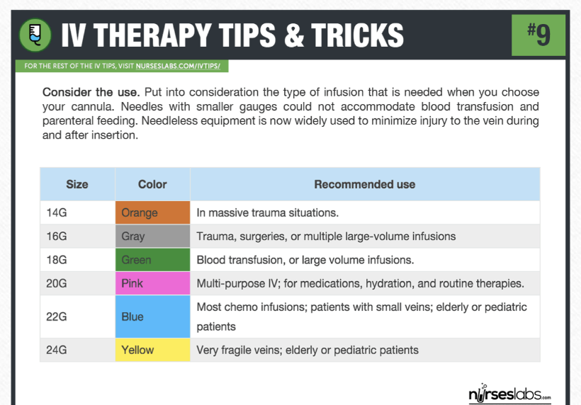
midline catheter
-placed in a peripheral vein of the upper arm; tip ends near axillary vein
-used with poor vein selection or IV needed longer time (2 weeks)
-Ultrasound guided
Do Nots
-use for vesicant meds
-avoid routine, frequent blood draws
-administer incompatible meds simultaneously through double lumen
Starting an IV
-verify order
-prime tubing to prevent air embolism
-tourniquet 2-4 inches above insertion site
-remove tourniquet before flushing (use 10 cc)
-assess for complications
-secure IV to skin
-document
IV documentation
-date+ time
-who inserted
-vein/location
-type of IV cath (gauge, length)
-number of attempts
-patient tolerance
-dressing type
-status (saline lock, med infusing)
-policy on how often to document
Assessing IV site
-observe redness, swelling, bruising, edema, patency, leaking of fluid
-palpate for pain, hardness at site, temperature
-ask if it’s painful
-document findings
-determine if removal is necessary
-assess often
infiltration
→ Non-vesicant IV fluid leaks into surrounding tissue.
Signs: Cool, pale, swollen skin; discomfort; slowed/stopped infusion.
Intervention: Stop the IV, remove the catheter, elevate the extremity, and restart the IV in a different site. Apply warm/cold compress as appropriate for the solution.
extravasation
→ Vesicant medication leaks into surrounding tissue, potentially causing tissue damage/necrosis.
Signs: Burning, pain, swelling, redness, blistering, or tissue damage.
Intervention: STOP the infusion immediately. Do NOT flush. Leave the catheter in place initially to aspirate remaining medication if indicated, then follow the medication-specific extravasation protocol. Notify the provider/pharmacy.
⭐ Remember: Extravasation = STOP + DON'T FLUSH
phlebitis
→ Inflammation of the vein, often from catheter irritation or medication.
Signs: Redness, warmth, tenderness, pain, and a hard/cord-like vein.
Intervention: Stop the infusion and remove the IV. Apply a warm compress as appropriate and restart at a different site if IV therapy is still needed.
thrombophlebitis
→ Inflammation of the vein + blood clot.
Signs: Pain, redness, warmth, swelling, and a firm/cord-like vein.
Intervention: Stop the infusion and remove the IV. Notify the provider; assess for worsening symptoms and follow orders for treatment of the thrombus.
hematoma
→ Blood leaks into surrounding tissue because the vein was punctured or damaged.
Signs: Bruising, discoloration, swelling, and tenderness.
Intervention: Stop the infusion and remove the IV. Apply pressure to the site; use a cold compress initially to reduce bleeding/swelling.
air embolism
→ Air enters the bloodstream and interferes with circulation.
Signs: Sudden dyspnea, chest pain, coughing, confusion, hypotension, or loss of consciousness.
Intervention: Stop the infusion and clamp the line. Place the patient on their left side with the head lowered as directed by protocol, administer oxygen, and notify the provider/rapid response.
IV site infection
→ Microorganisms enter through the IV site, potentially causing local or systemic infection.
Signs: Redness, warmth, swelling, tenderness, drainage, fever, or chills.
Intervention: Stop/remove the IV, notify the provider, and monitor vital signs. Cultures and antibiotics may be ordered.
Catheter related bloodstream infection (CRBSI)
→ A bloodstream infection caused by an IV catheter. This is more serious than a local IV-site infection because the infection has entered the bloodstream.
Signs: Fever, chills, hypotension, tachycardia, redness/drainage at the catheter site, and possible sepsis.
Intervention: Stop/remove the catheter as ordered, notify the provider, obtain blood cultures as ordered, monitor for sepsis, and administer prescribed antibiotics.
⭐ Remember: CRBSI = catheter + bloodstream infection
thrombosis
→ A blood clot forms inside the vein, often due to irritation or damage from the IV catheter.
Signs: Pain, swelling, redness, warmth, and a firm/cord-like vein.
Intervention: Stop the infusion and remove the IV. Notify the provider, assess the extremity, and monitor for complications such as embolism. Treatment may include anticoagulation if prescribed.
⭐ Remember: Thrombosis = clot
venous spasm
→ The vein suddenly contracts or spasms, often in response to irritation from the catheter, medication, or a cold solution.
Signs: Sudden pain, discomfort, resistance when flushing, slowed/stopped IV flow, and the vein may feel tight.
Intervention: Stop the infusion, check the IV site, and assess for infiltration or phlebitis. A warm compress may help relax the vein. Restart the IV at a different site if needed.
⭐ Remember: Spasm = vein tightens → IV won't flow
nerve damage
→ The catheter or needle injures or irritates a nearby nerve.
Signs: Sharp/electric or burning pain, numbness, tingling, weakness, or pain that radiates along the limb.
Intervention: Stop the procedure/infusion immediately and remove the catheter. Assess sensation and movement, notify the provider if symptoms persist, and document the findings.
⭐ Remember: Electric/shooting pain or numbness = think nerve damage
lumen occlusion
→ The IV catheter becomes blocked, preventing fluid or medication from flowing through the lumen.
Signs:
IV will not flush
Resistance when flushing
Infusion slows or stops
Pump may alarm for occlusion
No blood return (depending on catheter type)
Common causes:
Blood clot in the catheter
Medication precipitate
Catheter kinked or positioned incorrectly
Intervention:
→ STOP the infusion and check for mechanical problems (kinks, clamps, positioning).
→ Do NOT forcefully flush because this can damage the catheter or dislodge a clot.
→ If mechanical causes are ruled out, follow facility protocol for an approved catheter-clearing solution (such as a thrombolytic for a clot-related occlusion) and notify the appropriate clinician.
isotonic solutions
What is it?
→ Has a similar osmolarity to blood/plasma, so water stays mostly in the extracellular space rather than causing major fluid shifts into or out of cells.
Main purpose:
→ Expand intravascular volume and increase circulating fluid.
Common examples:
0.9% Normal Saline (NS)
Lactated Ringer's (LR)
D5W ⭐ technically isotonic in the bag, but acts hypotonic after glucose is metabolized
When used:
Hypovolemia/dehydration
Blood loss
Burns
Hypotension
Fluid resuscitation
Perioperative fluid replacement
Careful considerations:
Too much → fluid overload
Monitor for edema, crackles, dyspnea, increased BP
Use caution in heart failure and renal impairment
🧠 Remember:
ISO = same → stays in the bloodstream
hypotonic solutions
What is it?
→ Has a lower osmolarity than blood, so water moves from the extracellular space into cells.
Main purpose:
→ Hydrate cells.
Common examples:
0.45% NS (½ NS)
0.225% NS (¼ NS)
D5W (acts hypotonic after metabolism)
When used:
Hypernatremia (high Na⁺)
Cellular dehydration
Patients who need free water replacement
Careful considerations: ⚠
Can cause cellular swelling
Avoid/use extreme caution with increased ICP or brain injury → cerebral edema can worsen
Can worsen hypotension because fluid leaves the vascular space
Monitor sodium and neurologic status
avoid in trauma/burn victims
🧠 Remember:
HYPO = water goes IN → into the cells
hypertonic solutions
What is it?
→ Has a higher osmolarity than blood, so it pulls water out of cells and into the extracellular/intravascular space.
Main purpose:
→ Increase intravascular volume and pull excess water out of swollen cells.
Common examples:
3% NS
5% NS
D5NS
D5½NS
D10W
When used:
Severe symptomatic hyponatremia
Cerebral edema/increased ICP (particularly hypertonic saline)
Significant cellular dehydration in specific situations
Careful considerations: ⚠⚠
Can cause fluid overload
Can cause hypernatremia
Monitor sodium closely
Monitor neurologic status
Hypertonic saline is often given through a central line depending on concentration/protocol
Correcting sodium too quickly can cause serious neurologic injury
🧠 Remember:
HYPER = pulls water OUT of cells → into the bloodstream
IV additives
What are IV additives?
→ Medications, electrolytes, vitamins, or other substances added to IV fluids to treat or prevent a specific problem.
Examples:
Potassium (KCl)
Magnesium
Calcium
Sodium bicarbonate
Antibiotics
Vitamins
colloids
What are colloids?
→ IV fluids containing large molecules (usually proteins) that stay in the bloodstream and pull water into the intravascular space.
Main purpose:
→ Increase blood volume/intravascular pressure.
Examples
Albumin ⭐
Dextran
Hydroxyethyl starch (less commonly used due to safety concerns)
When are they used?
Hypovolemia when increased intravascular volume is needed
Severe fluid loss in certain situations
Low albumin / selected patients needing oncotic support
How do they work?
Think: Colloids = pull fluid INTO the bloodstream
Their larger particles remain in the vascular space longer than crystalloids, increasing colloid oncotic pressure.
⚠ Nursing Considerations
Monitor for fluid overload
Monitor BP, HR, lung sounds, edema, and I&O
Use caution with heart failure or renal impairment
Monitor for allergic reactions, particularly with some colloid products
Albumin may be used to increase oncotic pressure in specific clinical situations.
IV meds
-have a rapid therapeutic effect but can have adverse reactions
RN must know:
-indications, dosage, contraindications, precautions, dilution, rate of infusion, potential vesicant/irritant, necessary pt monitoring with med