GENETICS MODULE ONE/TWO
 Call Kai
 Learn
Learn Practice Test
 Spaced Repetition
 Match
Match Flashcards
 Knowt Play
1/140

Earn XP
Description and Tags
yes
Name | Mastery | Learn | Test | Matching | Spaced | Call with Kai | Chat |
|---|
No analytics yet
Send a link to your students to track their progress
141 Terms
module one!
revision:
DNA(template strand/anti sense is used) to make mrna is read as codons where anticodon on tuna makes proteins by combining amino acids
5’->3’ or also called N-terminus to c-terminus
Eukaryotic has methymini UAC
Protein-E-P-A, initiation, elongation, termination
The catalytic activity of the ribosomes is done by the rRNA not the protein
• ⁃ Trna: aminoacyl-tRNA-synthetase
Transfer RNAs (tRNAS) and the adaptor hypothesis and its structural shape and ISOACCEPTORS:
The adaptor molecule is the tuna, it is the adaptor between mrna codons and amino acids
Trna anticodon base-pairs antiparallel with the mrna codon
It has a ‘cloverleaf’ secondary structure
Amino acids covalently attached (esterified) to the 3’terminal adenosine (CCA end), so think of it like the 3’ end of every tuna ends in cca and the amino acid attaches to the last A. This is OF THE TRNA so what make it.
Esterified means when tRNA (empty) uses an atp to attach an amino acid onto it
• ⁃ Each amino acid has one or more tRNA genes that encodes tRNAS used specifically for the amino acid (isoacceptors). So many codons make one amino acid
ISOACCEPTORS:
A different tRNA that carries the same amino acid
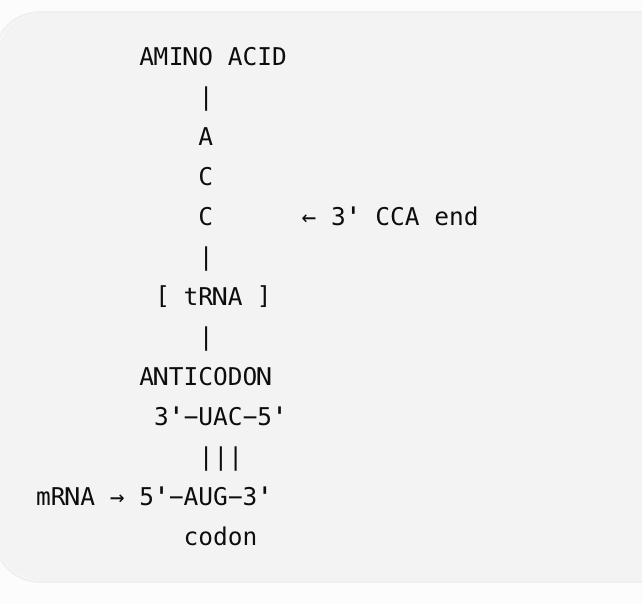
tRNA specificity there are two levels:
1. Aminoacylation = putting the correct amino acid onto the correct tRNA
Before tRNA can go to the ribosome, it needs to be loaded (charged) with an amino acid.
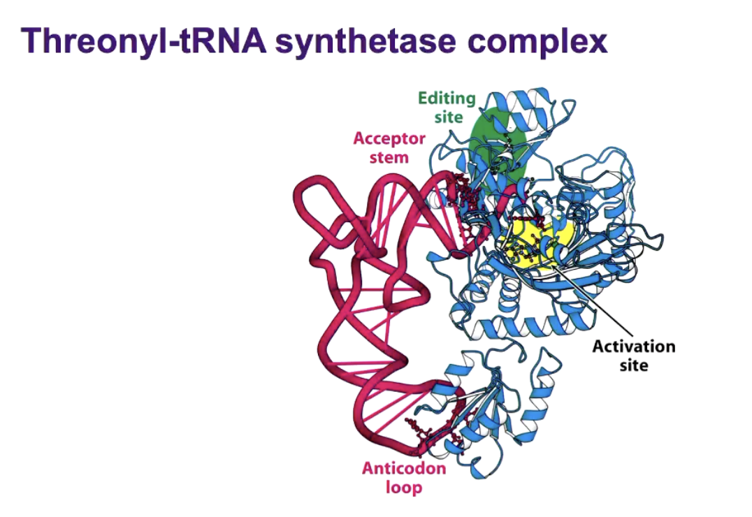
An enzyme called aminoacyl-tRNA synthetase does this.
There are generally 20 types of aminoacyl-tRNA synthetases, one corresponding to each standard amino acid.
For example:
Alanine → alanyl-tRNA synthetase → alanine tRNA
The synthetase has to recognise TWO things:
Correct amino acid
       ↓
aminoacyl-tRNA synthetase
       ↓
Correct tRNA
       ↓
tRNA—AMINO ACID
How does it know which tRNA is correct?
It recognises specific identity elements in the tRNA's structure. These can include:
the anticodon
bases in/near the acceptor stem
other characteristic bases/structural features
Then, using ATP, it attaches the amino acid to the 3′ CCA end of the tRNA.
So:
Aminoacyl-tRNA synthetase makes sure the correct amino acid is attached to the correct tRNA.
2. Decoding = putting that tRNA at the correct mRNA codon
Now imagine the tRNA has already been loaded:
       ALANINE
          |
       3′ CCA
          |
        tRNA
          |
      ANTICODON
The tRNA enters the ribosome.
Its anticodon base-pairs with the complementary mRNA codon.
For example:
mRNA: 5′- G C U -3′
| | |
tRNA anticodon: 3′- C G A -5′
|
ALANINE
GCU codes for alanine.
Therefore, when this tRNA's anticodon matches GCU, the ribosome accepts the tRNA and alanine gets added to the growing protein.
So there are TWO different types of specificity:
Aminoacylation:
"Does this tRNA have the correct amino acid attached?"
Controlled by → aminoacyl-tRNA synthetase
Decoding:
"Is this tRNA matching the correct mRNA codon?"
Controlled by → codon–anticodon base pairing in the ribosome
So the full process is:
amino acid → loaded onto correct tRNA → tRNA anticodon finds matching mRNA codon → ribosome incorporates that amino acid into the protein
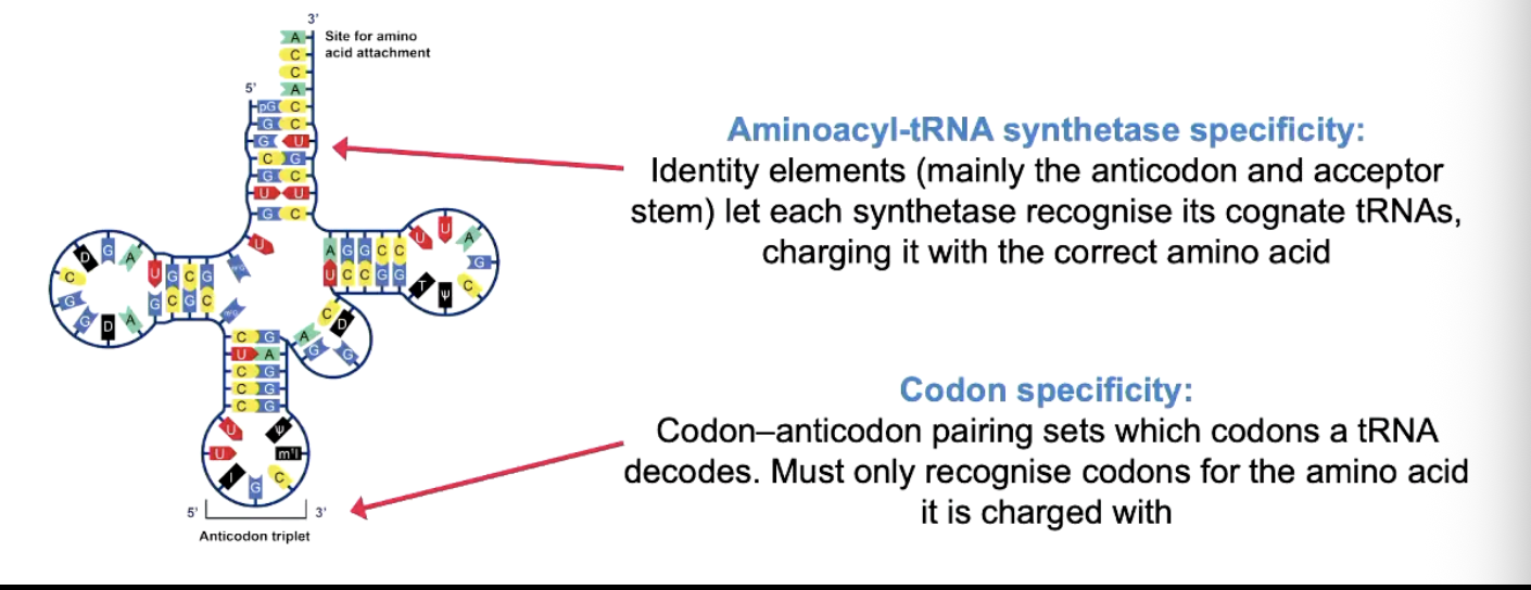
anticodon loop, draw a diagram
Remember 5’ to 3’
Interact through Hydrogen bonds
3rd base in codon is recognised by 1st base in anticodon
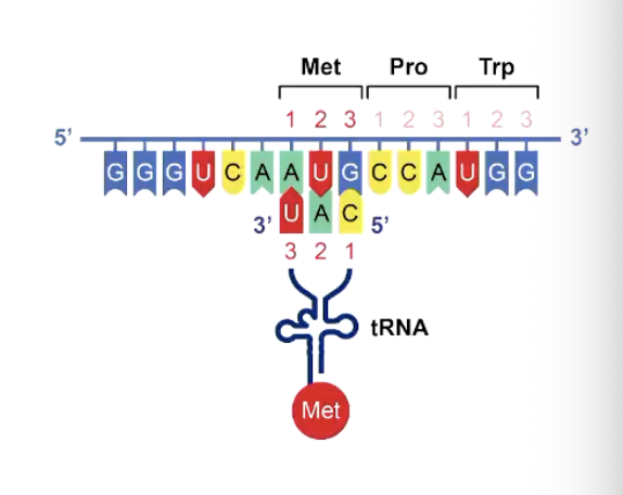
ABNORMAL BASES IN TRNAS:
Trnas have many abnormal bases/nucleotides that are created via post-transcriptional modification
Modified bases allows TRNA to be more flexible of function then only being able to use g,c,a,u
There are 80 known ones
And the best known one is INOSINE (I)
This is created by deamination of adenine into hypoxanthine
Forms hydrogen bonds with C,A, and U but not G (H, idk y h but its just to say not g…), this is important when in position 34 of a TRNA explain later…
Terriary TRNA STRUCTURE:
A common l shaped structure enables TRNAS to fit into ribosomes
• ⁃ Modified bases assist tertiary structure formation

HOW DID THEY GET EVIDENCE OF TRIPLET CODE??? as in that three account of one amino acid, In vitro transition studies
first s cientists know that dna→mrna→ protein
and then mrna told ribosome which amino acids to add.
But they didn't yet know: which 3-base codon = which amino acid?
The first evidence was trinucleotide binding of aminoacly-trnas to ribosomes
they did the bottom experiment when a short rna of only three cnucleotides ro one codon, so they kno whwat codon codes for what. they just put a lot of trna’s in it to see which will go to it.
The second was the cell-free (in vitro) protein synthesis experiment by nierenberg and matthael; khorana et al):
They wanted to make proteins outside a living cell, so they could control every ingredient and test their ideas.
They know protein-making needs: amino acids, tuna, ribosomes, energy (ATP/GTP), and an mRNA “instructions” template.
They did this:
Smashed open e-coli bacteria using an abrasive powder to release everything inside
Spin it in a centrifuge to remove solid junk like broken cell walls
Keep the leftover liquid (cell lysate), which sill has trns, ribosomes, and enzymes floating in it
They added an enzyme (DNase) to destroy any DNA so that the cell can’t make its own new RNA this was only the mRNA the scientists add will be used. Stops de novo RNA synthesis (which is when an rna ploymerase builds a new rna strand directly from indivisual nucleotide building blocks without a preesisiting primer
And any leftover natural RNA breaks down on its own cause RNA doesn’t last long
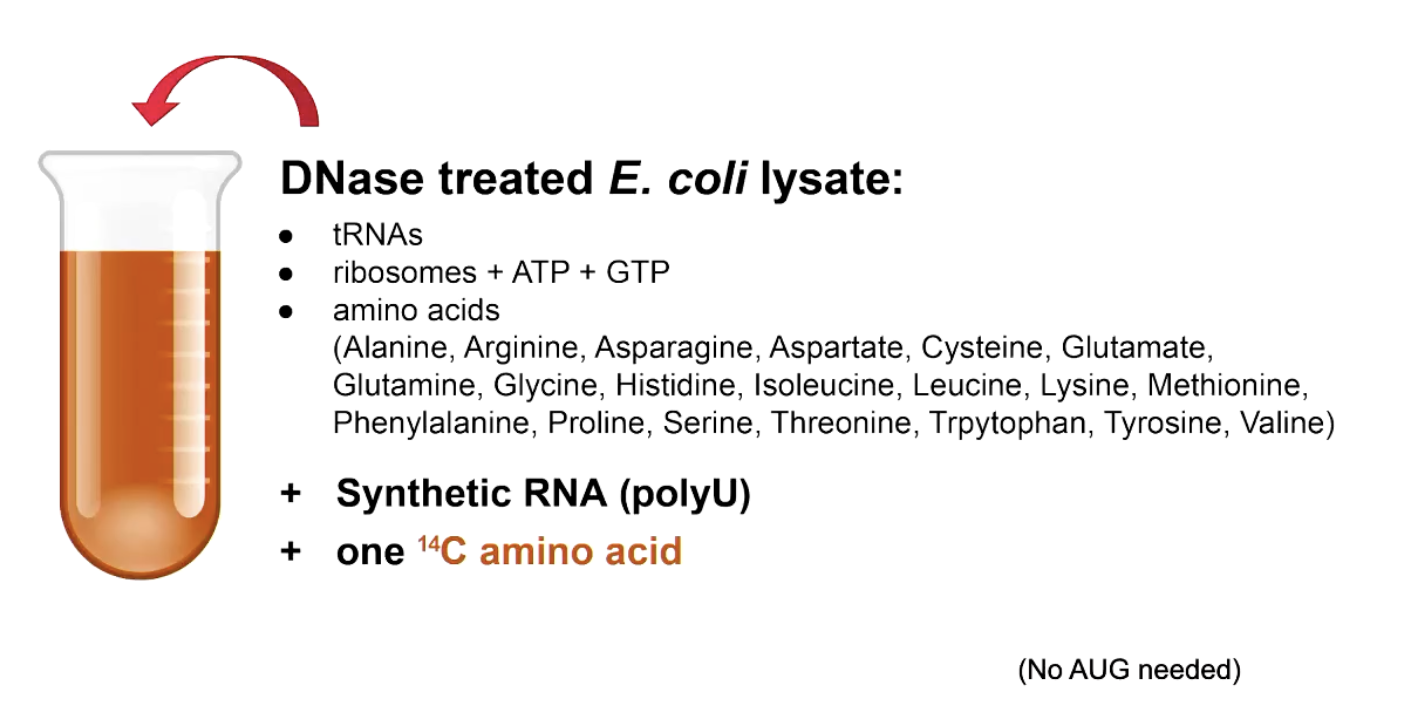
Now they added their own synthetic MRNA sequence, plus amino acids which one of is radioactively labeled (14C) so they can track it. so they did this experiment many times adding phenylalanine then alanine ttcetc and which ever one showed up uuu is what codes for it
• ⁃ For the first experiment they used this for was poly(U) RNA template (UUUU) where it had all 20 amino acids plus one 14C radioactively labeled. 20 seperate experiments conducted in parallel
so that helped find codon+trna used.

results
have one that worked and one that didnt
so basically the mrna only takes the right amino acid which are the ones we added in. hense if not radioactive then it wasnt used
Left side
The 14C is attached to alanine
Poly-u MRNA gets fed into the ribosome
The ribosome builds a protein but it turns out to be PHE, PHE, PHE all phenylalanine
Since no alanine was used at all, the radioactive tag never shows up in the final protein
So the purified protein is not radioactive which means poly-u does not code for alanine
Right side
• ⁃ Proteins made the the the and since phenylalanine is the labelled one every one made has a radioactive tag so this does code for it

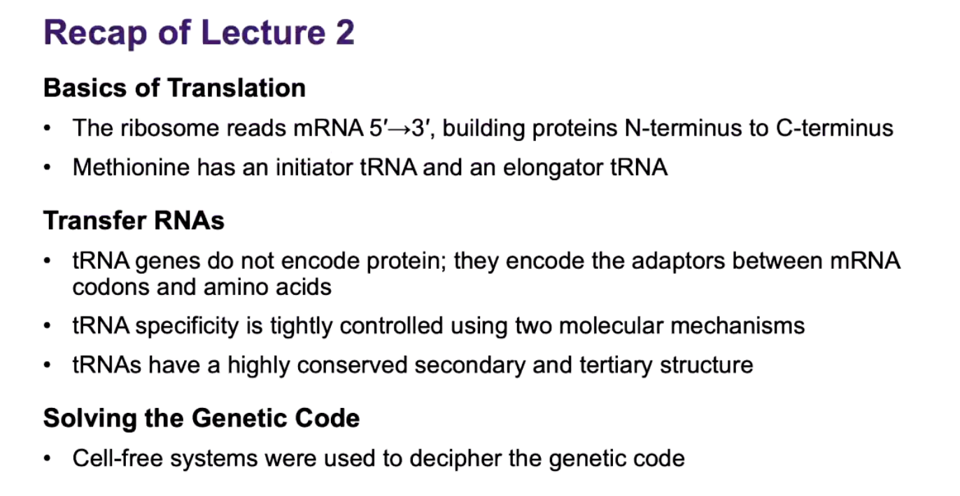
lecture two recap
nitiator tRNA = "I'm bringing Met to START the protein."
Elongator tRNA = "I'm bringing Met to ADD to an already-growing protein."
ADDING ONTO TO EXPERIMENT BUT THEN WHAT ABOUT THOSE WHICH ARE GUGUGUGU ETCETCETC

ac question
You dont know where it starts, at the g? Or the u?
So firstly you narrow it down to two options, so either valine (GUG OR UGU) or cysteine (GUG or UGU), do the three reading frames way
Then you try different repeating sequences with there codons instead gugguggug or ugguggugg or ggugguggu etc. valine showed up (GUG,UGG,GGU) AND TRYPTOPHAN (GUG,UGG,GGU) AND GLYCINE (GUG, UGG, GGU), you can just choose one gugguggug etc cause when u do the three reading frames it takes them all into account
Then if you combine the two.
Experiment one: GUG (valine r systeine)
Experiment two: valine, try, glycine
• ⁃ Only similar is valine so it has to be that
prac quesiton


• ⁃ It is using RNA and not DNA because the experiments during this time only knew it came from RNA
nswer should look liek this:

Initiation and termination codon:
Initiation start codon: for eukarAUG which codes for met but for prokay its fmet for other species
Termination stop codon: UAA, UAG, UGA
DEGENERACY:
Most amino acids are specified by 2-4 codons, and most degeneracy is in the 3rd base of the codon
Deviations from the standard genetic code:
In human mitochondria, AUG, AUA and AUU can all act as start codons. UGA encodes tryptophan instead of stop. AUA encodes methionine instead of isoleucine and AGA and AGG encode stop instead of arginine THIS ISNT MARKED!!!! THE OTHER TWO ARE
Prokaryotes use GUG or UUG too as an alternative for AUG doe fmet, but only at start
CUG encodes serine instead of leucine in candida cylindracea and some other candida species (Candida albicans)
WOBBLE:
Each amino acid has its only TRNA GENE
Standard genetic code has 64 codons
• ⁃ 61 of these specify the 20 amino acids (the other r stop/start). Does this mean that there must be 61 TRNA genes, no. Some TRNA can do many different codons

hypothesis:
THE WOBBLE HYPOTHESIS:
By Francis crick in 1966 to explain how one TRNA molecule could recognise one degenerate codon
The hypothesis: the base at the 5’ end of the anticodon (position 34) can wobble’ meaning can be different codons (explained better below)
• ⁃ Extra base-pair options compared to standard A—U AND G—C

consequences
WOBBLE CONSQEUNCES:
Theoretical minimum of 32 tuna species to read 61 CODONS (31 unique anticodons + 1 initiator TRNA)
E.COLI: 41 anticodons and around 45 TRNA species
mitochondria: 22 TRNA species; super wobble lets U34 sometimes pair with A,G,C,U
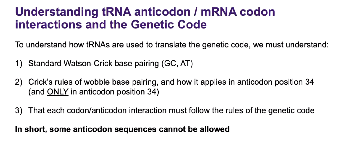
This is limited by the fact that TRNAS must only recognise codons for a single amino acid, otherwise translational fidelity would be compromised EXPLAINED BELOW
EXAMPLE OF BAD PART OF WOBBLE: REMEMBER TO LOOK AT THE WHOEL BOX!!!

The bottom ones can’t happen as it represents another amino acid!!!!
in exma:

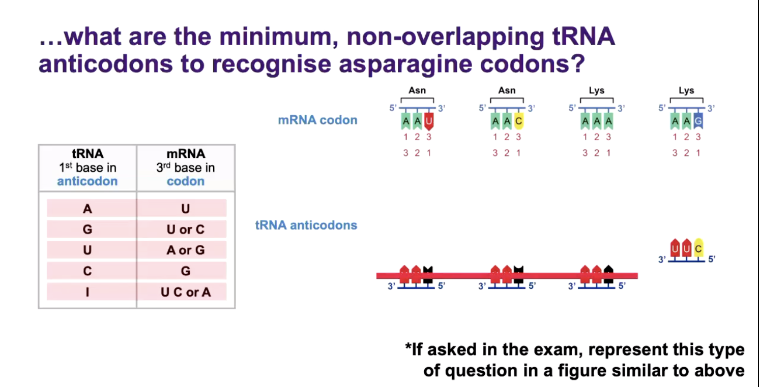
EXAMPLE OF WOBBLE:

things u need to know:
DNA STRANDS NAMING:
TOP STRAND OF DNA IS USUALLY SHOWN (CODING/non-template/crick/sense/forward STRAND) 5’ TO 3’
The other strand is called noncoding/reverse/antisense/watson/template, BOTTOM IS USED TO MAKE MRNA CAUSE IT TECHNICALLY IS A CODING STRAND
recap of lecture 3


READING FRAME:
Refers to the position at which you start reading nucleic acid in 3 nucleotide codons
If you start at the first nucleotide, that is reading frame +1, reading frame +2 starts at nucleotide 2 and reading frame +3 at nucleotidee 3, etcetcecet
• ⁃ As mrna is single stranded it has three reading frames which dan has 6
OPEN READING FRAME (ORF):
- traditional definition: a region in any reading frame that begins with a start codon (ATG) and ends with a stop codon
DNA start codon = ATG
If transcribed an ORF might be translate to produce a protein
IN EXAM!!!!!:
N-terminal amino acid means (fmet or met), which one? The one WITHOUT STOP!! If it said the ENITRE open reading frame then the one w start and stop
• ⁃ Base substitution (nonsense mutation), shorted

words to know:

MUTATION:
It is a change in the genetic material and the process producing it
Heritable at cell division; only offspring-founding lineages transmit to next gen
Ultimate source of new varitation for evolution
Recombination rearranges genetic variability into new combination
Natural selection raises the frequency of fitter variants in the current environment
MUTANT:
An organism with a non-wild-type genotype (and often phenotype)
Wild type (WT) = the standard/reference form normally used for comparison.
Mutant = has a genetic change compared with WT.
The mutation may or may not change the observable phenotype.
MUTATION IS INEVITABLE: two different ways DNA can get damaged spontaneously
Spontaneous cytosine deamination (C loses a amino group turning it into a U)-> uracil (U:G mismatch, cause u doesn’t normally belong under DNA)
so U SINT IN DNA BUT SOEMTIMES A C CAN TURN INTO U WHICH THEN WHEN TURNED INTO MRNA IS AN A→T
Errors occur during replication (unexcused uracil pairs with A) and repair. So c-g turns into u-g which replicates into a mutation of t-a (BASE SUBSTITUTION MUTATION)
Spontaneous hydrolysis of bond linking purines (G and A) to deoxyribose
Purines are a and g and normally these are attached to the deoxyribose sugar
A/G-SUGAR-PHOSPHATE
Sometimes the bond connecting the purine base to the sugar breaks spontaneously so the a or g is literally lost form the dan so now you have
Sugar-no base- sugar this is called an apurinic site
It is estimated 10,000 purines lost/day/human, and substitutions can occur when these uninformative apurinic sites are replicated
Since it is blank it may insert a incorrect nucleotide (substitution mutation)
REMEMBER DNA IS NOT TOALLY CHEMICALLY STABLE, PROOFREADING AND REPAIR MACHANISMS WILL REPAIRS MOST MISTAKES BUT NOT ALL. ALSO FIDELITY OF REPLICAITON IS NOT 100%
• ⁃ mutations are inevitable, mutagens just increase the frequency
FOUR TYPES OF MUTATIONS:
Spontaneous
Induced
Germinal
Somatic
Spontaneous:
Spontaneous: arise endogenously through replication errors, the intrinsic chemical instability of dan, OXIDATIVE DAMGE ETC
WE TALKED ABT TI ABOVE, it is internal/natural DNA damage
INDUCED:
ARISE WHEN LESIONS FROM MUTAGENS ESCAPE CORRECT REPAIR
Caused by something external
Uv radiation chemical etcetce
lesion= dna damage
Usually is repaired but sometimes isn’t
Mutagen->dna dmage-> dna repair-> fixed
Mutagen-> dan besoin-> Dailed to repair-> mutation
• ⁃ GERMINALL
OCCUR IN GERM-CELL PRECURSORS AND WILL BE TRANSMITTED THROUGH DERIVED GAMETES; ALL OFFSPRING WILL THEN CARRY THEM
This is where the mutation occurs
Occurs in germ-cell precursor, the cells which will produce sperm or eggs
Parents gremlin mutation-> gamete-> child-> mutation int he child’s cells
SOMATIC:
SOMATIC: occur in somatic cells; confined to the clone descended from that cell, not transmitted to offspring, but can drive cancer.
Normal body cells
Normal skin cell-> mutation-> ,utaed skin cell-> cell division-> Manu descendant cells
Can cause cancer
Call -> mutation0> uncontrolled divsiion-> cancer/tumour
PRE-EXISITING MUTANTS: THE EXPERIMENT
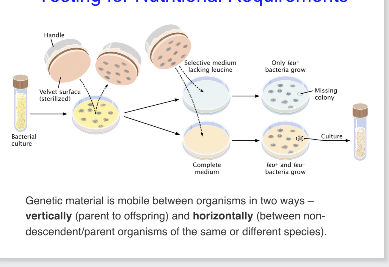
Replica plating was used to identify the appearance f resistance in proveiously anti-biotic-sensitive Esherichia coli prior to treatment with the drug
Stress does not direct which mutation occurs, it selects among variants already present
Start with culture: a bacteria culture (no streptomycin (antibiotic) exposure yet) is grown from a liquid tube
Master plate: the bacteria are spread onto plate with no streptomycin, so colonies grow in fixed, identifiable positions. A valvet-covered taped is pressed onto this ‘master plate’ the fine hairs of the velvet pick up cells from each colony like a rubber stamp
Cells stick to the velvet: because the velvet touched every colony in the exact same spatial layout, it now carries a ‘fingerprint’ copy of the last plates colony pattern
Replica stamping: the velvet is pressed onto new plates: one containing streptomycin, one without. Since the velvet preserves the spatial arrangement, each new plate ends up with colonies in the same positions as the original master plate.
Selection reveals pre-existing resistance: On the streptomycin plate, almost all colonies die, except one or two that happen to carry a resistance mutation. Critically, when researchers go back to the master plate (which was never exposed to streptomycin) and pick cells from that exact same spot, those cells are also resistant.
So what does that prove? The resistance wasn't caused by the antibiotic — it was already there, sitting in the population by random chance (mutation), before the antibiotic ever showed up. The antibiotic didn't create resistant bacteria; it just killed off everything that wasn't already resistant, revealing the ones that were.

STARCVATION, SLECTION, AND MUTAITON IN MIRCOBOES: DRAW A GRAPH AND LABEL
This is in a flask or plate with minted nutrients:
Lag phase: Bacteria are just settling in, adjusting to their environment. Population size is flat — barely any division happening yet.
Log phase: Explosive growth. Cells are dividing rapidly, doubling again and again, food is abundant. This is the steep upward curve.
Stationary phase: Growth stalls out and flattens. Nutrients are running low, waste products are building up, and the environment is getting stressful. New cell division roughly equals cell death, so the population size plateaus
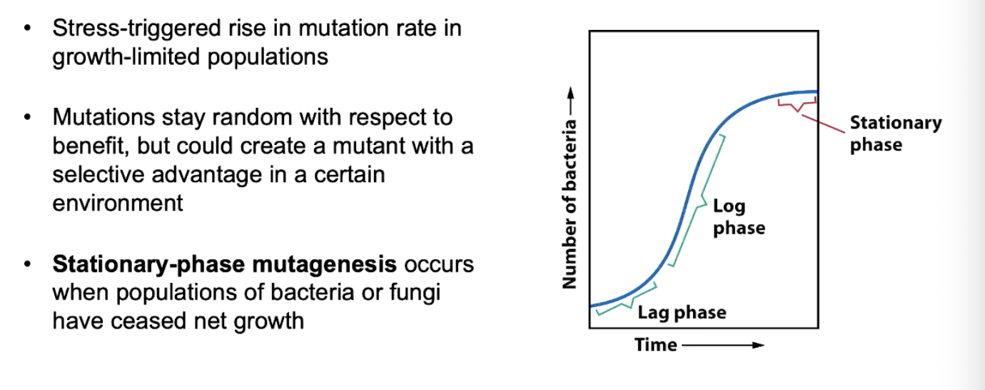
Here's the twist: in that stressful, nutrient-starved stationary phase, bacteria (and fungi) actually start mutating faster than normal. This is sometimes called "stress-induced mutagenesis."
Why is this interesting/important? It might sound like it contradicts the Lederberg experiment (where mutations were shown to happen randomly, independent of the stress/selection agent). But look closely at the second bullet — it clarifies that this isn't a contradiction:
"Mutations stay random with respect to benefit, but could create a mutant with a selective advantage in a certain environment."
This is the key distinction:
The rate of mutation can go up under stress (more mutations happening overall when times are tough).
• • But which mutations occur is still random — the stress doesn't "aim" the mutations toward a useful outcome. It's not that stress directs bacteria to evolve resistance specifically. It just cranks up the overall mutation dial, and by chance, some of those random mutations might happen to be useful in that environment
STRESS EFFECTS HOW MUCH NOT WHAT!!
REVERTING A MUTANT PHENOTYPE:
Forward mutation: wilf-type to mutant allele
Reverse mutation (reversion): a secondary mutation which restores wild-type phenotypes
Back mutation: restores the wild-type sequence
Suppressor mutation: compensating change elsewhere; first mutation persists
vocab

MULTIGENIC CONTROL OF PHENOTYPES: how to stop phenotypes from happening bad and good

CONDITIONAL LETEHAL MUTATIONS: permissive vs restrictive AND NEXT THREE SLIDES ARE TYPE OF CONDITION MUTANTS
Conditional lethal mutations: a normal mutation might make a cell unable to survive all the time/A conditional lethal mutation is a mutation that is only lethal under certain conditions.
Two conditions: permissive and restrictive. Permissive is the mutation is okay and cell survives//grows
restrictive condition is mutation causes a problem and cell dies/cannot grow
no growth or death in restrictive conditions
Viable in permissive conditions
Grown under the permissive condition, then shifted to stricture conditions to reveal the phenotype
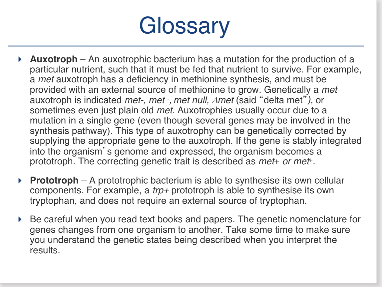
• ⁃ Auxotophes: vs proto
Auxotophes:cannot make an essential metabolite that prototrophs synthesise ; they grow only if it is supplied, so minimal medium is restrictive
An auxotroph has a mutation that means it cannot make an essential substance itself.
For example, imagine a mutant bacterium cannot make amino acid X.
So for an auxotroph:
X supplied = permissive
X not supplied = restrictive
• ⁃ Temperature-
Temperature- sensitive mutants: grow only at permissive temperatures
25°C → 🟢 grows
37°C → 🔴 doesn't grow
• ⁃ The lower temperature might be the permissive condition, while the higher temperature is the restrictive condition.
Suppressor
Suppressor- sensitive mutants are viable only when a second genetic factor (a suppressor) is present
Mutation A → causes lethal problem ❌
But:
Mutation A + suppressor mutation → problem is compensated → 🟢 survives
Therefore:
Suppressor present → permissive → survives
Suppressor absent → restrictive → dies
4 main types of mutations

wildtpye: IS THE original one
Substation : one is changed
Inversion-; flipped aourd 180
Insertion: put in
• ⁃ Deletion: taken out
mutation types again
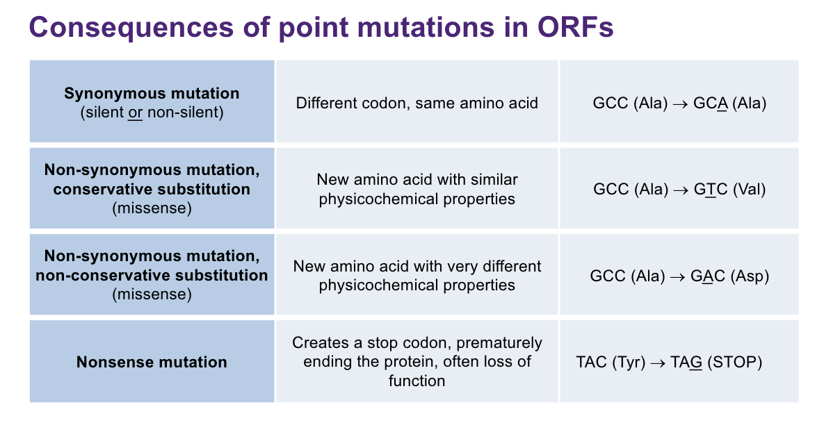
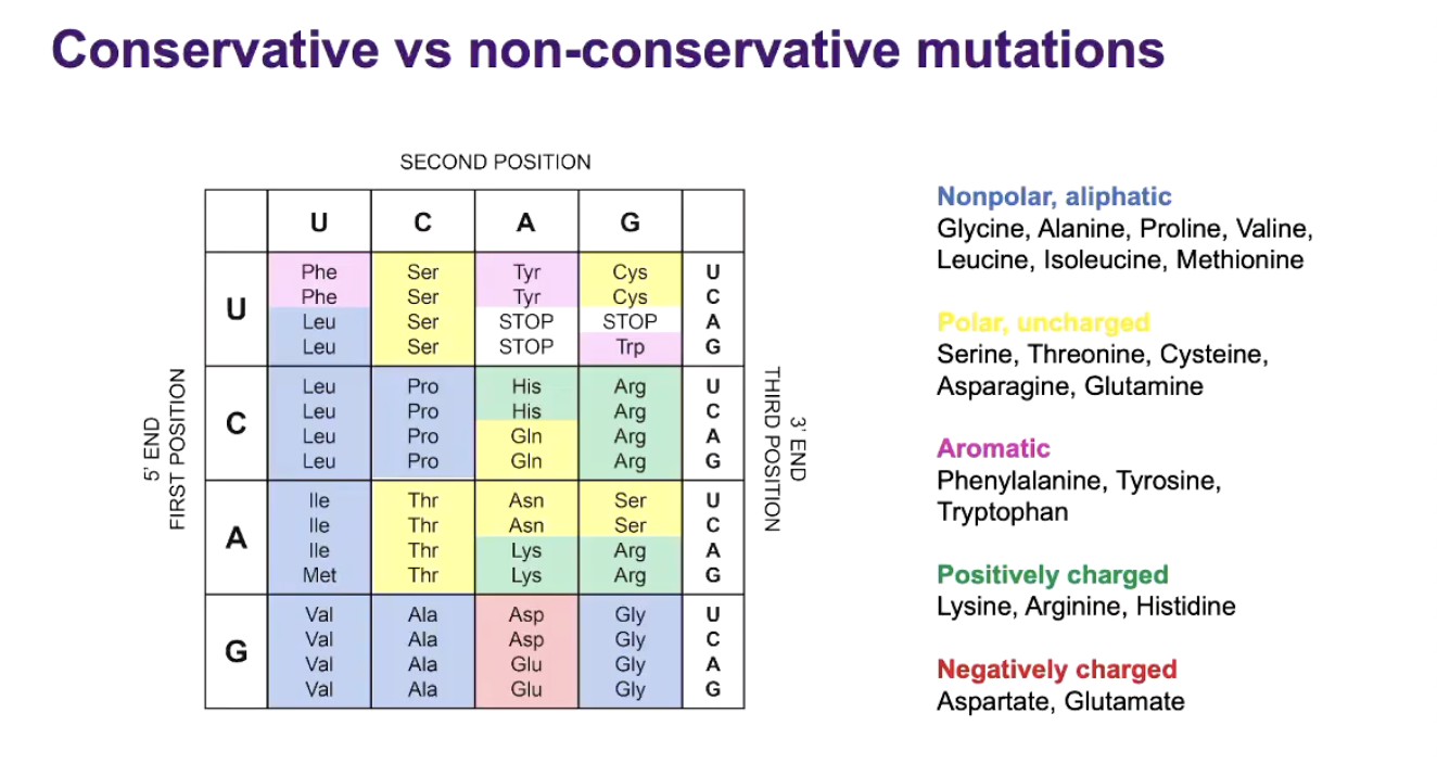
HOW TO EXPLAINC ONSERVATIVE VS NONE CONCERVATION:amino acids


review lectyre 4

TAUTOMERIC SHIFTS AFFACT: TYPE OF MUTATION
SPONETEOUS BASICALLY HYDROGEN ATOM BECOES WEIRD FORM AND BECOMES A DIFFERENT TAUTOMER, LIKE THE G BECOMES WEIRD SO IT CAN NOW PAIR W A now it is a t-a pair etc. THIS IS ISOMERISATION
This shift is when a hydrogen atom temporarily moves to a different position within a dan base
Cause the doubles bonds/electrons to change
So it become a different tautomer (different form of same molecule)
Means c can also pair w a etcetc which when replicated could turn into a mutaition of t-a
Isomerisation (which it changes to a similar one) caused by a reversible change in the location of hydrogen atom in a base
• ⁃ By modifying the pairing of nucleotides, tautomeric shift can cause substitution mutation

TYPES OF SUBSTITUTION: base substation where ne dan base is replaced by another, two types
transitions:
Pyrimidine-> pyrimidine
Purine-> purine
TRANSVERSIONS:
Pyrimindine->purine
Purine->pyrimidine
Although there are 8 possible transversions and only 4 possible transitions, transitions are more common!!
Transitions
There are only two possible same-group swaps:
A ↔ G
C ↔ T
That's 4 possible directional substitutions if you count each direction:
A → G
G → A
C → T
T → C
= 4 transitions
Transversions
Each purine can change into either of the two pyrimidines:
A → C
A → T
G → C
G → T
And each pyrimidine can change into either purine:
C → A
C → G
T → A
T → G
= 8 transversions
So:
4 transitions
8 transversions

core concepts

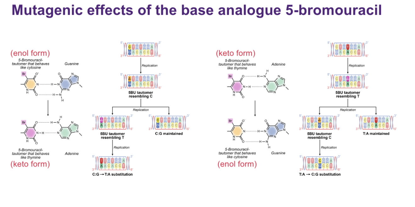
mutagens 5-BROMOURACIL
MUTAGENS: a base ANALOUGE (structure if like a normal dan base so it is mistaken as one) called 5-BROMOURACIL (chemical mutagen)
Turns in keto and enrol form
Keto form likes to pair with A (acts as t)
ENOL with G (acts as c)
• ⁃ When replicated it can switch into the other form meaning c-g turns into a-t which isn’t good
NIRTROUS ACID
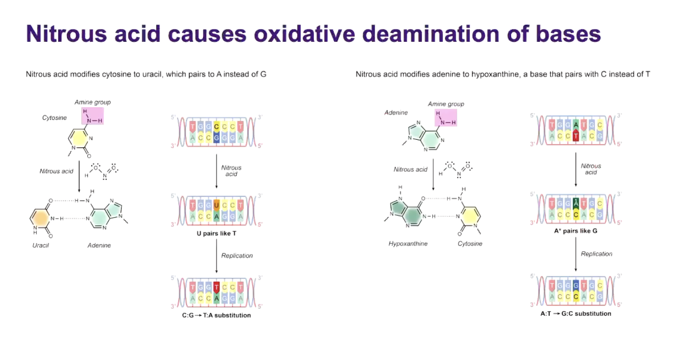
CAUSES OXIDATIVE DEAMINATION OF BASES:
Deamination is removing an amine group (-NH2) from a base
This acid does this, converting on base into a different one
Left side:
Cytosine turns into uracil
Uracil acts like thymine, meaning it pairs with a not guanine so in the next replication the c:G will turns into t:a
Right:
Adenine turns to hypoxanthine
Adenine also has an amine group that nitrous acid can remove turns into hypo.
• ⁃ So you get h:C but not he next replication Hypo acts like G directs a c to be inserted so the A:T pair becomes a G:C
Mutagenesis by ultraviolet radiation
Uv light can react with cytosine to form a photo hydrate, becomes chemically unstable and prone to deamination
Converts to uracil (like nitrous acid)
C:g-> t:a
Cytosine photo hydrate can deanimate to uracil, giving C->T transition
UV light hits two adjacent pyrimidines on the same DNA strand, that means nay of the TT,TC,CT,CC in sequence can cause a abnormal covalent bonds to form between each other making a cyclobutane pyrimidine dimer (CPD).This is the big one.
Pyrimidines are C and T.
So if two pyrimidines are next to each other on the SAME DNA strand, such as:
TTTCCTCC
UV can cause abnormal covalent bonds to form between them.
If the damage isn't repaired, the cell may use special translesion synthesis (TLS) polymerases.
These polymerases can:
replicate ACROSS the damaged site
which lets replication continue.
But some TLS polymerases are less accurate than normal replicative DNA polymerases, so there's a greater chance of inserting the wrong nucleotide opposite the damaged bases → mutation.
Two neighbouring bases get chemically welded together sideways forming a little four-sided ring.
Normal dna polymerase can’t read through the leision, to get past it translation synthesis polymerase are used these are sloppier and error prone polymerase. They often in sort wrong bases which is a big issue
• ⁃ Adjacent pyridines (TT,TC,CT,CC) form cyclobutane dimers that stall replicative polymerases and force translesion synthesis

INTERCALATION OF AN ACRIDINE DYE CAUSE FRAMESHIFT MUTATIONS:
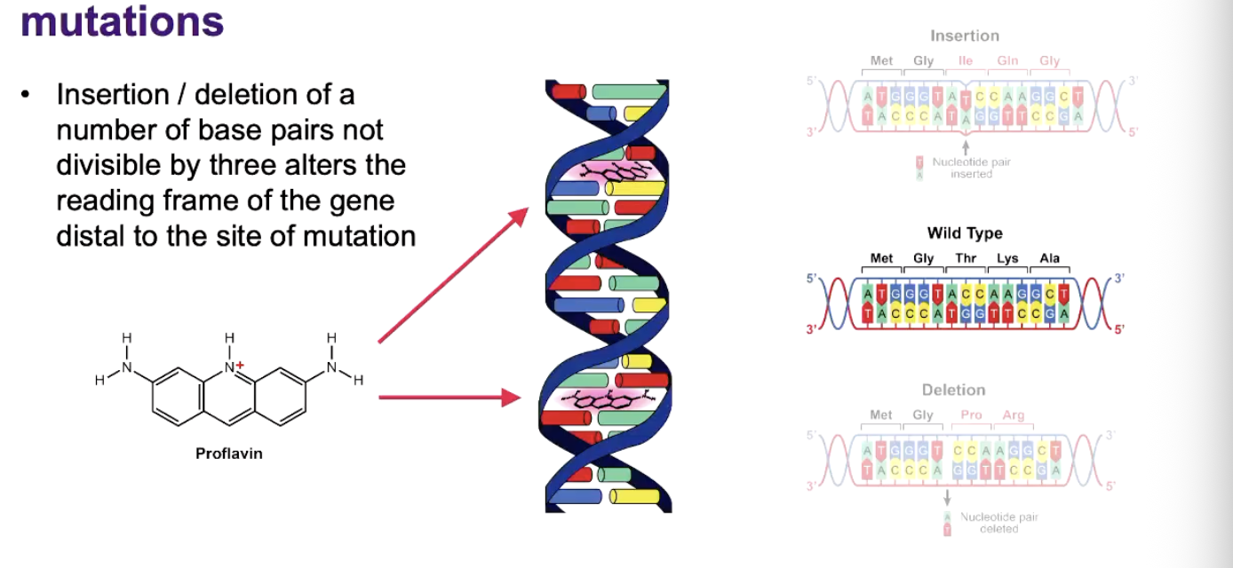
PROFLAVIN IS AN EXAMPLE:
The key is: proflavin itself doesn't become a DNA base. Instead, by sitting between bases, it can cause DNA polymerase to insert an extra nucleotide or skip/delete a nucleotide during replication.
So:
Proflavin → intercalates between bases → distorts DNA → replication error → insertion or deletion
Why does that cause a frameshift?
Remember that mRNA is read 3 bases at a time:
ATG | AAA | CCG | TTT
If you insert ONE base:
ATG | TAA | ACC | GTT | T...
Everything after the insertion is now grouped differently.
That's a frameshift.
Sightly positively charged size, which can slip in the middle of the bases as shown in photo. Influxes the helices, it leads to the addition and deletion of the bases-> changes reading frame. This happens when unwinding, it gets int he way and gets rid or adds bases
• ⁃ Insertion/deletion of a number of base pairs not divisible by three alters the reading frame of he gene distal to the site of mutation
HOW INSERTIONS AND DELETIONS (INDELS) AFFECT PHENOTYPE:
• ⁃ They cause frameshift mutations
Consider a single insertion in an ORF:
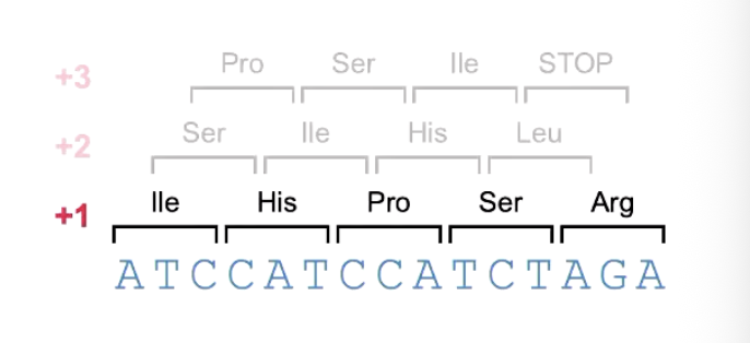

CALLED FRAMESHIFT MUTATIONS
Occur only in ORF’S
Usually give nonsense mutation (premature stop)
Usually encode nonsense proteins
EFFECT OF IONISING RADIATION, HYDROXYLAMINE AND ALKYLATING AGENTS
1. Ionising radiation
Ionising radiation has enough energy to remove electrons from molecules, which can severely damage DNA.
One major consequence is double-strand breaks:
DNA ========   ========
              ↑
        both strands broken
The cell tries to repair the break, but if the pieces are rejoined incorrectly, you can get:
Deletion → DNA piece is lost
Inversion → DNA piece is put back flipped
Translocation → DNA piece attaches somewhere else, often another chromosome
So:
Ionising radiation → DNA breaks → imperfect repair → large chromosome changes
2. Hydroxylamine
Hydroxylamine is a chemical mutagen that modifies cytosine.
Normally:
C pairs with G
After hydroxylamine modifies C → N4-hydroxycytosine, the modified C can mispair with A.
After replication, this can become:
G:C → A:T
That's a transition because C→T and G→A stay within their respective base classes.
So memorize:
Hydroxylamine → modifies C → G:C → A:T transition
3. Alkylating agents
These chemicals add alkyl groups onto DNA bases.
That can change how bases pair or damage DNA more extensively, so they can cause:
transitions, transversions, and chromosome aberrations.
So they're less specific than hydroxylamine.
Ionising radiation causes double stranded breaks, repair can cause deletions, inversions and translocations.
It has enough energy to knock electrons out of molecules, comes broken. Can try to repair it but it isn’t always perfect
Hydroxyl amine converts cytosine to N4-hydroxycytosine, pairing with A, results in G:C-> A:T transitions
Chemical mutagen that specifically modifies cytosine (C) so again G:C->A:T
Alkylating agents are chemicals that donate alkyl groups to other molecules, inducing transitions, transversions, and gross chromosome aberrations
Are chemicals that add an alkyl group to DNA bases
• ⁃ Transitions/transversions or Chromosome aberrations (changes in number of physical structure of chromosomes
SUPPRESSOR MUTATIONS: definitions and how it was discovered?
Definition:
When a second mutation event masks the deleterious effects of a first mutation
Discovery: from studies in bacterial genetics
means it can make leucine by itself
Start with wild-type bacteria that are Leu+
Leu+ means they can make leucine themselves.
Mutate the Leu+ bacteria and select a mutant that is Leu-
Leu- means it cannot make leucine itself.
This is an auxotroph.
Grow the Leu- mutant on medium containing leucine
It needs leucine supplied because it cannot produce its own.
Mutate the Leu- cells again
Plate the cells on medium without leucine
Most Leu- cells cannot grow
But if a second mutation restores the ability to grow, that cell now has a Leu+ phenotype
Select the Leu+ colonies that appear
These are called revertants because their phenotype has returned to Leu+.
Sequence/compare the relevant genes in:
wild-type Leu+
first mutant Leu-
second mutant/revertant Leu+
Compare the sequences:
If the original mutation has been corrected/gone → reversion/back mutation
If the original mutation is still there, but there is a second mutation that compensates for it → suppressor mutation
So the key sequence is:
Leu+ → first mutation → Leu- → second mutation → Leu+ phenotype again
And the definition:
Suppressor mutation = a second mutation that masks or compensates for the harmful effect of the first mutation, while the original mutation remains.
What can happen:

Intragenic:
Intragenic: means instead of fixing the one basic codon aadd another acidic somewhere else to cancel it

INTRAGENIC SUPESSORE: T4 rIIB
When a second mutagen happens at a different site in the same gene
Classical genetic evidence fro the triplet code:
Proflavin used to induce frameshift in phage TA rIIB
RiiB function function scored by plaque assay- if inavtictaed, cant grow on esceriachia coli K12(game)
After successive mutational events, it was found that gain or loss of 1 or 2 bases inactive rIIB but 3 couldd store funcition
Genetic code must be a triplet (or a multiple of 3), non-overlapping, read from a fixed starting point
One taken out and reading frame restored: works well if the codes still code for some similar
Classical genetic evidence for the triplet code: Proflavin, an intercalating mutagen, was used to induce frameshift mutations in the rIIB gene of bacteriophage T4, whose function could be assessed using plaque assays. RiiB function function scored by plaque assay- if inavtictaed, cant grow on esceriachia coli K12(game). Researchers found that the insertion or deletion of 1 or 2 bases usually inactivated the rIIB gene because it shifted the reading frame, changing all downstream codons. However, the insertion or deletion of 3 bases could restore the reading frame and therefore restore gene function. Similarly, a +1 insertion could be suppressed by a nearby −1 deletion (or vice versa), because the second mutation restored the original reading frame; although the sequence between the two mutations was altered, the protein could still function if those amino-acid changes were tolerated. These experiments provided strong evidence that the genetic code is read in non-overlapping groups of three nucleotides (triplet codons) from a fixed reading frame.

Intergenic supressor mutations:
Second mutation in a different gene: an extragenic supressor mutation
• ⁃ Example of 2 genes encoding a multimeric protein


TRNA INTERGENIC SUPRESSOR MUTATIONS: A DIFF TYP amber
TRNA INTERGENIC SUPRESSOR MUTATIONS: A DIFF TYPE
so basically turned into stop (amber mutation is the example) but then the trna mutates and instead of puttign a stop codon it bputs an amino acid so it turns back to normal!
First mutation in an ORF is deleterious to bacterium
Second (supressor) mutation nia anticodon region of a TRNA gene restores full ORF fuction
histroical: studies of nonsense mutants in E.coli:
Cause shown to be point mutation in ORF producing premature STOP
• ⁃ “Amber” mutation: tyr codon converted to stop codon


THE AMES TEST:
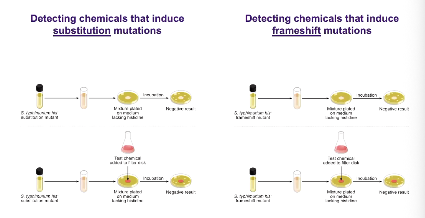
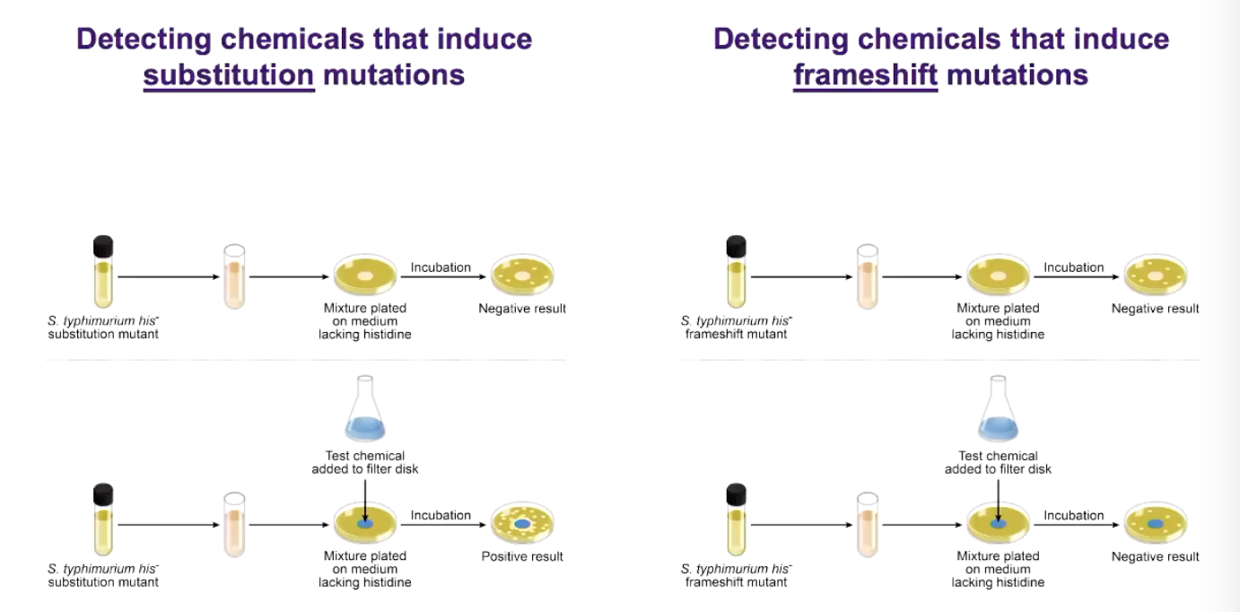
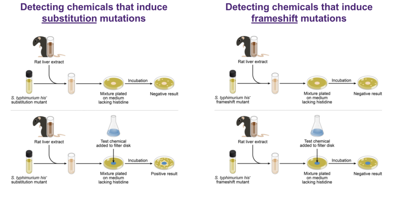
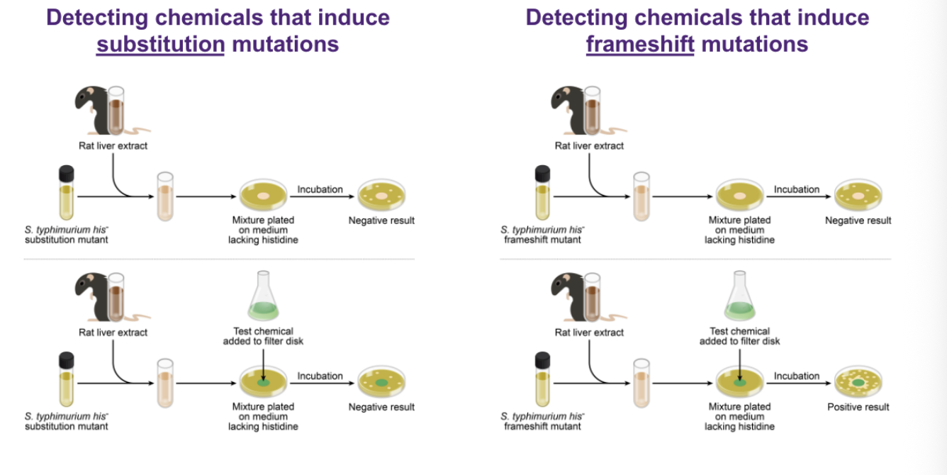
Ames test: The Ames test is a simple, inexpensive and highly sensitive bacterial screen for chemical mutagens, developed by Bruce Ames and coworkers, using histidine auxotrophic (His⁻) mutants of Salmonella Typhimurium. His⁻ bacteria cannot synthesise histidine and therefore normally cannot grow on medium lacking histidine. If a mutation restores a His⁺ prototrophic phenotype, the bacterium can synthesise histidine again and form a colony. Therefore, a chemical is considered mutagenic if it increases the number of His⁺ revertant colonies above the spontaneous/background reversion rate. Different strains are used to detect different types of mutations: TA1535 contains a base-substitution missense mutation (associated with a Leu→Pro loss-of-function change) and is used to detect base-substitution mutagens, whereas TA1537 contains a frameshift mutation and is used to detect frameshift mutagens. The tester strains are also made more sensitive to mutagens by having defective nucleotide-excision repair, so DNA damage is less efficiently repaired, and a defective lipopolysaccharide barrier, which increases the uptake of bulky mutagens. The test can also be performed with and without rat-liver S9 fraction. S9 contains liver metabolic enzymes because some chemicals are not directly mutagenic but are converted by mammalian metabolism into mutagenic metabolites. Therefore, an increase in His⁺ revertants only when S9 is present indicates that the chemical requires metabolic activation to become mutagenic.
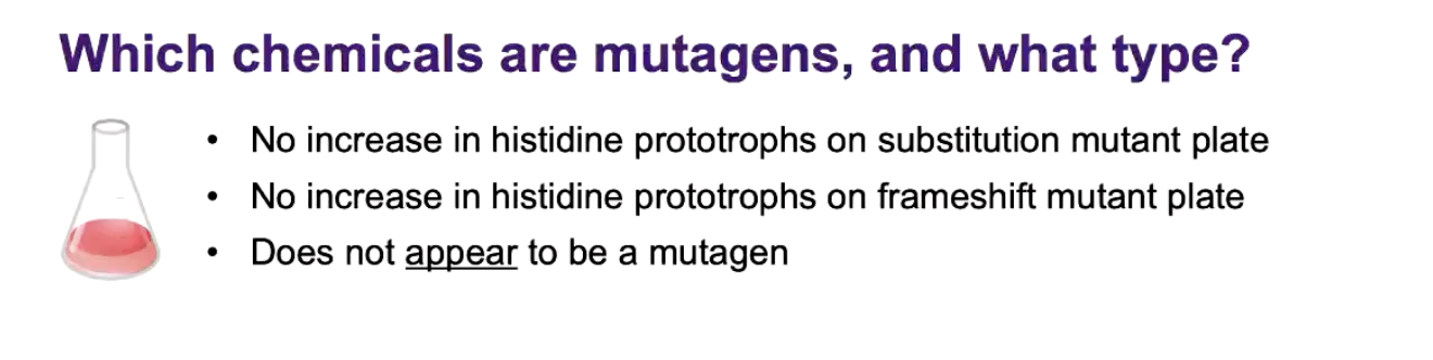
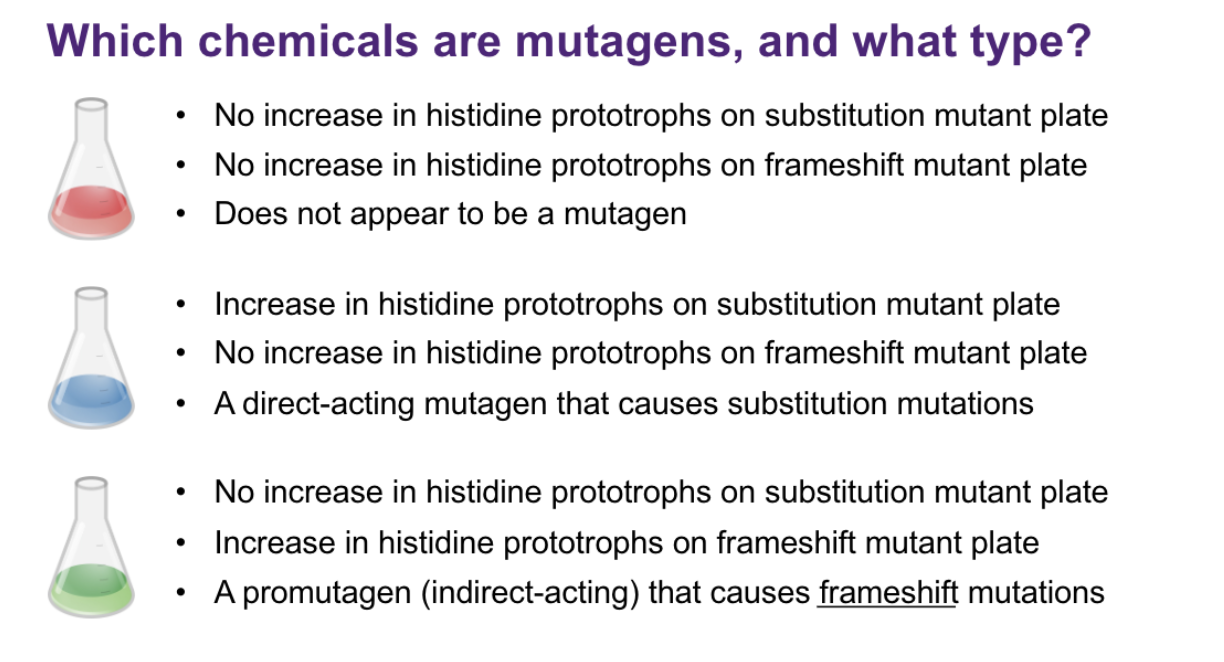
In the example results, the red chemical caused no increase in His⁺ colonies on either strain, so it does not appear to be a mutagen. The blue chemical increased His⁺ colonies on the substitution-mutant plate without S9, but not on the frameshift plate, so it is a direct-acting mutagen that causes substitution mutations. The green chemical caused no increase without S9, but when rat-liver S9 was added it increased His⁺ colonies on the frameshift-mutant plate, showing that it is a promutagen (indirect-acting mutagen) that must first be metabolically activated and then causes frameshift mutations. Thus, which strain shows increased His⁺ colonies tells us the mutation type, while whether S9 is required tells us whether the mutagen is direct- or indirect-acting.
indirect: it is mutagenic with liver
direct: is it mutagenic
A bacterial screen for chemical mutagens
Bruce Ames and coworkers developed a simple, inexpensive and extremely sensitive test for the mutagenicity of chemicals using histidine auxotrophic mutants of Salmonella Typhimurium
Mutagens raise reversion to His⁺ prototrophy above background
Strain TA1535 has a missense mutation (Leu→Pro, a loss of function substitution) (case substitution)
Strain TA1537 has a frameshift mutation
Nucleotide excision-repair mutant (⇧ mutagen sensitivity)
Defective lipopolysaccharide barrier (⇧uptake of bulky mutagens)
THIS IS HWY TWO DIFFERNET ONES ARE USED:

ADD S9: FROM RATS INTO IT



recap 5

DNA REPAIR MECHANISMS IN ECOLI AS OUR EXAMPLES: photo reactivation
Photo reactivation
cyclobutane dimmers (LIKE Thymine dimers) are caused by the excitation energy of UV radiation
Fixed using light dependant repair
Photolayse absorbs blue light for energy
Cleaves thymine dimers, restoring original state
• ⁃ Doesn’t work in the dark.
To fix it, cells that have this pathway use an enzyme called photolyase. Photolyase:
1. Recognises and binds the pyrimidine dimer
↓
2. Absorbs blue/visible light for energy ☀
↓
3. Uses that energy to break the abnormal covalent bonds joining the two bases
↓
4. Restores the bases to their normal structure ✅
The important thing is that photolyase doesn't cut the bases out and replace them. It directly reverses the UV-induced bond.

Base excision repair:
BER VS NER
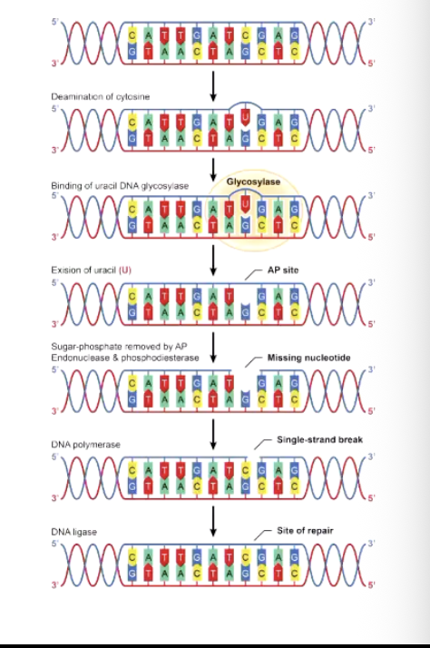
Excision repair: DNA repair systems recognise damaged or abnormal DNA and excise (remove) the damaged region. In base excision repair (BER), an abnormal or chemically modified individual base is recognised and removed, followed by processing of the resulting site. In nucleotide excision repair (NER), a larger stretch of nucleotides surrounding a bulky, helix-distorting lesion, such as a UV-induced thymine/pyrimidine dimer, is removed by an endonuclease-containing repair complex. After the damaged DNA has been removed, DNA polymerase fills the resulting gap using the undamaged complementary DNA strand as the template, ensuring the correct sequence is restored. Finally, DNA ligase seals the remaining nick in the sugar-phosphate backbone, completing the repair.
A dna repair endonuclease binds to an excises the damaged base or bases
This recognises the error
Base excision repair pathways remove abnormal or chemically modified bases with a single enzyme
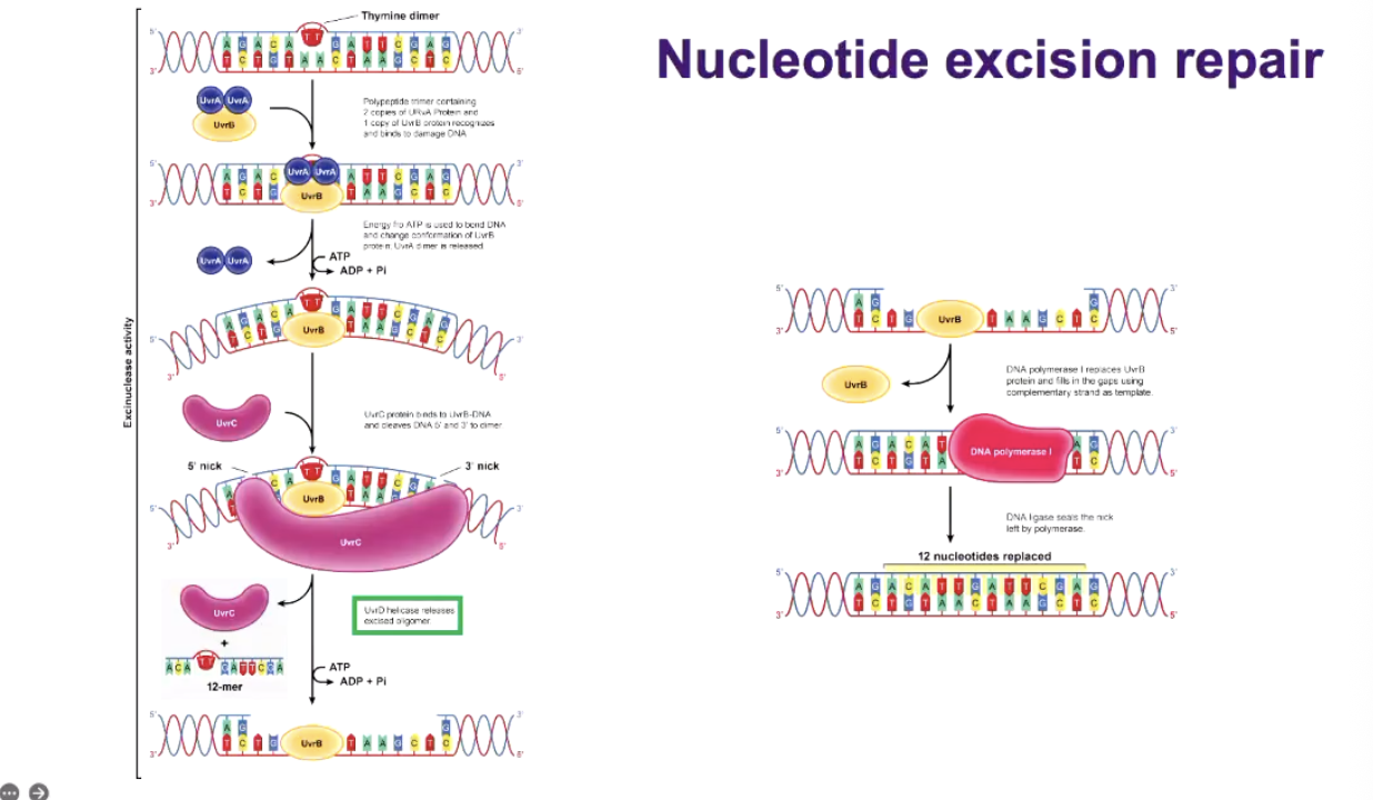
Nucleotide excision repair pathways remove larger defects, such as thymine dimers and uses an endonuclease-containing complex (two two next slide)
A DNA polymerase fills in the gap, suing undamaged complementary strand of DNA as a template. This fixes it
• ⁃ Dna ligase seals the break left by dan polymerase. This glues it back
• 3. NUCLEOTIDE EXCISION REPAIR:
NUCLEOTIDE EXCISION REPAIR:
UVRB AND UVRA , recognises the big error, then it bends it
UVRC cuts it, four bases in one direction, 8 in other direction
Then with uvrD it will unwind it and throw it away with the big error
Can cell can use it to make other stuff
Then DNA polymerase fills it in up
• ⁃ And liagse glues it back
AAAA | BBBBBBBB-XX-BBBB | CCCCC
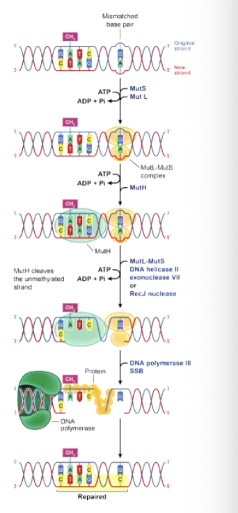
Mismatch repair in E.coli: BAE IS WRONG A IS A C BUT SHOUDLNT ETC
Mismatch repair in E.coli: BAE IS WRONG A IS A C BUT SHOUDLNT ETC
This is for if a is in the spot of a c, they know which one it is, there is GATC thing on the base so it knows that the original strand. basically the orginal is methalated which the mutation base isnt so it reconigess that its not methalyted
MUTS recognises mismatches and binds to them to initiate the repair process
MUTL joins and activates MUTH
MUTH (an endonuclease) cuts the unmethylated strand at nearest hemimethylated (half methylated) GATC sequence; on either side of the mismatch
Excision requires MUTS, MUTL, UVRD (DNA helices 2, also known as MUTU, and an exonuclease
• ⁃ Dna polymwerase 3 fills in the gap and DNA ligase seals the nick
DEALING WITH THYMINE DIMERS USING POSTREPLICATION REPAIR
A thymine dimer in the template strand blocks replication
DNA polymerase 3 restarts DNA synthesis past the dimer, leaving a gap in the nascent strand
RecA binds to the single strand (this is bad btw that’s y we NEED the other options to work) of DNA at the gap and meditates base pairing with the homologous segment of the sister double helix to fill the gap
• ⁃ Dna polymerase fills the gap in the problematic strand using information from the sister double helix, and dan ligase seals the nick
so it goes back to normal one to see what it was meant to be

• 7. THE SOS RESPONCE IN E.COLI
7. SOS response in E. coli
Replication-blocking DNA damage causes DNA polymerase III to stall at a lesion.
Stalling leaves/exposes single-stranded DNA (ssDNA).
RecA binds/coats the ssDNA, forming an activated RecA* filament.
RecA* stimulates the self-cleavage/inactivation of LexA.
Normally, LexA dimers bind SOS boxes in the promoters of SOS genes and repress their transcription.
When LexA is inactivated, repression is removed → SOS genes are expressed.
The SOS response includes translesion DNA polymerases, such as DNA polymerase V, which can replicate across lesions that normally block DNA polymerase III.
DNA Pol V is low-fidelity and lacks proofreading, and damaged bases may provide poor/no reliable coding information. Therefore, it can insert incorrect nucleotides.
Translesion synthesis (TLS) allows replication to continue despite the lesion, increasing cell survival, but it can also introduce mutations.
Replication-blocking DNA damage (a lesion) activates the SOS respond
Dna polymerase V bypasses lesions that stall DNA polymerase 3; Pol V lacks proofreading and lesions are non-intructive
Translation synthesis fills these gaps but tolerates lesions, leaving mutations
lexA dimers normallin bind SOS boxes in SOS gene promoters, repression transcription
Stalled replication exposes single-stranded DNA which recalls coats to form a filament (ReA*)
This activates RECA which stimulates LEXA to inactivate itself, turning on SOS
RECOMBINATION: HOMOLOGOUS
Recombination between homologous DNA molecules involves numerous enzymes, that clease, unwind. Stimulate single-strand invasions of double helices, repair and join strands of DNA
In most eukaryotes crossovers mature within the synaptonemal complex during prophase of meiosis I
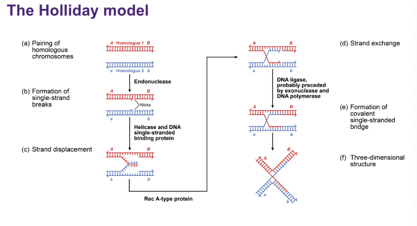
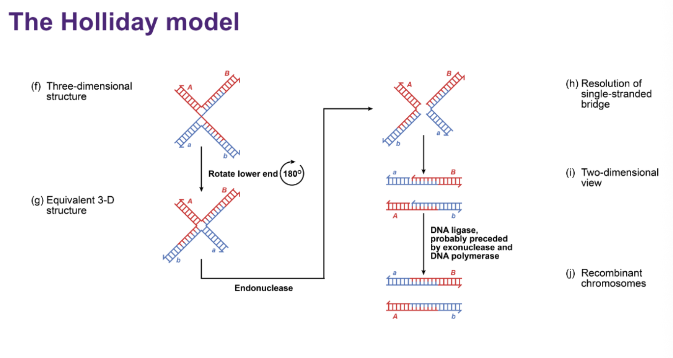
Crossing over involves the breakage of parental chromosomes, rejoining the parts in new combinations through the formation and resolution of a Holliday junction
The Holliday model is one of the most widely supported explanations of the molecular basis of recombination
• ⁃ LOOK VIDEO LECTURE 6: 00:29:00
holiday model/
CHI STRUCTURE: OR HOLLIDAY INTERMEDIATE:
Order of the Holliday model: Homologous chromosomes first pair and align with one another. An endonuclease makes single-strand breaks (nicks) at corresponding positions in the two homologous DNA molecules. Helicase unwinds the DNA, exposing the cut single strands, and a RecA-type protein promotes strand invasion, where each exposed strand pairs with the complementary sequence on the other homologous DNA molecule. This causes strand exchange, and DNA ligase joins the exchanged strands, producing a crossed DNA structure called a Holliday junction. The Holliday junction can then be resolved by an endonuclease cutting the crossed strands, followed by DNA polymerase filling any gaps and DNA ligase sealing the nicks. The DNA molecules separate, producing recombinant chromosomes containing new combinations of parental DNA.

EVOLUTION OF GENE CONCEPT:
A mutation in a particular gene can cause a defect in a particular enzyme, blocking a specific step in a metabolic pathway and causing characteristic metabolites to accumulate or become deficient.
GARROD: ONE GENE- ONE METABOLIC BLOCK:
In born errors of metabolism is the first book made
This is common is inbreding: the mutations below
Mutations in homogentisic acid oxidase leads to the production of black urine, kidney stones, cartilage damage and heart disease
Discovery of the first recessive allele in humans
Mutations in phenylalanine hydroxylase cause mental impairment, light hair color, and the presence of metabolites in blood and urine

BEADLE AND TATUM: ONE GATE- ONE ENZYME
Proposed that biosynthesis of essential metabolites is under genetic control
Mutations in genes involved in biosynthesis of metabolites would produce strains with additional growth- factor requirements
Beadle and Tatum demonstrated that one mutation resulted in the loss of one enzyme activity
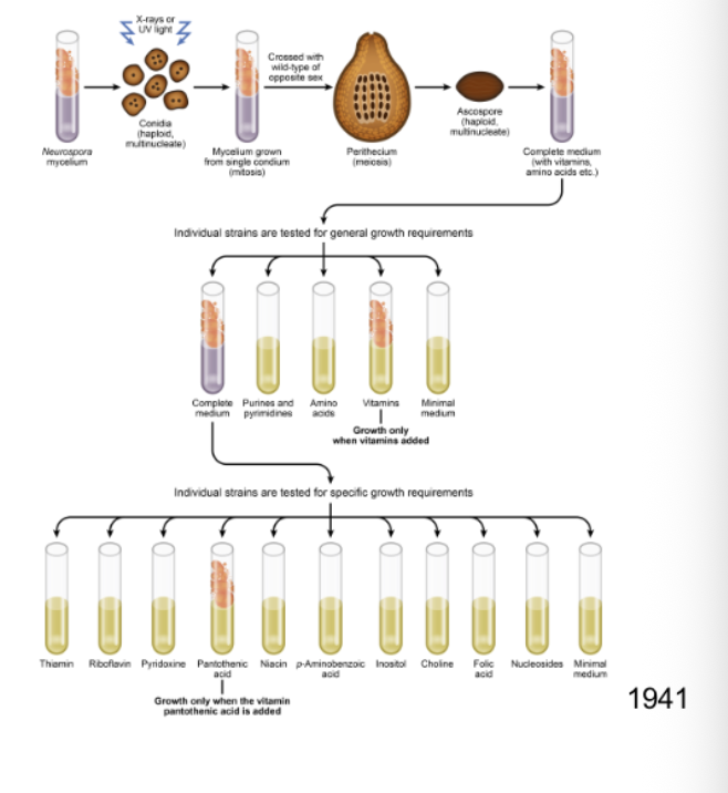
The experiment:
1. They used the fungus Neurospora crassa.
Normal wild-type Neurospora can grow on minimal medium because it can synthesise all the metabolites it needs.
2. They exposed the fungus to a mutagen.
This created random mutations in its genes.
3. They allowed the fungus to undergo sexual reproduction and collected haploid spores.
The fact that the spores were haploid was really useful because each gene only had one copy. So if that copy contained a recessive mutation, its effect could be seen immediately.
4. They first grew the mutants on complete medium.
Complete medium contains lots of nutrients, vitamins and amino acids, so even mutants that couldn't synthesise something themselves could still survive.
5. They then tested each mutant on minimal medium.
Most could grow:
Minimal medium → grows ✅ → can make everything it needs.
But some couldn't grow:
Minimal medium → no growth ❌ → mutation has probably blocked a metabolic pathway.
6. They added specific nutrients to figure out what was missing.
For example, suppose a mutant couldn't grow on minimal medium:
Minimal medium ❌
But then:
Minimal + vitamin X → grows ✅
That tells you the mutation prevented the fungus from making vitamin X (or something in that pathway).
By studying many mutants, Beadle and Tatum found that particular mutations could eliminate particular enzyme activities.
The conclusion
This led to the one gene–one enzyme hypothesis:
One gene controls the production/function of one enzyme, and that enzyme controls a particular step in a metabolic pathway.
So:
Gene mutation → enzyme doesn't work → metabolic step blocked → required product isn't produced → organism needs that product supplied externally.
Why was this such a big deal?
At this time, scientists didn't yet know that DNA was the genetic material. Proteins were still considered strong candidates for the hereditary material.
• ⁃ Fungus used: mutengenised it, grew it up, get them to make the sexual structure (like a volcano), pulled out haploid the spores from int here, plate it out, and see if they have phenotypes, tested after it grew to see if it’d grow properly even tho mutenegenised, one dint work it needed vitamin, when one is added it worked. They did this again and again again
REMEMBER AT TGIS TIME NOONE KNEW DNA WAS THE GENES WE THOGUTH PROTEIN
The pre-1940 beads-on-a-string concept
The gene was thought to control the inheritance of one attribute or phenotype
The gene was not thought to be subdivisible by recombination, and was therefore the smallest unit of genetic material that could be mutated
The modern concept of the gene
Controls synthesis of one polypeptide chain or RNA molecule
The nucleotide pair is the smallest unit that can be mutated; it cannot be subdivided by recombination
RECOMBINATION WITHIN A GENE
Intragenic recombination: Pete Oliver demonstrated recombination between different mutant sites within the lozenge (lz) gene of Drosophila. Mutations such as spectacle (lzs) and glassy (lzg) affect the compound eye. When flies carrying different mutations within the same gene were crossed, rare wild-type recombinants could be recovered, demonstrating that recombination can occur within a gene. This showed that the gene is not an indivisible unit of recombination. Decades later, Charles Yanofsky, studying the trpA gene of E. coli, demonstrated genetic resolution down to extremely closely spaced nucleotide sites, further establishing that genes consist of many independently mutable and recombinable sites.
Pete Oliver described recombination within the lozenge gene of Drosophila, done in male flies. They could make a female with BOTH the LZg mutant
Shows genes can have recombincation
Mutants have severe compound eye perturbations
Can you get recombination within a gene? If so, WT recombinants should be obtainable from a cross of spectacle (lzs) and glassy eye (lzg) mutants
• ⁃ Charles Yanofsky described recombination between adjacent nucleotide pairs in the trpA gene of E. coli decades later

How protein made:
Collinearity means that the linear order of coding sequences in a gene corresponds to the linear order of amino acids in its polypeptide product.

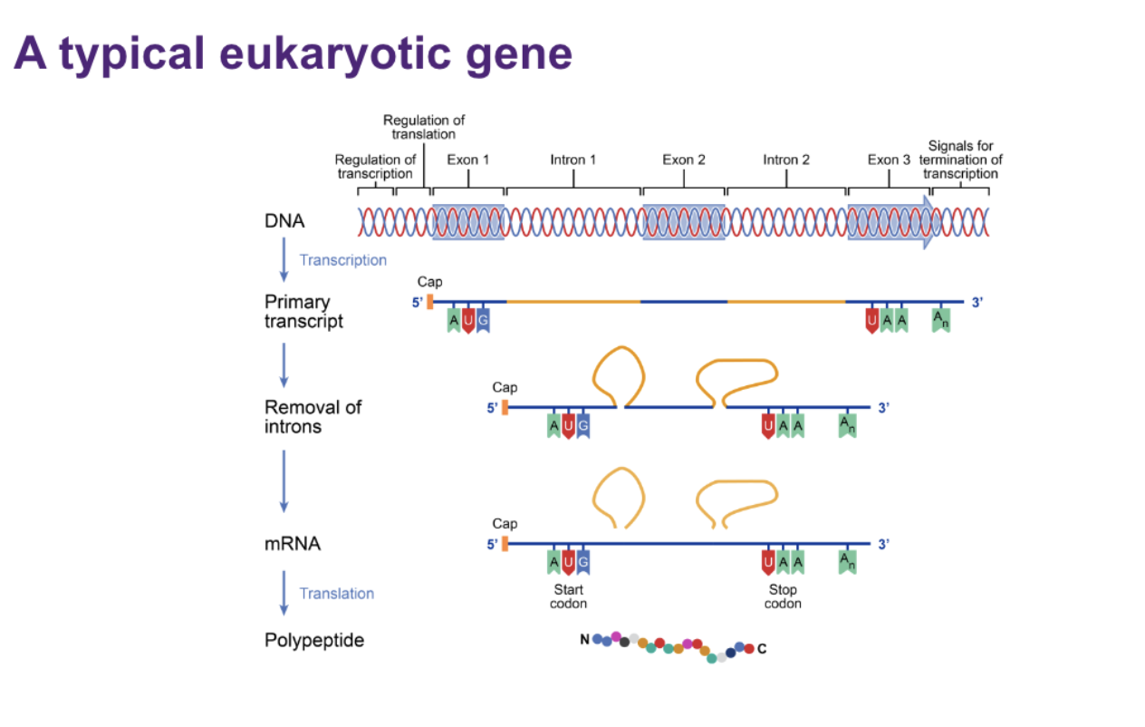
PROKARYOTIC GENE:
• ⁃ Polysistzonic mrna, each has start stop, start stop. So does one by one
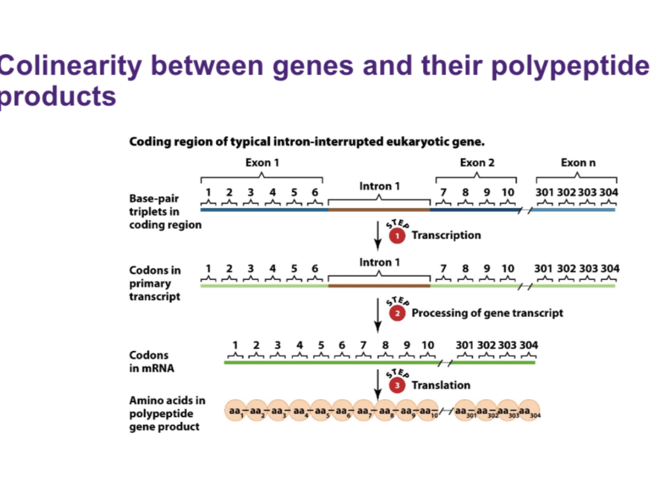
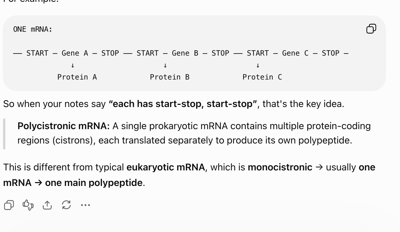
EUKARYOTIC GENE:
One single but with introns. No one knows where introns come from

GENES WITHIN GENES IN BACTERIOPHAGE X174:

shows overlapping

Circular, single stranded DNA
5386 nucleotides
Encodes 11 proteins that together contain 2300 amino acids (2300*3)
The genome contiains overlapping genes and genes within genes
Never say never!
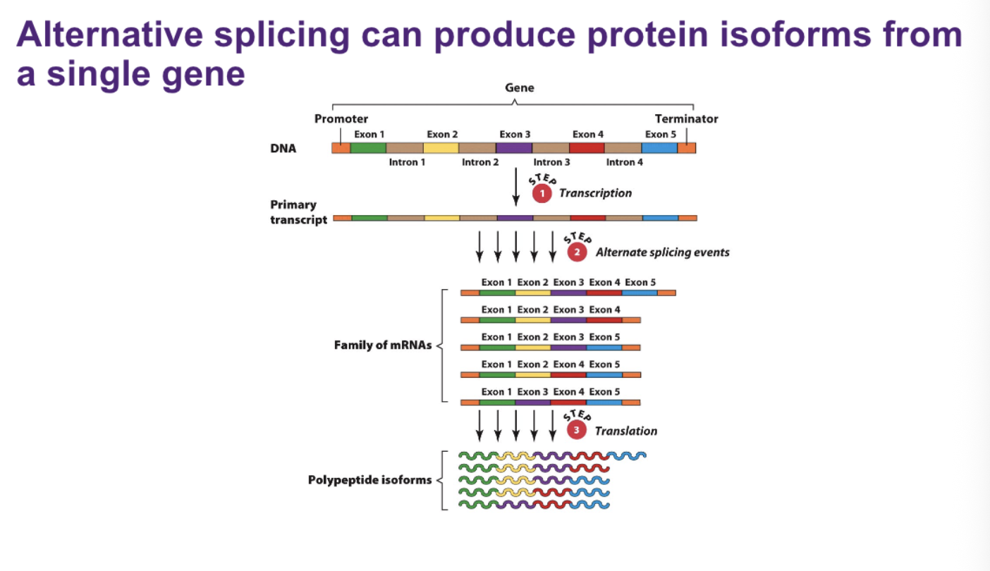
ALTERNATIVE SPLICING CAN PRODUCE PROTEIN ISOFORMS FROM A SINGLE GENE:
Alternative splicing: A single eukaryotic gene can produce multiple different protein isoforms through alternative splicing. The gene is first transcribed into a primary RNA transcript containing its exons and introns. During RNA processing, the introns are removed, but different combinations of exons can be retained and joined together, producing a family of different mature mRNAs from the same gene. These different mRNAs are then translated into different polypeptide isoforms. Therefore, one gene can produce multiple different protein products, increasing protein diversity without requiring additional genes.
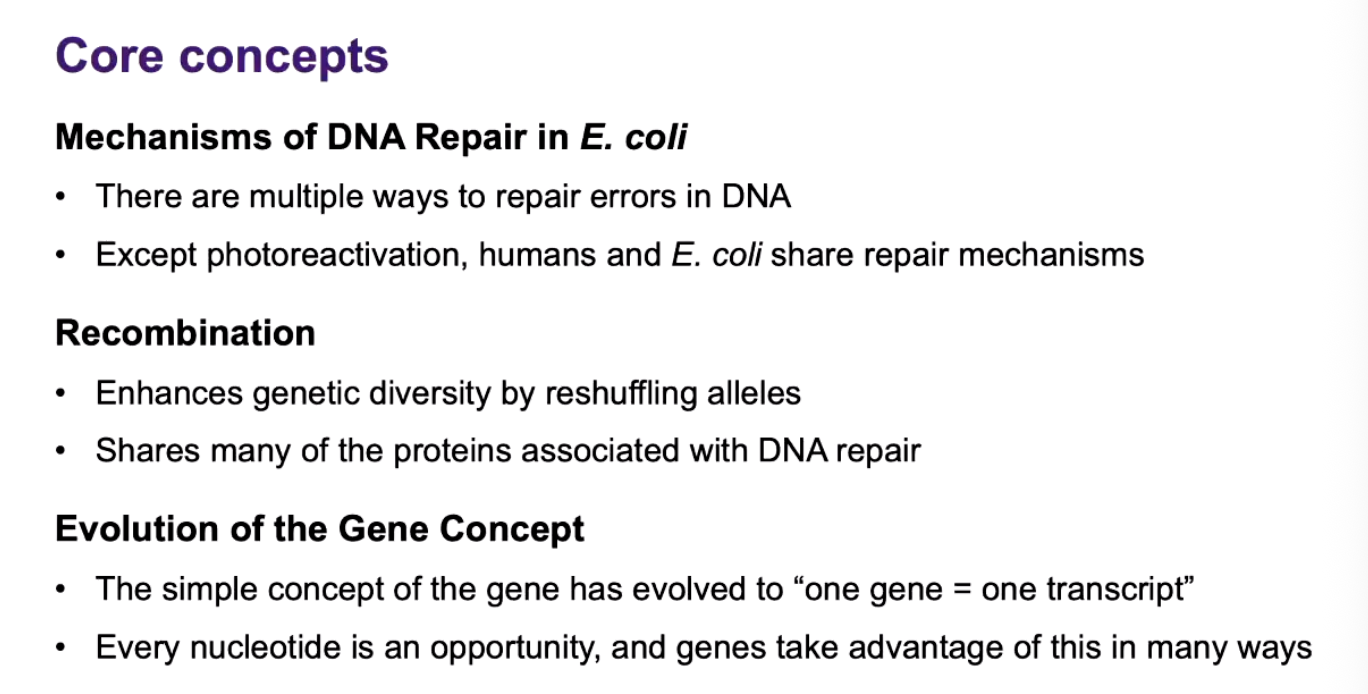
core concept
MODULE TWO!
WHO HAS A GENOME?
All living entities
Bacteria
Archaea
Eukaryotes (fungi, plants, animals, protists)
Some non-living entities
Viruses
Plasmids
Entire communities (“metagenome”)
FUNCTIONS OF THE GENOME:
It is the storage unit
The genetic material must replicate, control the growth and development of the organism, and allow the organism to adapt to changes in the environment
Genotypic function:
Replication
Phenotypic function
Gene expression
Evolutionary function
Mutation
• ⁃ Adaptations etcetc if + change
DOES SIZE MATTER?
No.
A flower has more than a lizard and baby.
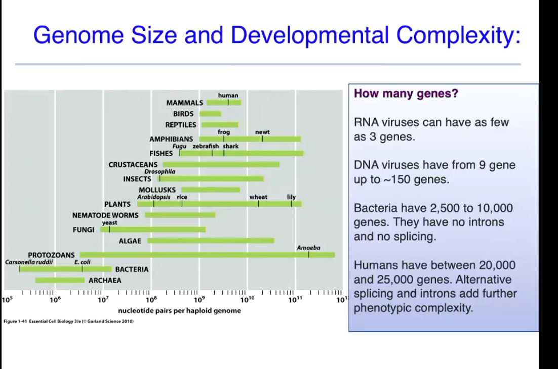
THE GENOMES OF PROKAY. AND VIRUSES. mono vs haploid vs doploid
Bacterial and viral genomes are monoploid
What is the difference between monoploid, haploid and diploid?:
Bacterial and viral genomes are monoploid
Most viruses and bacteria have single set of genes stored in a single chromosome
This means that bacteria and viruses dont have the allelic complexity that comes with inheritance of chromosomes from two different parents
Bacteria also contain plasmids and transposable DNA elements

E.coli: genome plasmids:
Fertility, resistance, col
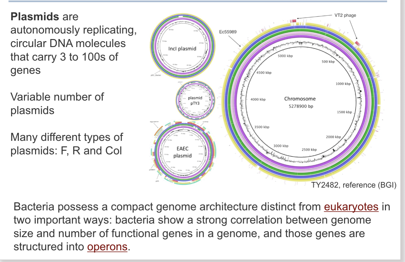
Structure of bacterial genomes:
Just shows the many changes bacterias go through.
• ⁃ Operon is helps sit all these genes. It controls it all (genes), operon makes it all on or all off, it depends but if an operon is on it, that is what it is for, more efficient

Gene content in bacterial genes:
Virus genomes:
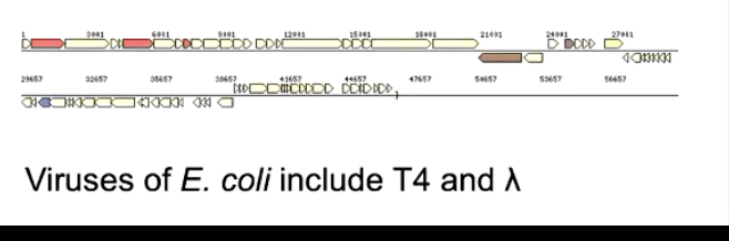
Can only reproduce by infecting living host cells
Outer capsid protein enclosing genetic materials- DNA, ssDNA, ds RNA, ssRNA
Linear and circular genomes
Most viruses are bacteriophages a virus that infects bacteria and arches
• ⁃ Very high coding density

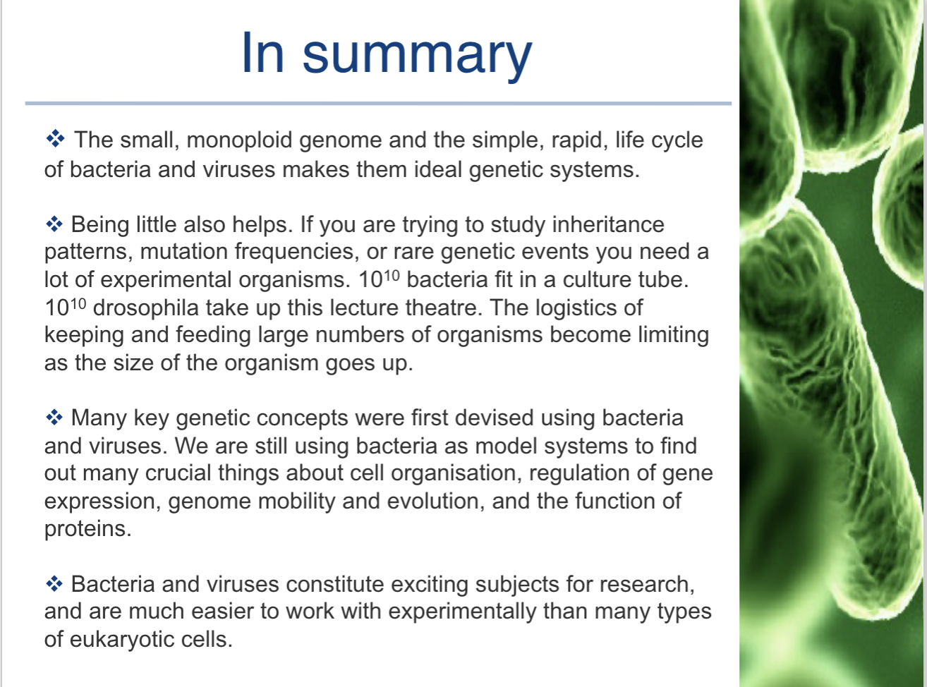
Why should we use viruses and bacteria to study for genetics?
Why should we study viruses and bacteria for genetics?
Such a system would also be useful for finding out fundamental things about gene expression, DNA replication, intra
cell DNA strand exchange events, DNA repair, transcription, and translation
High repilicaiton rate
No ethics issue
Simple genetic structure (not rlly why but yeah)
Can culture them

BACTERIAS CLONAL REPLICATION AND A SIMPEL CELL CYCLE:

phenotypes in bacteria are:
(i) defined by the genome
(i) ways to see that genes have been inherited and expressed
(ii) things that can be used for selection in genetic screens
WHAT ARE SOME USEFULL PHENOTYPIC CHARACTERICS THAT WE CAN USE FOR SELCTION OR OBSERVATION

Is bacterial genuine static or labile (mobile and plastic)?
Know how to write genes vs phone

PROTOTROPHS VS AUXOTROPHIC

Testing for nutritional requirements:
Horizontal vs vertical transfer
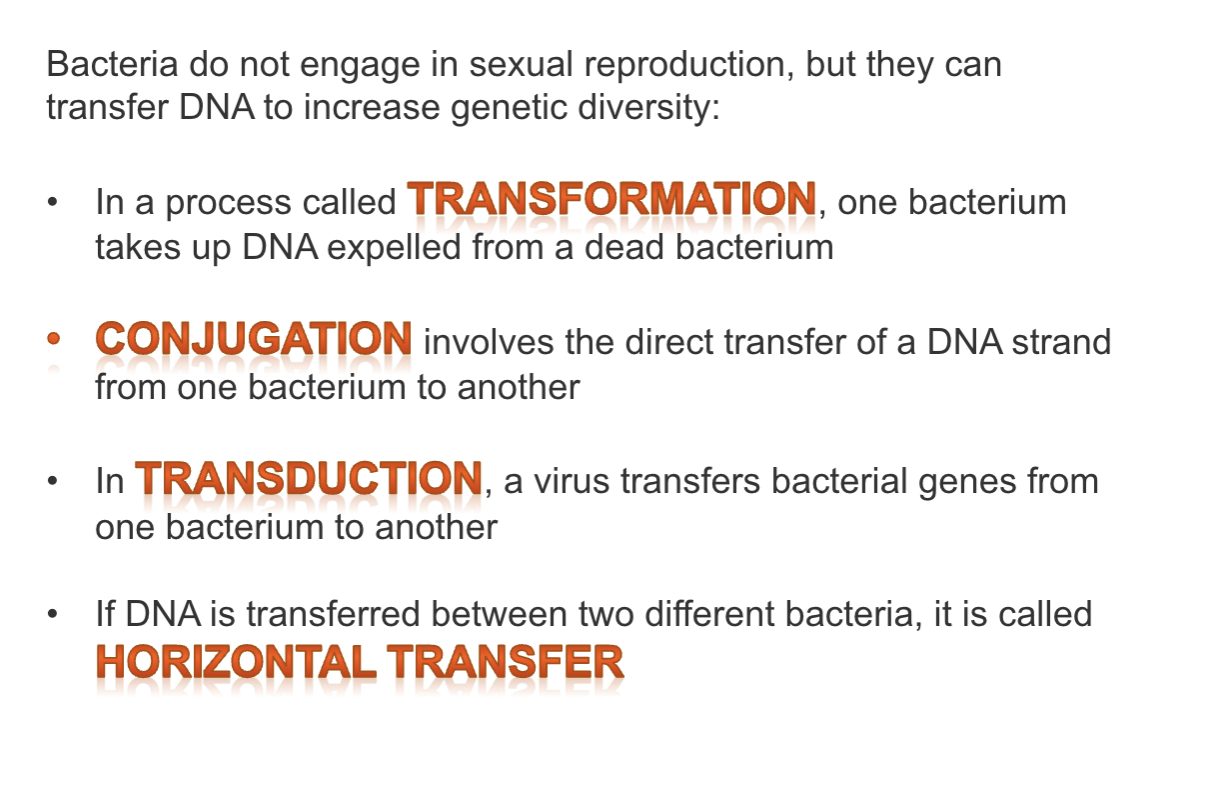
PANASEXUAL PROCESSES: FORU TYPES
lecture seven review


Review three parasexual mechanisms of dna:\ in bacteria:

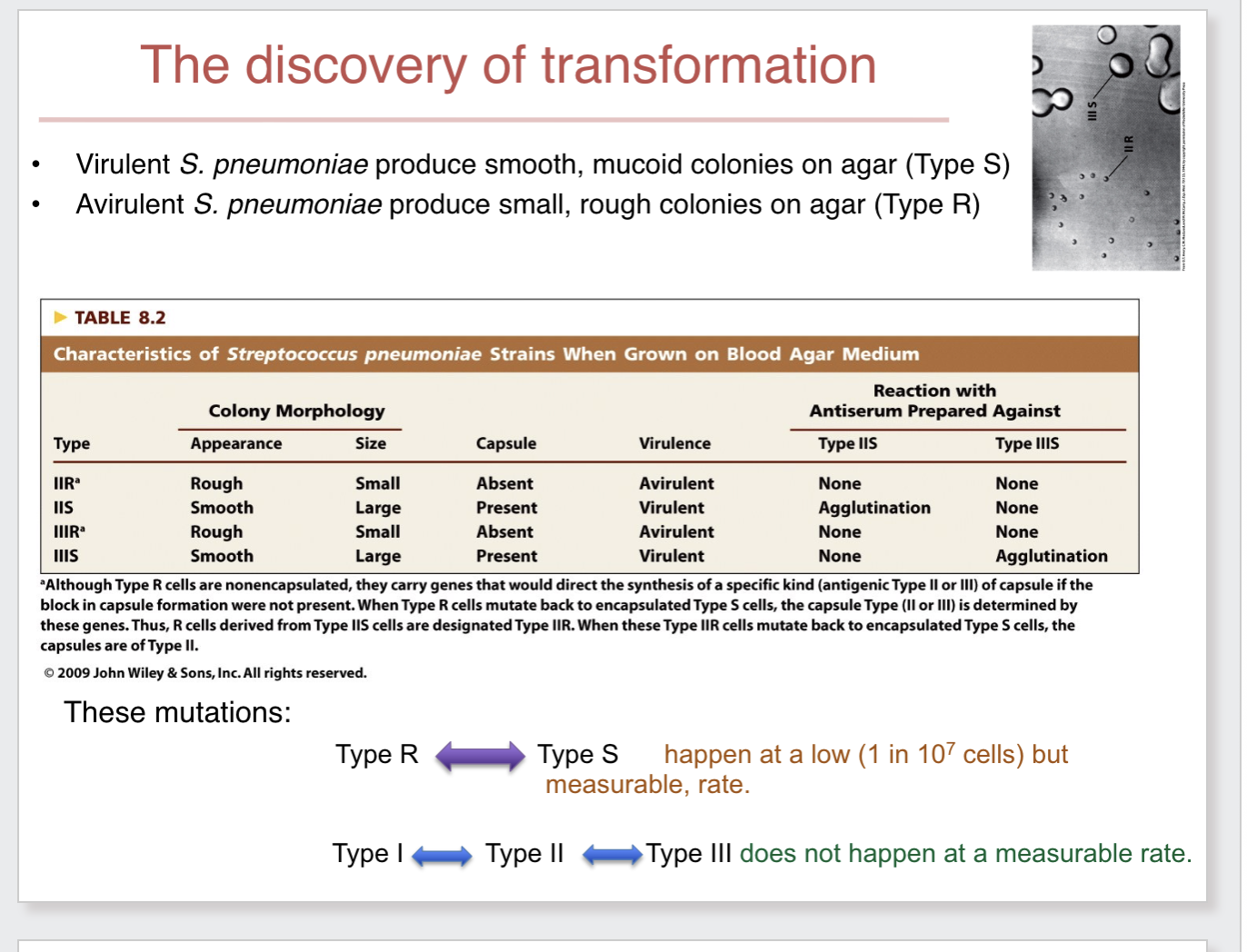
The discovery of transformation:

How know what it is:
Agglutination: