Macular holes + VMT
 Call Kai
 Learn
Learn Practice Test
 Spaced Repetition
 Match
Match Flashcards
 Knowt Play
1/23

Earn XP
Description and Tags
Week 1 Tri A
Name | Mastery | Learn | Test | Matching | Spaced | Call with Kai | Chat |
|---|
No analytics yet
Send a link to your students to track their progress
24 Terms
Why do macular holes occur
As a result of a combination of antero-posterior traction from the vitreous, and tangential traction from the internal limiting membrane
Average diameter of a macular hole
About 400 microns, less than a third of the optic disc diameter
Staging for macular holes
Stage 1 - impending FTMH
Stage 2 - FTMH
Where should the hole diameter be measured
-At the narrowest point of the hole
-Should be diameter and not a chord
Where is the basal diameter measured from
Where the retina joins the RPE
Natural history for macular holes
Stage 1 - majority do not progress
Stage 2 - some will close
Stage 3 - spontaneous closure rare, not unknown
Presentation of macular holes
-May be an incidental finding
-Loss of central vision, almost ever better than 6/18
-Metamorphopsia
-F>M, 4:1
-Approx 1:6 risk of bilateral hole
-Prevalence 3/1000 in >50's
Differential diagnosis of macular hole
Lamellar hole
-Clearer classification, degenerative lamellar hole and ERM foveoschisis
-Vision usually 6/18 or better
-No treatment shown to be beneficial
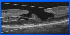
Degenerative lamellar hole
-Irregular foveal contour
-Lamellar hole-associated epiretinal proliferation (LHEP) 1
-Foveal cavitation and loss of foveal tissue 2
-Foveal bump 3
-Ellipsoid zone disruption 4
Top to bottom 1-4
Epiretinal membrane foveoschisis
-Epiretinal membrane contraction
-Thickening and splitting of the retina
-Outer retina remains intact
Pseudohole
-Hole in an epiretinal membrane over the fovea
-VA 6/18+
-Edges are not elevated
-Treat as an epiretinal membrane
-Only refer if symptomatic
Referral for full thickness macular hole
-Refer all
-Urgent if
Do you refer a lamellar hole
-Do not refer
-Monitor in community
-Non-urgent referral only if symptomatic and documented loss of VA due to macular change
Management of FTMH
-Observe as unlikely to improve, delay reduces visual recovery
-Surgery (PPV, ILM peel and gas +/- phaco and IOL) very effective
Risks of FTMH
-Cataract, will 100% develop, may do lens surgery at the same time
-Retinal detachment
-Poor outcome, unable to predict final VA, some patients improve less than 10 letters, some patients do not improve for 6-12 months
Current outcomes for FTMH surgery
-95% of holes close with one operation
-VA improvement averages 0.5 logMAR but can be unpredictable
-Improvement of VA is greater for near than distance
-Adverse effects and RD are rare
VMT
-Vitreous adheres to macular, causing elevation plus or minus distortion
-Usually asymptomatic, may lead to FTMH in a few patients
-May cause metamorphopsia/blurring
Management of VMT
If asymptomatic
-None, as does not need treatment
-Do not refer
If symptomatic
-Vitrectomy and peel
Epiretinal membrane
Glial tissue on surface of retina which contracts, distorting the retina and results in retinal elevation and theickening
Symptoms of epiretinal membrane
Metamorphopsia and reduced VA
Signs of an epiretinal membrane
-Pre-macular sheen, shiny appearance
-Retinal folds
-Distortion of blood vessels, straightened/wrinkled
-Visible white/grey pre-retinal membrane
Causes of epiretinal membrane
-PVD, damage to ILM allows glial proliferation
-Retinal breaks, RPE cells migrate through break and proliferate over macula
-Retinal vein occlusion
-Diabetes
-Unknown
Management of epiretinal membrane
-Nil, unless symptomatic
-If symptomatic, vitrectomy and membrane peel +/- cataract surgery
-80% report improved, metamorphopsia can improve even if VA unchanged, VA improvement may take 6 months
Do you refer an epiretinal membrane
Only if they are symptomatic
-Do they close ERM eye to see better with good eye? if so refer lowk
-Non-urgent referral, as ERM progress very slowly, if at all
-Name, DoB, VA, refraction, previous eye history