Learn: Intro to Kines Unit 1
 Call Kai
 Learn
Learn Practice Test
 Spaced Repetition
 Match
Match Flashcards
 Knowt Play
1/91

There's no tags or description
Looks like no tags are added yet.
Name | Mastery | Learn | Test | Matching | Spaced | Call with Kai | Chat |
|---|
No analytics yet
Send a link to your students to track their progress
92 Terms
Gross Anatomy: Macro structures
Structures that can be seen with the eye (via cadavers, xrays, MRI
Superficial Anatomy
Study of External features, links what you can see on the outside to the internal organs underneath
Regional Anatomy
How different structures work together in one specific part of the body
Medial vs Lateral
Medial: Closer to the midline of the body
Lateral: Further from the midline of the body
Anterior vs. Posterior
Anterior: closer to front of the body
Posterior: Closer to back of the body
Proximal vs Distal
Proximal: Towards the base of a limb
Distal: Further from the base of a limb
Systemic Anatomy
Study of organ systems in the body
Histology
Study of anatomy on the micro level (can see plant and animal tissue)
Noncontractile tissue
Soft tissue that is unable to contract and relax (common in older people)
Why do children often have more than 206 bones
As a child develops, some bones fuse together into one
Cortical vs Cancellous Bones
Cortical = Harder, exterioir
Cancellous = Spongy or softer (more ability to absorb force)
Structure of epithelial tissue
Sheets of tightly packed cells with minimal extracelluar matrix, can be simple (single layered) or stratified (multiple layers)
Function of Epithelial tissue
Protection, absorbtion, secretion, filtration and sensation
Examples of Epithelial Tissue
Simple squamous epithelium (lining of blood vessels), Transitional epithelium (urinary bladder), pseudostratified epithelium (skin, mouth, esophagus)
Structure of connective tissue
Diverse cells dispersed in an abundant extracellular matrix containing different fibers
Function of connective tissue
(Support, protection, transport, storage, insulation)
Examples of Connective Tissue
Loose connective tissue (areolar, adipose, reticular), Dense connective tissue (tendons, ligaments), cartilage, bone, blood, lymph
Structure of Muscle Tissue
Elongated cells containing contractile proteins (actin, myosin)
Function of muscle tissue
Contraction for movement, posture, heat production
Examples of muscle tissue
Skeletal muscle, cardiac muscle, smooth muscle
Which types of muscles are voluntary?
Skeletal muscle
Which types of muscle are involuntary
Cardiac muscle, smooth muscle
Structure of nervous tissue
Neurons and glial cells
Function of nervous tissue
Conducting electrical impulses, communication, regulation of body functions
Examples of nervous tissue
Central nervous system (brain, spinal cord), nerves
The skeletal system is composed of bones, cartilage and connective tissues. Main functions of the skeletal system include:
Rigid levers for motion, protection to internal organs and to produce and store nutrients and blood cells
Axial skeleton vs Appendicular skeleton
Axial- Skull, Rib Cage, Vertebrae
Appendicular- Appendage Bones (arms, legs), Pelvis, Pectoral
The shaft of long bones is called:
diaphysis
The ends of long bones are called:
epiphysis
Reasons bones are important to walking:
Act as levers that allow muscle to propel us forward, help to absorb ground force reaction forces, help to store energy and make walking more efficient, optimize stability
3 planes of movement:
Frontal, Sagittal, Transverse
Examples of an exercise on the frontal plane
Lateral raises/jumping jacks, side shuffles (any side to side movements)

Examples of exercises along the sagittal plane
Running/walking, bicep curls, rows, (any forward and backward motion)

Examples of exercises along the transverse plane
Russian twists/rotational medicine ball throws (any movement that involves twisting)
What are fibrous joints:
Immovable, such as the fusion of bones (skulls bones in adults)
Cartilaginous joints:
Strong, slightly moveable joints often surrounded by large cartilage structures (joints between vertebrae and the pubic bones)
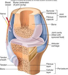
Synovial joints:
Freely moveable with large, fluid filled capsules that can move in a variety of directions (shoulder, hip, elbow, knee). The following joints all fall under synovial joints
Hinge joint:
permits movement in one plane, usually flexion and extension (elbow, knee)

Saddle joint:
opposing articular surfaces with a concave-convex shape

Plane joint:
surfaces are flat and adjacent to each other, allowing bones to glide across one another (subtalar joint)

Pivot joints:
Allows for rotation only, formed by a central bony pivot (proximal and distal radioulnar joints)

Condyloid Joint:
convex surface which joins with a concave elliptical cavity (wrist joint)
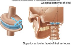
Ball and socket joint:
ball shaped surface fits into a cup life depression, permits free movement on multiple axes (hips, shoulders)

Anterior
Front or towards the front
Posterior
Behind, towards the rear
Ventral
stomach side
Dorsal
back (spine) side
Cephalic
towards the head
Caudal
Towards the tail bone
Superior
above/at a high lever
Inferior
Below, at a lower level
Medial
Towards the midline of the body
Lateral
Away from the midline
Distal
Away from the attached base of a limb
Proximal
Towards the attached base of a limb
Superficial
Towards or near the surface
Deep/profound
Away from the surface
Flexion:
anterior surfaces being brought together
Extension:
anterior surfaces are moved away from each other
Abduction
joint rotating away from the midline of the body
Adduction
joint rotating towards the midline of the body
internal rotation
anterior portion moving towards the midline of the body (internal rotation of the forearm is known as pronation)
External rotation
anterior portion moves away from the midline of the body (external rotation of the forearm is known as supination)
Accelerates the person while walking
Concentric contraction
Slows down the person walking
Eccentric contraction
Stabilizes a person while walking
Isometric contraction
Afferent flow:
Carries information towards the brain and spinal chord (information arrives)
Efferent flow:
Carries information way from the brain and spinal chord (exiting)
Function of Skeletal muscle
Produces body movement, maintains posture, generates heat
Advantages of Skeletal muscle
Voluntary and allows for precise movement, generates large amounts of force, supports posture and joint stability, major contributor to thermoregulation (produces heat)
Limitations to skeletal muscle
Easily fatigued, requires neural input for any action, susceptible to strains, tears and overuse injuries
Function of cardiac muscle
Pumps blood throughout the body
Advantages of cardiac muscle
Fatigue resistant, contracts continuously throughout life, highly efficient and possesses auto rhythmicity (generates its own electrical impulses)
Limitations of cardiac muscle:
Cannot be controlled consciously, limited regenerative capabilities, damage can permanently reduce function
Function of smooth muscle:
Move substances through internal organs
Advantages of smooth muscle
Extremely energy efficient, can maintain contractions for long periods of time (not easily fatigued), functions automatically, can stretch substantially while still maintaining function
Limitations of smooth muscle
Produces low force compared to skeletal muscle, slower contraction speed, limited precision compared to skeletal muscle
Sarcomere
contractile unit of a muscle fiber
actin and myosin
contractile proteins in muscle (contraction results from the sliding of these two filaments)
How muscles attach to bones
Tendons
Origin of muscle
the less moveable attachment point
insertion
the moveable attachment point of a muscle
Fast Fatiguable motor units
Muscle type 2B, produce quick, short bursts of power but fatigue quickly
Fast Fatigue resistant motor units
Muscle Type 2A, produce slightly less energy but are less susceptible to fatigue
Slow twitch motor units
Muscle type 1, fatigue very slowly and are designed for prolonged activities
Size principle of motor unit recruitment
Small motor units are recruited first (small = slow twitch), and as the demand for force increases, large motor units (fast twitch) are recruited
Crossbridge cycle
cyclical formation of links between actin and myosin resulting in the sliding of thin filaments along the thick filaments, producing/holding the muscle contraction
Titin
A protein responsible for allowing the sarcomere to stretch and recoil (acts as a spring, uses free energy we think)
Non-contractile elastic fatures
Fascia around the muscles, titin at the ends of sarcomere, tendons
Contractile tissue
Contains actin and myosin, uses crossbridge cycle
Muscular Endurance
ability of a muscle or muscle group to perform repeated contractions over a prologned period without becoming fatigued
Muscular Strength