First Aid - Hem/Onc - Pathology - FULL
 Call Kai
 Learn
Learn Practice Test
 Spaced Repetition
 Match
Match Flashcards
 Knowt Play
1/670

There's no tags or description
Looks like no tags are added yet.
Name | Mastery | Learn | Test | Matching | Spaced | Call with Kai | Chat |
|---|
No analytics yet
Send a link to your students to track their progress
671 Terms
What is hemolytic disease of the fetus and newborn also called?
Erythroblastosis fetalis
Which blood group antigens most commonly cause hemolytic disease of the fetus and newborn?
The major blood group antigens, especially Rh and ABO
Can minor blood group incompatibilities cause hemolytic disease of the fetus and newborn?
Yes — minor incompatibilities such as Kell can also cause disease ranging from mild to severe
ABO hemolytic disease classically occurs with what maternal and fetal blood types?
Type O pregnant patient; type A or B fetus
What is the mechanism of ABO hemolytic disease?
Preexisting maternal anti-A and/or anti-B IgG antibodies cross the placenta → attack fetal and newborn RBCs → hemolysis
How does ABO hemolytic disease typically present?
Mild jaundice in the neonate within 24 hours of birth
Can ABO hemolytic disease occur in the firstborn baby?
Yes — unlike Rh hemolytic disease, it can occur in firstborn babies
Is ABO hemolytic disease usually more or less severe than Rh hemolytic disease?
Usually less severe
How is ABO hemolytic disease treated?
Phototherapy or exchange transfusion
Rh hemolytic disease classically occurs with what maternal and fetal Rh types?
Rh-negative pregnant patient; Rh-positive fetus
What happens during the first pregnancy in Rh hemolytic disease?
The pregnant patient is exposed to fetal blood, often during delivery → formation of maternal anti-D IgG
Why is Rh hemolytic disease usually a problem in subsequent pregnancies rather than the first?
The first pregnancy commonly causes maternal sensitization and anti-D IgG formation; later Rh-positive fetuses can then be attacked by these antibodies
What are the major presentations of Rh hemolytic disease?
Hydrops fetalis; jaundice shortly after birth; kernicterus
How is Rh hemolytic disease prevented?
Administration of anti-D IgG to Rh-negative pregnant patients
At which stage of pregnancy is anti-D IgG given for prevention of Rh hemolytic disease?
During the third trimester and early postpartum period
What additional situations call for anti-D IgG prophylaxis?
Ectopic pregnancy; miscarriage; abdominal trauma; antepartum hemorrhage, if the fetus is Rh-positive
How does anti-D IgG prevent Rh hemolytic disease?
It prevents maternal anti-D IgG production
(Goal is to attempt to sequester whatever fetal D antigens may have gotten int the mother’s circulation during delivery)
ABO vs Rh hemolytic disease: can it affect the firstborn?
ABO = yes
Rh = usually no
ABO vs Rh hemolytic disease: which is usually more severe?
Rh hemolytic disease
ABO vs Rh hemolytic disease: classic presentation
ABO = mild jaundice within 24 hours of birth
Rh = hydrops fetalis, jaundice shortly after birth, kernicterus
ABO vs Rh hemolytic disease: prevention/treatment
ABO = phototherapy or exchange transfusion
Rh = prevent with anti-D IgG
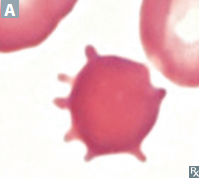
What are acanthocytes?
"Spur cells" — RBCs with irregularly spaced projections of varying size
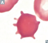
Acanthocytes are associated with what conditions?
Liver disease
Abetalipoproteinemia
Vitamin E deficiency
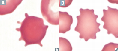
What are echinocytes?
"Burr cells" — RBCs with smaller, more uniform, evenly spaced projections than acanthocytes

Echinocytes are associated with what conditions?
Liver disease
ESRD
Pyruvate kinase deficiency

How do acanthocytes differ from echinocytes?
Acanthocytes = irregular projections of varying size and spacing;
Echinocytes = smaller, more uniform, evenly spaced projections
(A = Acanthosis / B = Echinocytes)
What are dacrocytes?
"Teardrop cells"
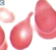
Dacrocytes are associated with what pathology?
Bone marrow infiltration, e.g. myelofibrosis

Why do dacrocytes become teardrop-shaped?
RBCs are mechanically squeezed out of abnormal/infiltrated bone marrow, deforming them into a teardrop shape

What are schistocytes?
"Helmet cells" — fragmented RBCs

Schistocytes are associated with what conditions?
Microangiopathic hemolytic anemia, e.g. DIC; TTP/HUS; HELLP syndrome; mechanical hemolysis such as prosthetic heart valves
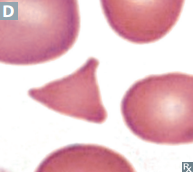
Why do schistocytes form?
RBCs are physically sheared apart as they pass through abnormal small vessels or mechanical surfaces

What are degmacytes?
"Bite cells"

Degmacytes are associated with what condition?
G6PD deficiency
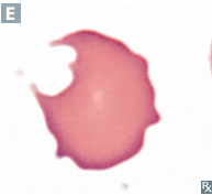
Why do Degmacytes form in G6PD deficiency?
Oxidative damage causes Heinz bodies; splenic macrophages remove the Heinz bodies and "bite" out part of the RBC membrane

What are elliptocytes?
Elongated, oval/elliptical RBCs

Elliptocytes are classically associated with what condition?
Hereditary elliptocytosis
Caused by mutation in genes encoding RBC membrane proteins (eg, spectrin)
What are spherocytes?
Small, spherical RBCs without central pallor
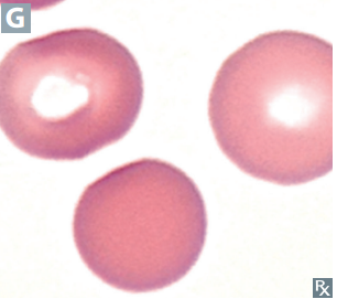
Spherocytes are associated with what conditions?
Hereditary spherocytosis; autoimmune hemolytic anemia
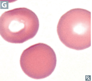
Spherocyte SA/V
Less than normal
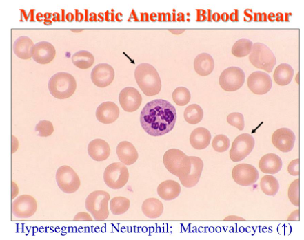
What are macro-ovalocytes?
Large, oval RBCs

Macro-ovalocytes are classically associated with what condition?
Megaloblastic anemia

What WBC finding may accompany macro-ovalocytes in megaloblastic anemia?
Hypersegmented neutrophils
What are target cells?
RBCs with a central area of hemoglobin surrounded by pallor and an outer hemoglobin ring, giving a bullseye appearance

Target cells are associated with what conditions?
HbC disease; splenia; liver disease; thalassemia
HALT

Target cell SA/V
Greater than normal
What are sickle cells?
Crescent/sickle-shaped RBCs caused by HbS polymerization
Sickle cells are associated with what condition?
Sickle cell anemia
What conditions promote sickling?
High altitude; acidosis; high HbS concentration; dehydration
Why does low oxygen cause RBC sickling?
Deoxygenated HbS polymerizes and distorts the RBC membrane into a sickle shape
How can you distinguish the main spiky/fragmented RBC morphologies?
Acanthocyte = irregular spikes, liver disease/ abetalipoproteinemia/ vitamin E deficiency
Echinocyte = uniform evenly spaced spikes, ESRD/pyruvate kinase deficiency/liver disease
Schistocyte = RBC fragments, MAHA/mechanical destruction
Degmacyte = bite taken out, G6PD deficiency
What are iron granules in the bone marrow?
Perinuclear mitochondria containing excess iron

Iron granules in the bone marrow are associated with what conditions?
Sideroblastic anemias, including:
Lead poisoning
Myelodysplastic syndromes
Chronic alcohol overuse

What stain is required to visualize iron granules in the bone marrow?
Prussian blue stain

What are ringed sideroblasts?
Erythroid precursors with iron-loaded mitochondria arranged in a ring around the nucleus
Why do ringed sideroblasts form in sideroblastic anemia?
Iron accumulates within mitochondria around the nucleus because it cannot be properly incorporated into heme
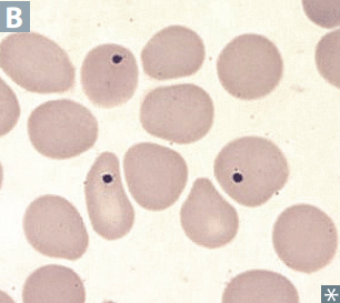
What are Howell-Jolly bodies?
Basophilic nuclear remnants within RBCs
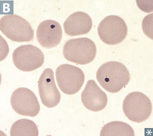
Do Howell-Jolly bodies contain iron?
No
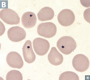
Howell-Jolly bodies are associated with what conditions?
Functional Hyposplenia (such as sickle cell disease)
Asplenia

Why do Howell-Jolly bodies appear in asplenia or functional hyposplenia?
The spleen normally removes nuclear remnants from circulating RBCs; when splenic function is absent or impaired, these remnants remain
What is basophilic stippling?
Basophilic ribosomal precipitates within RBCs

Does basophilic stippling contain iron?
No
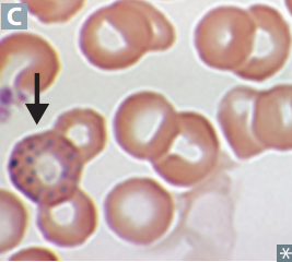
Basophilic stippling is associated with what conditions?
Sideroblastic anemias and thalassemias
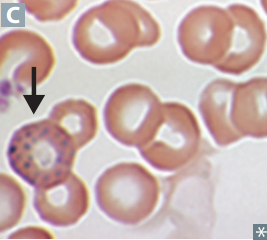
What cellular material produces basophilic stippling?
Aggregated rRNA
What are Pappenheimer bodies?
Basophilic iron-containing granules within RBCs

Do Pappenheimer bodies contain iron?
Yes

Pappenheimer bodies are associated with what condition?
Sideroblastic anemias

Differentiate between Pappenheimer bodies and basophilic stippling?
Pappenheimer bodies contain iron; basophilic stippling consists of ribosomal precipitates and does not contain iron
What are Heinz bodies?
Denatured and precipitated hemoglobin within RBCs

Heinz bodies are classically associated with what condition?
G6PD deficiency

Do Heinz bodies contain iron?
Yes, because they consist of precipitated hemoglobin
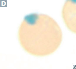
Why do Heinz bodies form in G6PD deficiency?
Oxidative stress damages and denatures hemoglobin, causing it to precipitate inside RBCs

What happens to Heinz bodies in the spleen?
Splenic macrophages remove them from RBCs, producing bite cells (Degmacytes)
What stain is required to visualize Heinz bodies?
Supravital stain, such as crystal violet
Howell-Jolly bodies vs basophilic stippling vs Pappenheimer bodies vs Heinz bodies
Howell-Jolly = nuclear remnants, no iron
Basophilic Stippling = ribosomal precipitates, no iron;
Pappenheimer = iron-containing granules
Heinz = denatured precipitated hemoglobin
What defines a microcytic, hypochromic anemia?
MCV < 80 fL with decreased hemoglobinization of RBCs
What are common causes of iron deficiency?
Chronic bleeding (eg, GI loss, heavy menstrual bleeding)
Malnutrition
Malabsorption disorders
GI surgery such as gastrectomy
Increased demand such as pregnancy

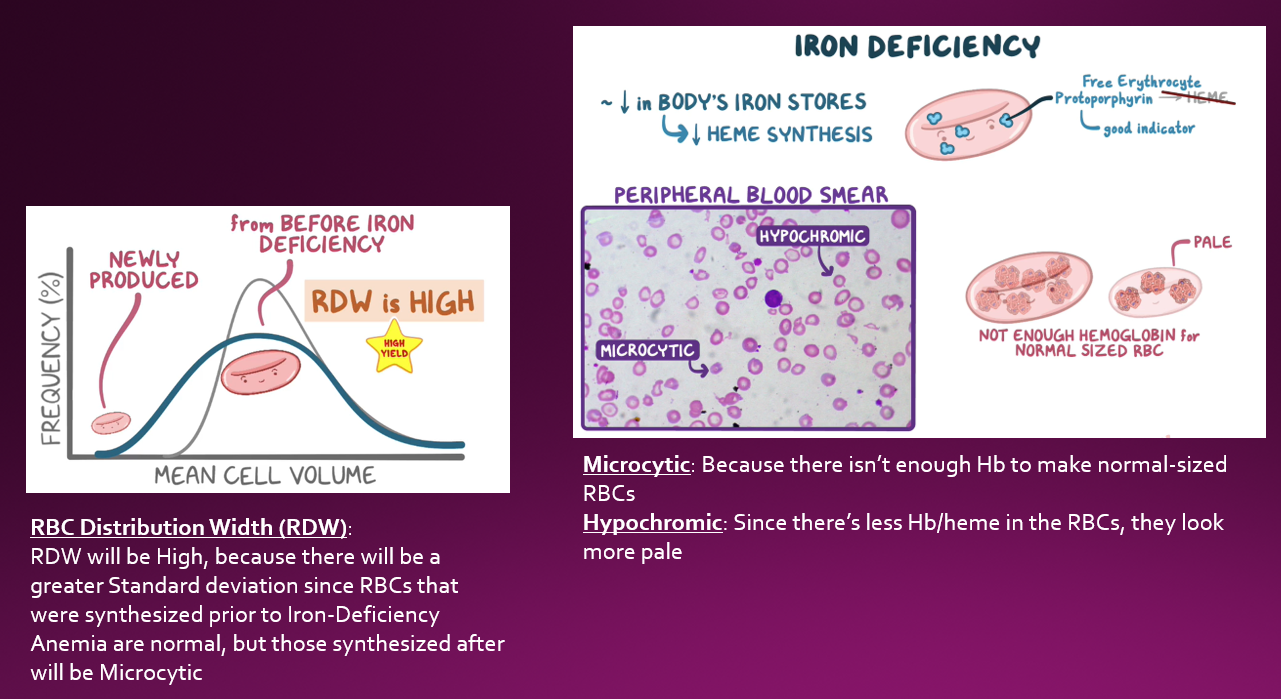
Iron Deficiency Anemia Iron studies
Low iron
High TIBC
Low ferritin
High free erythrocyte protoporphyrin
High RDW
Low RI.
(→ Microcytosis & hypochromasia & central pallor)
What happens to RDW in iron deficiency anemia?
Increased RDW
What happens to the reticulocyte index in iron deficiency anemia?
Decreased reticulocyte index

What RBC morphology is seen in iron deficiency anemia?
Microcytosis and hypochromasia with increased central pallor

What symptoms may be seen in iron deficiency anemia?
Fatigue;
Conjunctival pallor
Restless leg syndrome
Pica
Spoon nails (koilonychia)
What is pica?
Persistent craving and compulsive eating of nonfood substances
What nail abnormality can occur in iron deficiency anemia?
Koilonychia (spoon nails)
What oral finding may occur in iron deficiency anemia?
Glossitis
What esophageal findings may occur in iron deficiency anemia?
Esophageal webs
What causes alpha-thalassemia?
Alpha-globin gene deletions on chromosome 16 → decreased alpha-globin synthesis
How many alpha-globin genes are normally present?
4
In which populations is alpha-thalassemia more prevalent?
People of Asian and African descent
What RBC morphology may be seen in alpha-thalassemia?
Target cells
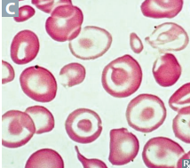
What is alpha-thalassemia minima?
Deletion of 1 alpha-globin gene
What is the clinical outcome of alpha-thalassemia minima?
No anemia; silent carrier
What is alpha-thalassemia minor?
Deletion of 2 alpha-globin genes
What is the clinical presentation of alpha-thalassemia minor?
Mild microcytic, hypochromic anemia
What are the two possible deletion patterns in alpha-thalassemia minor?
Cis deletion or trans deletion
What is a cis deletion in alpha-thalassemia?
Two alpha-globin gene deletions on the same chromosome
What disease results from deletion of 3 alpha-globin genes?
Hemoglobin H disease
What hemoglobin forms in Hemoglobin H disease?
Excess beta-globin forms β4 tetramers
What is the clinical severity of Hemoglobin H disease?
Moderate to severe microcytic hypochromic anemia
What disease results from deletion of all 4 alpha-globin genes?
Hemoglobin Barts disease