OMFPR 4: Making a Radiographic Image
 Call Kai
 Learn
Learn Practice Test
 Spaced Repetition
 Match
Match Flashcards
 Knowt Play
1/49

There's no tags or description
Looks like no tags are added yet.
Name | Mastery | Learn | Test | Matching | Spaced | Call with Kai | Chat |
|---|
No analytics yet
Send a link to your students to track their progress
50 Terms
Detail
Contrast resolution
Magnification and distortion
Diagnostic radiograph parameters
Image quality
Reliability of image to represent true state of anatomic region
Small
X-rays should originate from a _____ focal spot.
Long
SOD should be as ____ as possible.
Short
ORD should be as ____ as possible.
Parallel
Long axis of object should be _________ to receptor/film.
Perpendicular
Central ray should be ____________ to receptor.
Small
Does small or large focal spot produce sharper images with higher spatial resolution?
SOD
___ should be as long as possible.
Intra-oral radiography: collimated extensions: 8 inches, 12 inches, or 16 inches
How is a long SOD clinically achieved?
ORD (object to receptor distance)
___ should be as short as possible.
Closest
Place anatomy of interest ______ to receptor.
Parallelism
___________ between object and receptor decreases distortion and improves image sharpness.
Perpendicular
The central ray of the x-ray beam should be __________ to the long axis of the tooth to reduce geometric distortion.
Foreshortening
Central ray is perpendicular to the image receptor but the object is not parallel with the image receptor
Elongation
Central ray is perpendicular to the object but not to the image receptor
Foreshortening
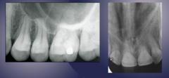
Foreshortening
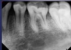
Elongation
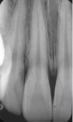
Paralleling technique
The central ray is directed at right angle to the central axes of object and receptor
Bisecting-angle technique
Central ray is directed at a right angle to the imaginary plane that bisects the angle formed by the image receptor and the central axis of the object
Paralleling
Preferred method for minimizing distortion
Sharpness
How well a boundary between two areas of differing radiodensity is revealed
Spatial resolution
Ability of an imaging system to differentiate between two nearby objects
Spatial resolution
How well a radiograph reveals small objects that are close together
Detail
Sharpness and resolution
Blurry vs. sharp
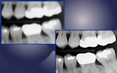
Density
Overall degree of darkening of a radiograph
Radiolucent
Radiolucent or radiopaque:
Air space, foramen, canal, suture, fossa, soft tissue
Radiopaque
Radiolucent or radiopaque:
Cortical bone, lamina dura, dentin, enamel, metal restorations
Cancellous bone
What structure falls between radiolucent and radiopaque?
mA
Exposure time
Filtration
Collimation
Distance
Factors affecting density
mA
Exposure time
Factors that increase density / darker image
Filtration
Collimation
Distance
Factors that decrease density / lighter image
Lighter
Artifacts (lead aprons, film reversal, earrings, thyroid collars) lead to _______ areas in the film.
Contrast
Difference in densities between the light and dark regions in a radiograph
Low vs. high contrast
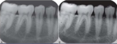
Long scale (low contrast)
Long or short scale:
Small gradual change among different optical densities
High kVp
Short scale (high contrast)
Long or short scale:
Large or abrupt differences among optical densities
Low kVp
Good vs. poor contrast
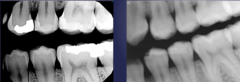
Fog
Scatter radiation (Compton or coherent interactions) produces ___.
Use a 3D imaging modality
CBCT
How to gather 3D radiologic information?
Right angle technique
Tube shift technique (SLOB rule, BOR / Clark's rule)
Special studies
Radiographic techniques in localization
Right angle technique
Obtain 2 radiographs at 90 degrees to each other of the area of interest e.g. periapical and occlusal radiographs, lateral cephalometric and postero-anterior view of skull
Same
Lingual
Opposite
Buccal
For tube shift technique, what is the SLOB rule for localization?
Lingual
Object moves in same direction as x-ray tubehead. Is it lingual or buccal?
Buccal
Object moves in opposite direction as x-ray tubehead. Is it lingual or buccal?
Lingual
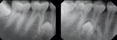
Is the impacted tooth buccal or lingual?
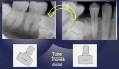
Buccal
Where is the impacted premolar located relative to the first molar?
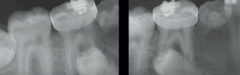
Lingual
Evaluate the position of the impacted premolar.