Anatomy 2300 Unit 2: Muscles of the Leg and Foot
 Call Kai
 Learn
Learn Practice Test
 Spaced Repetition
 Match
Match Flashcards
 Knowt Play
1/33

There's no tags or description
Looks like no tags are added yet.
Name | Mastery | Learn | Test | Matching | Spaced | Call with Kai | Chat |
|---|
No analytics yet
Send a link to your students to track their progress
34 Terms
Dorsiflex
Movement that bends the foot upward at the ankle.
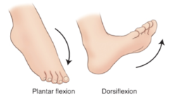
Plantar flex
Movement that points the toes downward away from the shin.

Tibialis
Muscle of the lower leg involved in inversion and ankle movement.

Fibularis
Muscle of the lower leg involved in eversion and ankle movement.

Crural Fascia
Deep fascia of the leg that encases and protects the lower leg muscles.

Hallux
The anatomical term for the big toe.

Tibialis anterior
- Action & Innervation?
Definition: A muscle located on the front (anterior) side of the lower leg, along the lateral side of the tibia. It is the main muscle responsible for lifting the foot upward and helps control the foot during walking.
- Action: Dorsiflex ankle (foot); Invert foot
- Innervation: Deep fibular nerve

Extensor hallucis longus
- Action & Innervation?
Definition: A long, slender muscle in the anterior compartment of the lower leg that runs from the fibula to the big toe, deep to the tibialis anterior. It primarily moves the big toe and assists with lifting the foot.
- Action: Dorsiflex ankle (foot); Extend hallux
- Innervation: Deep fibular nerve
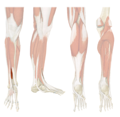
Extensor digitorum longus
- Action & Innervation?
Definition: A long, slender muscle located in the anterior compartment of the lower leg. It lies along the lateral side of the tibialis anterior and extends from the upper part of the tibia/fibula region down toward the ankle. Near the ankle, it divides into four tendons that travel across the top of the foot and attach to digits 2-5 (the four lesser toes).
- Action: Dorsiflex ankle (foot); Extend digits 2-5
- Innervation: Deep fibular nerve

Fibularis (peroneus) tertius
- Action & Innervation?
Definition: A small, thin muscle located in the anterior compartment of the lower leg, along the lower part of the fibula. It lies lateral to the extensor digitorum longus, and its tendon passes down the front of the ankle and attaches to the base of the 5th metatarsal on the top of the foot.
- Action: Dorsiflex ankle (foot); Evert foot
- Innervation: Deep fibular nerve
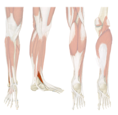
Fibularis (peroneus) longus
- Action & Innervation?
Definition: A long, superficial muscle on the lateral side of the lower leg, running along the fibula. Its tendon passes behind the lateral malleolus and underneath the foot to attach on the medial side of the foot.
- Action: Plantarflex ankle (foot); Evert foot
- Innervation: Superficial fibular nerve
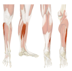
Fibularis (peroneus) brevis
- Action & Innervation?
Definition: A shorter, deep muscle in the lateral compartment of the lower leg, located deep to the fibularis longus along the lower fibula. Its tendon passes behind the lateral malleolus and attaches to the base of the 5th metatarsal.
- Action: Plantarflex ankle (foot); Evert foot
- Innervation: Superficial fibular nerve

Gastrocnemius
- Action & Innervation?
- Posterior leg layer?
Definition: A large, superficial muscle on the back of the lower leg (calf) with two heads—medial and lateral—that originate above the knee. It joins the soleus to form the calcaneal (Achilles) tendon, which attaches to the heel.
- Action: Flex knee (leg); Plantarflex ankle (foot)
- Innervation: Tibial nerve
- Superficial Posterior leg

Soleus
- Action & Innervation?
- Posterior leg layer?
Definition: A broad, flat muscle located deep to the gastrocnemius in the posterior compartment of the lower leg. It runs from the upper tibia and fibula to the heel through the calcaneal (Achilles) tendon.
- Action: Plantarflex ankle (foot)
- Innervation: Tibial nerve
- Superficial Posterior leg

Plantaris
- Action & Innervation?
- Posterior leg layer?
Definition: A small, thin muscle located in the posterior compartment of the lower leg, between the gastrocnemius and soleus. Its long tendon runs down the leg toward the heel.
- Action: Flex knee (leg); plantarflex ankle (foot)
- Innervation: Tibial nerve
- Superficial Posterior leg

Popliteus
- Action & Innervation?
- Posterior leg layer?
Definition: A small, deep muscle located at the back of the knee, deep to the gastrocnemius. It runs from the lateral femoral condyle to the posterior tibia and helps "unlock" the knee from a fully extended position.
- Action: Flex knee (leg) "unlocks knee"; Medially rotate tibia
- Innervation: Tibial nerve
- Deep Posterior leg
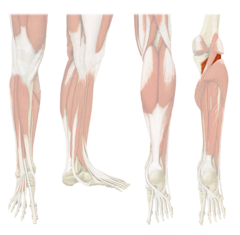
Flexor digitorum longus
- Action & Innervation?
- Posterior leg layer?
Definition: A long, deep muscle in the posterior lower leg, located along the medial side of the tibia. Its tendon passes behind the medial malleolus and divides into four tendons that attach to the distal phalanges of toes 2-5.
- Action: Plantarflex ankle (foot); Flex digits 2-5
- Innervation: Tibial nerve
- Deep Posterior leg

flexor hallucis longus
- Action & Innervation?
- Posterior leg layer?
Definition: A long muscle in the deep posterior lower leg, running along the lateral, back side of the fibula and passing behind the ankle to the big toe. It primarily flexes the big toe and also assists with plantarflexion of the foot.
- Action: Plantarflex ankle (foot); Flex hallux
- Innervation: Tibial nerve
- Deep Posterior leg

Tibialis posterior
- Action & Innervation?
- Posterior leg layer?
Definition: A deep posterior leg muscle located between the flexor digitorum longus and flexor hallucis longus; it runs behind the medial ankle and helps plantarflex and invert the foot while supporting the medial arch.
- Action: Plantarflex ankle (foot); Invert foot
- Innervation: Tibial nerve
- Deep Posterior leg

Calcaneal (Achilles) tendon
Powerful tendon formed by the gastrocnemius and soleus that inserts into the calcaneus.

Tarsal Tunnel
A narrow passageway on the medial side of the ankle, located behind the medial malleolus, that allows important tendons, blood vessels, and the tibial nerve to pass from the lower leg into the foot.
*From anterior to posterior
- Tendon of tibialis posterior (Tom)
- Tendon of flexor digitorum longus (Dick)
- Posterior tibial artery (And)
- Tibial nerve (Nervous)
- Tendon of flexor hallucis longus (Harry)
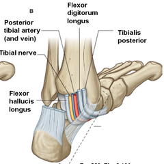
"Tom, Dick, And Nervous Harry" Mneumonic
The structures pass through the tarsal tunnel in this order from anterior to posterior:
*from anterio to posterior
- Tendon of tibialis posterior (Tom)
- Tendon of flexor digitorum longus (Dick)
- Posterior tibial artery (And)
- Tibial nerve (Nervous)
- Tendon of flexor hallucis longus (Harry)
Extensor hallucis brevis
- Action & Innervation?
Definition: The extensor hallucis brevis is a small muscle on the dorsum (top) of the foot that extends the big toe at the metatarsophalangeal joint and helps lift the toe upward.
- Action: Extend hallux
- Innervation: Deep fibular nerve

Extensor digitorum brevis
- Action & Innervation?
Definition: The extensor digitorum brevis is a small muscle on the top (dorsum) of the foot that extends the toes 2-4, helping lift them upward at the toe joints.
- Action: Extend digits 2-4
- Innervation: Deep fibular nerve

Abductor hallucis
- Action & Innervation?
Definition: The abductor hallucis is a muscle along the medial side of the foot that abducts (moves away from the other toes) and flexes the big toe and helps support the medial arch of the foot.
- Action: Abduct hallux
- Innervation :Medial plantar nerve

Flexor digitorum brevis
- Action & Innervation?
Definition: The flexor digitorum brevis is a muscle on the bottom (plantar surface) of the foot that flexes toes 2-5, especially at the proximal interphalangeal joints, and helps support the arches of the foot.
- Action: Flex digits 2-5
- Innervation :Medial plantar nerve

Abductor digiti minimi
- Action & Innervation?
Definition: The abductor digiti minimi is a muscle along the lateral side of the foot that abducts (moves away from the other toes) and flexes the little toe and helps support the lateral arch of the foot.
- Action: Abduct digit 5
- Innervation: Lateral plantar nerve

Sciatic Nerve
Main nerve of the posterior thigh, dividing into the tibial and common fibular nerves; it primarily supplies the posterior thigh muscles.
Tibial Nerve
Primarily supplies the posterior leg (superficial and deep compartments) and the plantar foot through its medial and lateral plantar branches.
Lateral plantar nerve
Supplies primarily the lateral plantar foot muscles and sensation to much of the sole.
Medial plantar nerve
Supplies primarily the medial plantar foot muscles and sensation to part of the sole.
Common Fibular Nerve
Primarily supplies the anterior and lateral compartments of the leg as well as the dorsum of the foot through its two main branches.
Superficial fibular nerve
Primarily supplies the lateral compartment of the leg.
Deep fibular nerve
Primarily supplies the anterior compartment of the leg and the dorsal foot muscles.