9 Secondary Glaucomas
 Call Kai
 Learn
Learn Practice Test
 Spaced Repetition
 Match
Match Flashcards
 Knowt Play
1/205

There's no tags or description
Looks like no tags are added yet.
Name | Mastery | Learn | Test | Matching | Spaced | Call with Kai | Chat |
|---|
No analytics yet
Send a link to your students to track their progress
206 Terms
What defines a secondary glaucoma, and how is it related to POAG?
Secondary glaucomas produce the characteristic optic neuropathy seen in POAG, but the elevated IOP is due to an identifiable secondary cause rather than typical POAG or PACG mechanisms.
What are the major categories of causes of secondary glaucoma?
Secondary glaucoma can result from:
Another acquired ocular condition
An acquired systemic condition
Medication use
Trauma
What is the key mechanism shared by secondary glaucomas?
Secondary glaucomas involve increased IOP through mechanisms outside of typical POAG or PACG, ultimately causing glaucomatous optic nerve damage.
What are examples of secondary glaucomas caused by ocular syndromes involving material or pigment deposition?
Pigmentary glaucoma, secondary to pigment dispersion syndrome
Pseudoexfoliation glaucoma, secondary to pseudoexfoliation syndrome
What inflammatory or uveitic conditions can lead to secondary glaucoma?
Inflammatory glaucoma can occur with regular uveitis or special uveitic episodes such as:
Glaucomatocyclitic crisis, also called Posner-Schlossman syndrome
Fuchs heterochromic iridocyclitis
UGH syndrome
What are other major examples of secondary glaucoma?
Other secondary glaucomas include:
Steroid-induced glaucoma
Traumatic glaucoma
Neovascular glaucoma
Phacolytic and phacomorphic glaucoma
Developmental glaucomas, although these are mostly managed by OMDs and not covered in this lecture
What is the main mechanism of pigmentary dispersion syndrome?
Pigmentary dispersion syndrome involves liberation of pigment granules from the posterior pigmented epithelium on the back surface of the iris.
What is the relationship between pigmentary dispersion syndrome and pigmentary glaucoma?
Pigmentary dispersion syndrome becomes pigmentary glaucoma when optic nerve head damage develops from the pigment-related increase in IOP.
What are the key epidemiologic features of pigmentary dispersion syndrome?
PDS may occur in up to 2.5% of Caucasians, with unclear prevalence in patients of African ancestry. Onset can begin as early as the mid-teens, but diagnosis usually occurs between ages 20 to 40.
How does sex distribution differ between pigmentary dispersion syndrome and pigmentary glaucoma?
PDS has no clear male or female predilection, but more males convert from PDS to pigmentary glaucoma than females.
How does pigmentary dispersion syndrome typically present between the two eyes?
Like other glaucomas, PDS is typically bilateral but asymmetric.
What anterior segment anatomy is commonly associated with pigmentary dispersion syndrome?
Patients with PDS are typically myopic and have a deep anterior chamber.
What is the mechanism causing pigment release in pigmentary dispersion syndrome?
Posterior bowing of the iris causes the iris to rub against the zonules and anterior lens surface during normal iris movement, mechanically liberating pigment from the posterior iris pigment epithelium.
Why does a concave iris configuration increase risk for pigment dispersion?
A concave or posteriorly bowed iris increases contact between the posterior iris and zonules, leading to repeated friction and pigment liberation.
After pigment is liberated in PDS, where does it go and why does that matter clinically?
Liberated pigment enters the aqueous humor and can accumulate in the trabecular meshwork, increasing resistance to aqueous outflow and raising IOP.
What are the two main ways pigment affects the trabecular meshwork in PDS?
Pigment can:
Physically block the trabecular meshwork, mechanically resisting aqueous outflow
Compromise TM endothelial cells by forcing phagocytosis of pigment granules instead of maintaining active transport/outflow functions
What is the mechanism by which PDS can progress to pigmentary glaucoma?
Posterior iris bowing causes pigment liberation → pigment accumulates in the trabecular meshwork → aqueous outflow resistance increases → IOP rises → optic nerve head damage develops.
What classic iris finding is associated with pigmentary dispersion syndrome?
Radial midperipheral iris transillumination defects caused by loss of pigment from the iris.
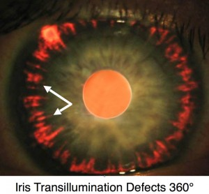
How are iris transillumination defects in PDS best visualized?
They are best seen with retroillumination.
What is important to remember about early iris transillumination defects in PDS?
They can be subtle early in the syndrome, so careful retroillumination is needed to detect the classic radial midperipheral pigment loss.
What is the classic sequence of findings in pigmentary dispersion syndrome?
Myopia/deep anterior chamber → posterior iris bowing → iris-zonule friction → pigment liberation → pigment in aqueous/TM → radial midperipheral iris transillumination defects ± elevated IOP.
What is a Krukenberg spindle in pigmentary dispersion syndrome?
A Krukenberg spindle is vertically oriented pigment accumulation on the posterior corneal endothelium from liberated iris pigment circulating in the aqueous.
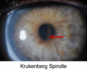
Besides the corneal endothelium, where else can liberated pigment accumulate in PDS?
Pigment can also accumulate on the iris surface, in addition to the corneal endothelium and trabecular meshwork.
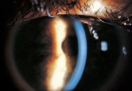
What anterior segment finding should make you suspect pigment dispersion syndrome on slit lamp exam?
A Krukenberg spindle on the posterior corneal endothelium should raise suspicion for PDS and prompt further evaluation, especially gonioscopy.
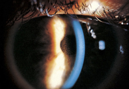
How does pigment accumulation in the angle contribute to elevated IOP in PDS?
Pigment accumulates in the trabecular meshwork, obstructing aqueous outflow and increasing IOP.
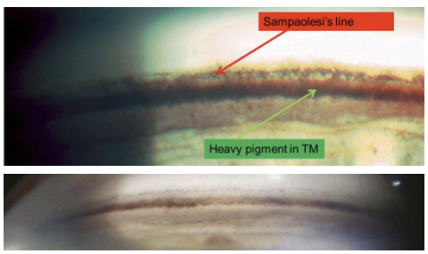
What is Sampolesi’s line?
Sampolesi’s line is extra pigment deposited anterior to Schwalbe’s line.
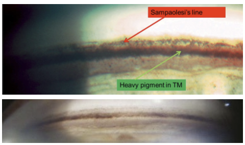
What exam is necessary when Krukenberg spindle or iris transillumination defects are seen?
Gonioscopy is necessary to evaluate angle pigmentation, trabecular meshwork obstruction, and signs such as Sampolesi’s line.
What is Zentmayer line in pigmentary dispersion syndrome?
Zentmayer line is pigment deposition on the zonules or peripheral anterior lens surface.
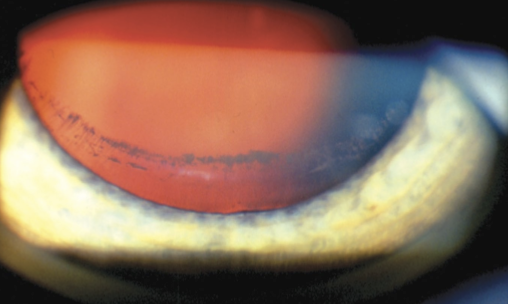
Why is Zentmayer line clinically important in PDS?
Zentmayer line is pathognomonic for pigmentary dispersion syndrome.
When can Zentmayer line be seen?
Zentmayer line is only seen during dilation, because the peripheral lens surface and zonules must be visible.
What are the classic pigment deposition signs of PDS and where are they located?
Krukenberg spindle: posterior corneal endothelium
Sampolesi’s line: anterior to Schwalbe’s line
Heavy TM pigment: trabecular meshwork/angle
Zentmayer line: zonules or peripheral anterior lens surface
Iris transillumination defects: radial midperipheral iris pigment loss
When does pigmentary dispersion syndrome become pigmentary glaucoma?
PDS becomes pigmentary glaucoma when pigment-related IOP elevation causes glaucomatous optic neuropathy.
What percentage of PDS patients are estimated to progress to pigmentary glaucoma?
About 25% of patients with PDS progress to pigmentary glaucoma.
How common is pigmentary glaucoma among all glaucomas?
Pigmentary glaucoma accounts for about 4.4% of all glaucomas.
What can heavy angle pigmentation do during gonioscopy in PDS or pigmentary glaucoma?
Heavy pigment can obscure all angle structures, making gonioscopic assessment more difficult.
Why does the risk of IOP spikes and conversion to pigmentary glaucoma decrease with age in PDS?
Risk decreases due to “burnout”:
The iris has less pigment left to release
Age-related lens growth pushes the posterior iris away from the zonules
Less iris-zonule friction means less pigment liberation
How does lens anatomy affect pigment release in younger versus older PDS patients?
Younger patients have a thinner lens, allowing more posterior iris bowing and iris-zonule friction. Later in life, the thickened lens pushes the iris away from the zonules, reducing friction and pigment release.
What age is clinically important when estimating conversion risk from PDS to pigmentary glaucoma?
Patients with PDS who pass age 45 without significant IOP elevation have a relatively low likelihood of converting to pigmentary glaucoma.
What are the key components of the clinical workup for pigmentary dispersion syndrome?
Workup includes:
Corneal evaluation for Krukenberg spindle
Iris retroillumination for radial transillumination defects
Gonioscopy for heavy TM pigment/Sampolesi’s line
IOP measurement to detect spikes
How often should newly diagnosed PDS patients have IOP checked?
Newly diagnosed PDS patients need IOP checks every 3 to 4 months during the first year, then every 6 months thereafter.
Why should young or newly diagnosed PDS patients have IOP checked immediately after vigorous exercise?
Vigorous exercise can cause adrenaline-induced pupillary dilation, which may increase iris-zonule friction and trigger IOP spikes.
What IOP measurement is recommended for all PDS patients after dilation?
All PDS patients should have a post-dilation IOP measurement because dilation can provoke pigment release and IOP elevation.
How is pigmentary glaucoma generally treated initially?
Pigmentary glaucoma is generally treated like POAG, using IOP-lowering medications and escalating therapy as needed.
Which medication class works well for pigmentary glaucoma despite heavy pigment involvement?
Prostaglandin analogs (PGAs) work well in the presence of pigment and are commonly used to lower IOP.
Why can laser trabeculoplasty be effective in pigmentary glaucoma?
ALT and SLT can be effective because pigment in the trabecular meshwork improves laser target absorption, especially in younger patients. Pigment is a predictor of success for ALT.
What is a limitation of laser trabeculoplasty in pigmentary glaucoma compared with POAG?
Laser effects may wear off faster than in POAG. After 3 years, fewer than 50% of pigmentary glaucoma patients continue to show IOP reduction after initial laser therapy.
Why might pilocarpine help young patients with active pigment liberation?
Pilocarpine can reduce pigment liberation by decreasing iris concavity, which reduces iris-zonule friction. It also increases aqueous outflow.
What mechanism makes pilocarpine useful specifically in pigmentary dispersion syndrome?
Pilocarpine causes miosis, which can flatten the posteriorly bowed iris and reduce mechanical rubbing between the iris and zonules.
What ocular side effects limit pilocarpine use in young or pre-presbyopic patients?
Pilocarpine can cause significant side effects, including:
Accommodative spasm
Miosis
Blurred vision
What serious retinal risk is increased with pilocarpine, and why is this especially relevant in PDS?
Pilocarpine increases the risk of retinal detachment. This is especially relevant because PDS patients are often myopic, and myopia already increases retinal detachment risk.
How can laser peripheral iridotomy theoretically help pigmentary dispersion syndrome?
LPI may reduce iris concavity, decreasing posterior iris bowing and reducing iris-zonule friction that causes pigment liberation.
Which PDS patients may benefit most from LPI?
LPI may be most helpful in patients younger than 45 years with extreme peripheral iris concavity who are still actively liberating pigment.
Why does the potential benefit of LPI decrease after about age 45?
After age 45, many patients have already liberated much of their pigment, and age-related lens thickening naturally reduces iris-zonule friction. Therefore, LPI benefit likely diminishes.
How do pilocarpine and LPI both target the mechanism of PDS?
Both aim to reduce posterior iris concavity and decrease iris-zonule friction, reducing pigment release.
Pilocarpine: pharmacologically flattens iris via miosis
LPI: anatomically reduces iris concavity by altering pressure dynamics
What is pseudoexfoliation syndrome, and what gene is associated with it?
Pseudoexfoliation syndrome (PXF/PXE) is a systemic condition involving abnormal basement membranes and “elastosis,” associated with mutation in the LOXL1 gene.
What systemic diseases are associated with pseudoexfoliation syndrome?
PXF is associated with systemic vascular and neurodegenerative conditions, including:
Vascular insufficiency
TIAs
Heart attack
Stroke
Alzheimer’s disease
What are the key epidemiologic features of pseudoexfoliation syndrome?
PXF affects over 70 million people worldwide, is most common in Caucasians of Scandinavian descent, and is usually diagnosed between ages 60 and 80.
How important is pseudoexfoliation syndrome as a cause of open-angle glaucoma worldwide?
PXF causes about 20 to 25% of open-angle glaucoma worldwide, making it a major secondary glaucoma etiology.
What is the main ocular mechanism of pseudoexfoliation syndrome?
Weak abnormal basement membranes lead to loss/sloughing of epithelial cells, primarily from the iris and ciliary body, causing exfoliative material to accumulate in the anterior chamber.
Where is pseudoexfoliative material most classically observed in the eye?
Exfoliative cellular debris collects on nearby anterior segment tissues and is most classically seen on the anterior lens surface.
What are the classic zones of pseudoexfoliative material on the anterior lens capsule?
The classic pattern includes:
Central disc of material
Clear zone
Peripheral band of exfoliative material
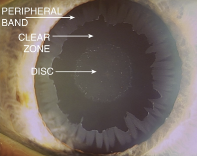
Why is pupillary dilation necessary when evaluating for pseudoexfoliation syndrome?
Dilation is needed to detect the classic bull’s-eye deposition pattern on the anterior lens surface, especially the peripheral exfoliative material.
What causes the clear zone in the pseudoexfoliation bull’s-eye pattern?
The clear zone forms because normal pupillary movement of the iris sweeps away exfoliative debris from that portion of the anterior lens surface.
What is the classic anterior lens appearance in pseudoexfoliation syndrome?
A bull’s-eye pattern on the anterior lens capsule, with a central disc, surrounding clear zone, and peripheral band of pseudoexfoliative material.
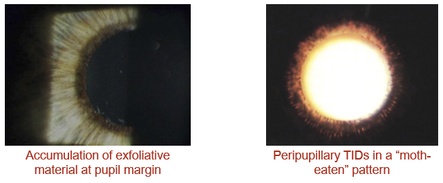
What pupil-margin finding is associated with pseudoexfoliation syndrome?
Pseudoexfoliative material can accumulate at the pupillary margin, appearing as flaky or dandruff-like material around the pupil.
What iris transillumination defect pattern is associated with pseudoexfoliation syndrome?
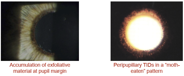
PXF can cause peripupillary transillumination defects in a “moth-eaten” pattern.
How do iris transillumination defects differ between pigment dispersion syndrome and pseudoexfoliation syndrome?
PDS: radial midperipheral iris transillumination defects
PXF: peripupillary “moth-eaten” transillumination defects
How does pseudoexfoliation syndrome affect the zonules?
Exfoliative material can accumulate on the zonules, causing them to become frayed, weak, or broken.
What is the main mechanism of IOP elevation in pseudoexfoliation syndrome?
Fibrin-like, sticky pseudoexfoliative material clogs the trabecular meshwork outflow channels, increasing outflow resistance and raising IOP.
Besides trabecular outflow obstruction, what other outflow pathway is impaired in PXF?
Uveoscleral outflow is also impaired.
When does pseudoexfoliation syndrome become pseudoexfoliation glaucoma?
PXF becomes pseudoexfoliation glaucoma (PXG) when elevated IOP causes glaucomatous optic neuropathy.
What gonioscopic findings can be seen in pseudoexfoliation syndrome?
Gonioscopy may show excessive pigment in the angle and Sampolesi’s line.
How often do patients with pseudoexfoliation syndrome convert to glaucoma?
Patients with PXF may convert to glaucoma up to 60% of the time, with conversion reported more often in women than men.
How does pseudoexfoliation glaucoma compare with POAG at presentation?
Compared with POAG, PXG often presents with:
Higher IOP
Greater IOP fluctuations
Worse optic nerve head damage
Worse visual field damage at diagnosis
Why is pseudoexfoliation glaucoma considered more aggressive than POAG?
PXG tends to progress more rapidly after diagnosis, has greater IOP fluctuation, worse baseline ONH/VF damage, and a higher risk of treatment failure.
What is the treatment implication of pseudoexfoliation glaucoma being more aggressive than POAG?
The target IOP needs to be lower than in typical POAG because PXG has faster progression and greater risk of treatment failure.
Why does pseudoexfoliation glaucoma require more aggressive IOP management than typical POAG?
PXG has large IOP fluctuations, pressure spikes, and rapid visual field loss, so treatment usually requires a lower target IOP than typical POAG.
What is the recommended first-line medication class for pseudoexfoliation glaucoma?
Prostaglandin analogs (PGAs) are recommended first-line because PXG responds best to therapies that enhance outflow.
What role do ALT and SLT play in pseudoexfoliation glaucoma treatment?
ALT or SLT can be effective initial treatment options and may delay the need for medical therapy by up to 8 years, but success gradually declines over time.
What is a limitation of laser trabeculoplasty in PXG over time?
The IOP-lowering effect can wear off. After about 6 years, the effect wears off in roughly 50% of patients.
What general treatment strategy is preferred for medically treating pseudoexfoliation glaucoma?
Outflow enhancement is preferred because PXG involves impaired aqueous outflow from sticky fibrin-like pseudoexfoliative material clogging the trabecular meshwork.
When may pilocarpine be used in pseudoexfoliation glaucoma?
If PGA therapy fails, pilocarpine may be used, often dosed as 2% QHS, especially in older patients.
Why is pilocarpine often better tolerated in PXG than in pigmentary glaucoma?
PXG patients are typically older, so accommodative side effects are often less problematic than in young pre-presbyopic PDS patients. However, miosis can worsen vision in patients with dense nuclear sclerosis.
Why should pilocarpine and prostaglandin analogs theoretically not be used together?
Pilocarpine contracts the ciliary body muscle, limiting extracellular space between muscle fibers where PGAs act to increase uveoscleral outflow.
What are the possible outcomes if pilocarpine and PGAs are combined?
Results are variable:
IOP lowering may be additive
IOP may increase
The combination may have no additional effect
Why are some POAG treatments less effective in pseudoexfoliation glaucoma?
PXG has a different mechanism than POAG, with sticky fibrin-like material impairing outflow, so treatments effective in POAG may not produce the expected IOP reduction in PXG.
Why is cataract surgery not curative for pseudoexfoliation glaucoma?
Cataract surgery does not eliminate the underlying pseudoexfoliative process or reliably normalize IOP, so it is not curative for PXG.
Why is cataract surgery higher risk in patients with pseudoexfoliation syndrome?
PXF causes weak zonules, compromised capsules, and poor pupillary dilation, increasing surgical complication risk.
Why are aqueous suppressants not recommended as initial therapy for PXG?
Lowering aqueous production may reduce the pressure-driven movement of aqueous through the trabecular meshwork, causing aqueous to become more stagnant rather than clearing obstructive material.
What is the treatment contrasts between POAG and PXG?
POAG: aqueous suppressants and outflow enhancers can both be standard options
PXG: prioritize outflow enhancement with PGAs or laser because the main problem is impaired outflow from exfoliative material in the TM
What proportion of patients on topical steroids develop a clinically significant IOP rise?
About 3 to 30% of individuals treated with topical steroids develop an IOP increase of about 6 to 15 mmHg.
When does steroid-induced IOP elevation typically occur after starting topical steroids?
It typically occurs after at least 10 days of steroid use, but can appear anywhere from 1 to 4 weeks after initiation.
Who is at highest risk for steroid-induced glaucoma?
Risk is highest in:
Older adults
Patients with existing glaucoma
Patients with a family history of glaucoma
What is the main mechanism of steroid-induced glaucoma?
Steroids decrease aqueous outflow despite an open angle by altering the microstructure of the trabecular meshwork, increasing outflow resistance and raising IOP.
What genes are associated with steroid-induced glaucoma susceptibility?
Steroid-induced glaucoma is associated with alterations in MYOC, GLC1A, and/or TIGR genes.
What determines how much IOP rises with steroid use?
The IOP rise is proportional to the steroid’s:
Potency
Intraocular availability/penetration
Which topical steroids are more likely to cause larger IOP increases?
More potent and more penetrative steroids cause larger pressure rises. High-yield examples include:
Dexamethasone 0.1%: about 22 mmHg average rise
Durezol/difluprednate: about 17.8 mmHg rise
Prednisolone 1%: about 10 mmHg rise
What is the first step in treating steroid-induced glaucoma?
Stop or taper the topical steroid if clinically possible.
What should be done if a steroid responder still needs anti-inflammatory steroid treatment?
Consider switching to a less penetrative or lower-risk steroid, while monitoring IOP closely.
How should IOP be treated if steroid-induced pressure elevation persists?
Treat with topical beta-blockers or other aqueous suppressants until IOP normalizes.