Lecture Final - 231 - Lectures 14, 15, 16
 Call Kai
 Learn
Learn Practice Test
 Spaced Repetition
 Match
Match Flashcards
 Knowt Play
1/44

There's no tags or description
Looks like no tags are added yet.
Name | Mastery | Learn | Test | Matching | Spaced | Call with Kai | Chat |
|---|
No analytics yet
Send a link to your students to track their progress
45 Terms
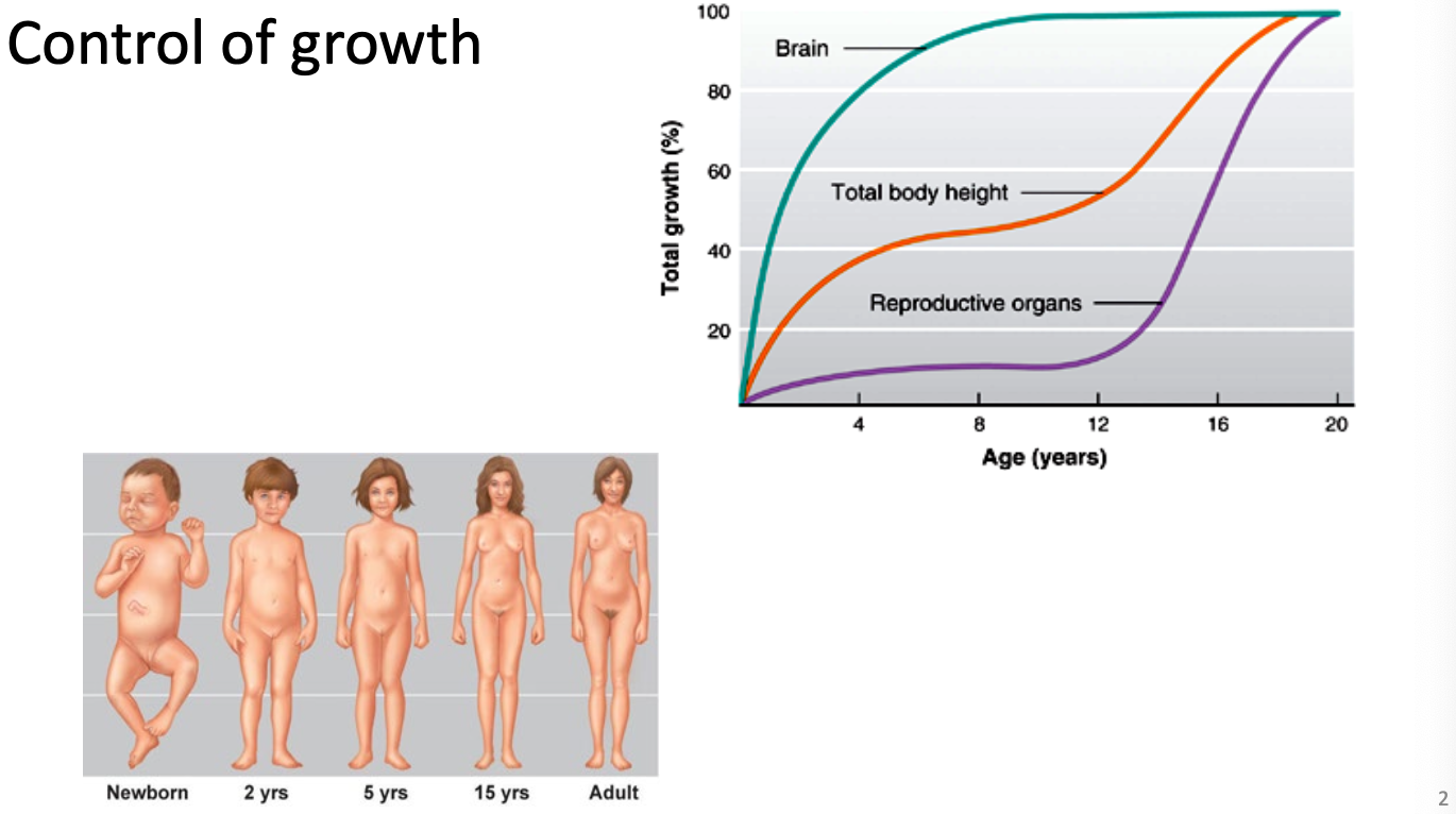
Control of growth
Two growth spurts
Bran, total body height, and reproductive organs do not grow at same rate
Control of growth- factors
Endocrine factors = promotes or inhibits growth
Growth hormone
thyroid hormone
sex steroids
cortisol
Environmental factors = need proper nutrition to grow
Nutrition
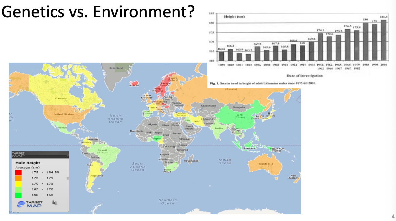
Genetics vs environment
Height mostly due to genetics but better acces to food and nutrition increases height
Growth hormone
ANTERIOR PITUITARY GLAND secretes GROWTH HORMONE to the LIVER to produce INSULIN-LIKE GROWTH FACTOR
Insulin-like growth factor does most of growth for cartilage and bone
Growth hormone has an anti-insulin effect that shifts the body toward using fat for energy
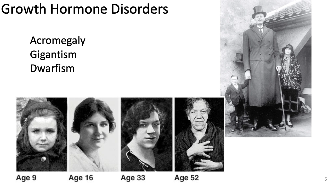
Growth Hormone Disorders
Acromegaly
Growth hormone released in adulthood thickens cartilage but do not grow taller
Gigantism
Excess growth hormone during childhood
Dwarfism
Decreased Growth hormone in childhood
*Usually caused by pituitary tumor which hyper secretes
Steriod Hormones and Thyroid Hormone
Glucocorticoids (cortisol)
decreased bone productoin
cell death of osteocytes
Estradiol and Testosterone
Increased bone formation
decreases osteoclast activity
Important during puberty
Ovaries = estrogen faster at closing epiphyseal plates
Testosterone slower at closing epiphyseal plates
Thyroid hormone
permissive effects for other hormones
stimulates release growth factors
How are bones held together to form joints?
Structural classification (BASED ON TYPES OF CT)
Fibrous
Cartilaginous
Synovial
Functional classification (AMOUNT OF MOVEMENT)
Synarthrosis
Amphiarthrosis
Diarthrosis
*Typically go together
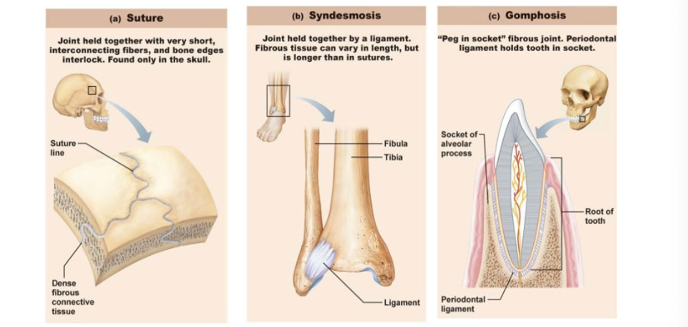
Structural type: FIBROUS
Suture = HELD TOGETHER BY FIBERS
Only in skull
Bones overlap, and fibers overlap
Syndesmosis
JOINT HELD BY LIGAMENT
Gomphosis
HOLD TOOTH
Structural Type: CARTILAGINOUS
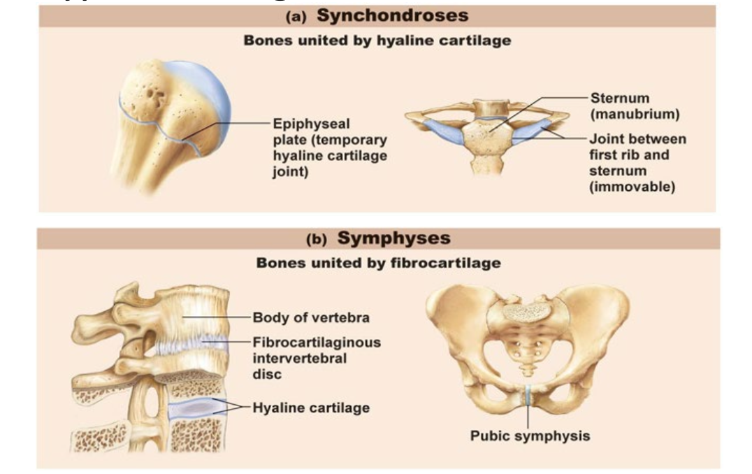
Synchondroses
United by hyaline cartilage
Symphyses
United by fibrocartilage
Structural Type: SYNOVIAL
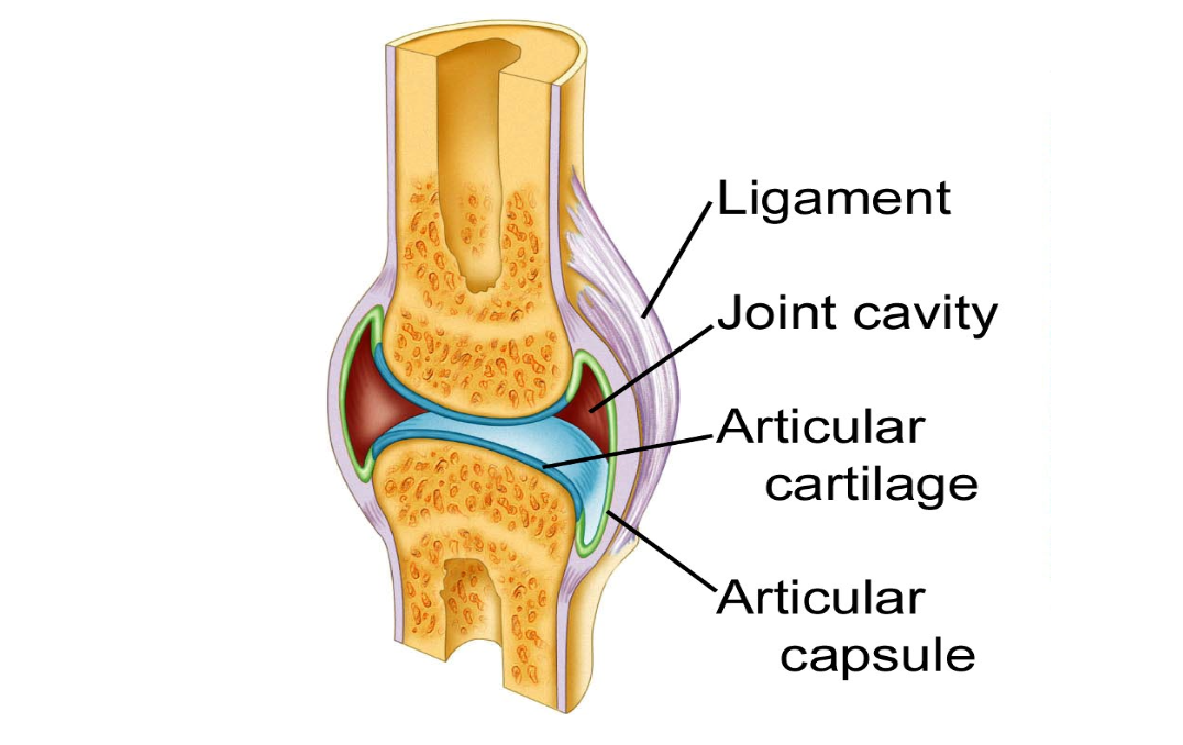
Held by ligament, but has synovial membrane around joint
Gap between bones
Other synovial joint structures:
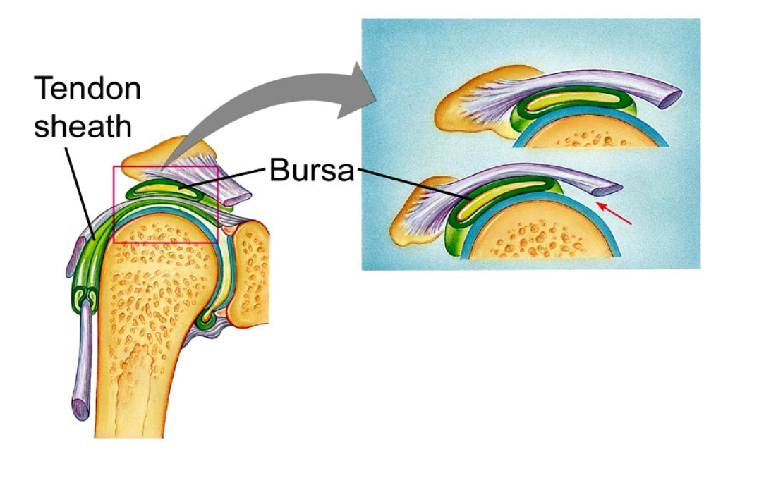
Burse = rolls along with movement (like a ball) which stops friction
Planes of Movement
Sagittal
Bicep curl, lunges
Frontal
Jumping hacks
Transverse
Spinning
Types of Synovial joints - Plane and Hinge
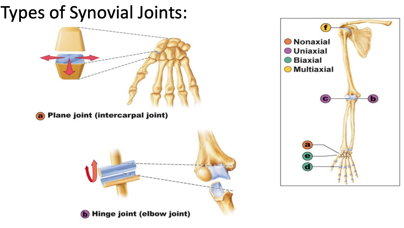
Plane joint
Non-axial
Only glide
Hinge joint
Uniaxial
Action in one plane
Flexion, extension
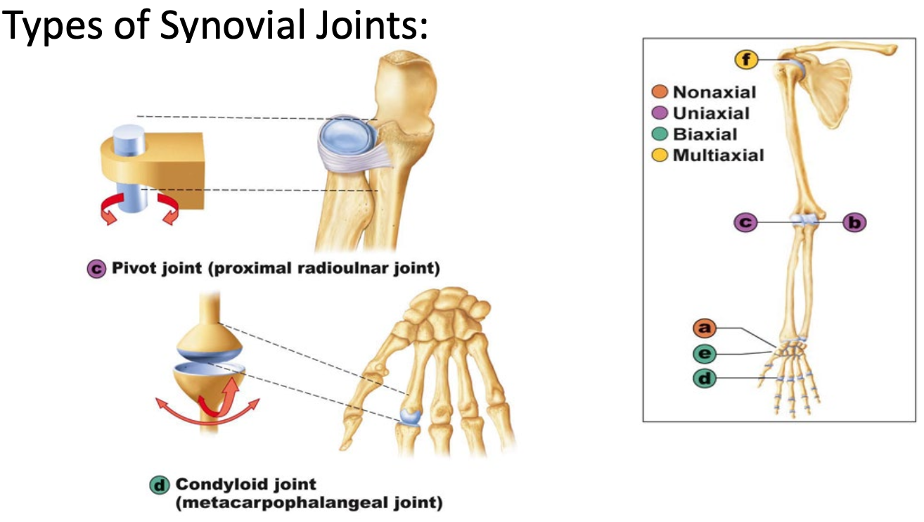
Types of Synovial Joints - Pivot and Condyloid
Pivot
Uniaxial
Pronation, supination
Condyloid joint
Biaxial
Flexion, extension, adduction, abduction
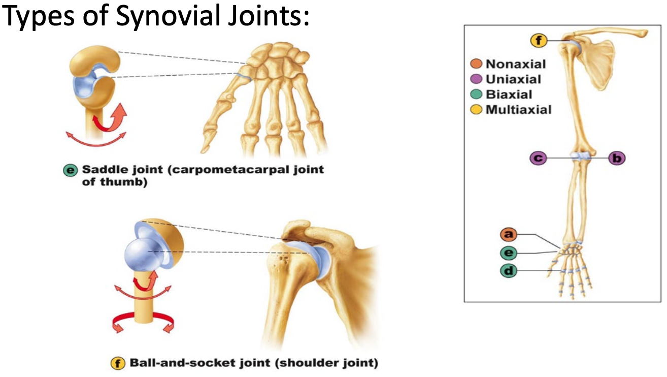
Types of synovial joints - Saddle and Ball-and-socket
Saddle joint
Saggital, frontal
Biaxial
Ball-and-socket
Multiaxial
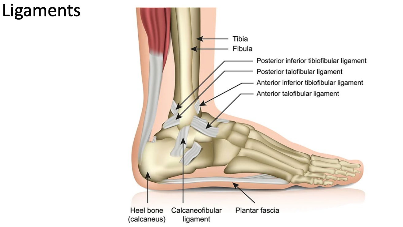
Ligaments
Can stop movement in a different plane
Limits movement
Ligaments not vascular (hard to heal)
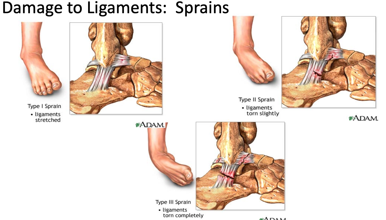
Damage to Ligaments: Sprains
Hard to put ligament together
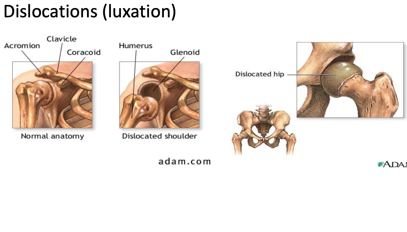
Dislocations (luxation)
Ball-and-socket susceptible
Inflammatory and Degenerative Conditions
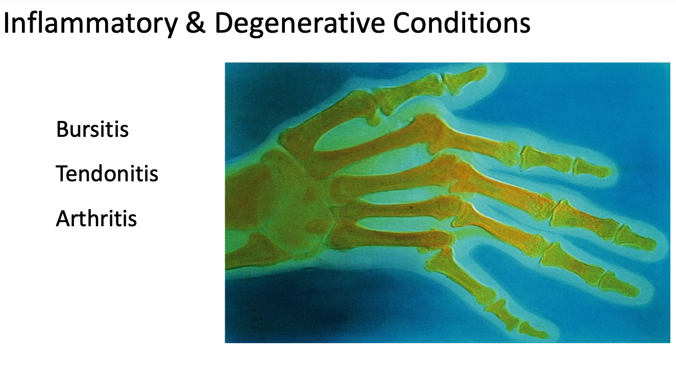
Inflammation = Immune response
Bursitis
Irritation to bursa
Tendonitis
Irritation to tendons
Artritis
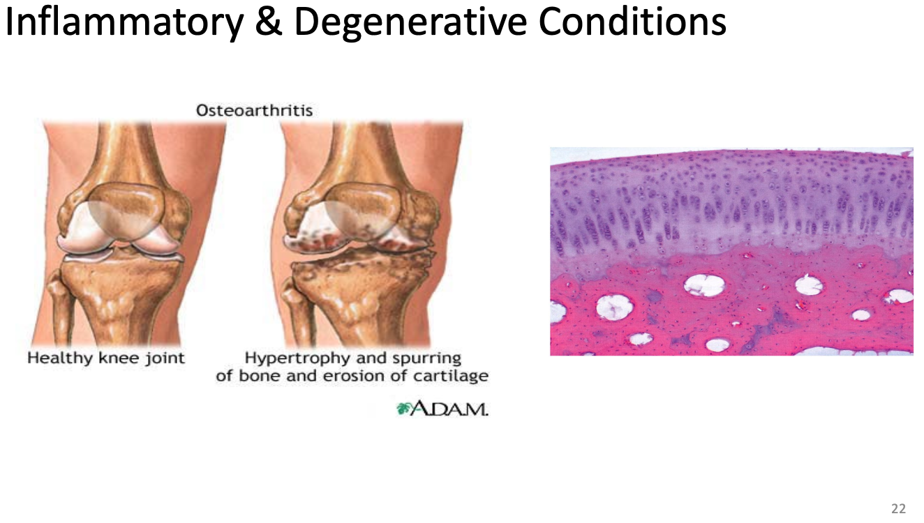
Hyaline cartilage degrading at ends of bones
Inflammatory and Degenerative Conditions
Glucosamine, Chondroitin Sulfate, and Combination
Primary response dropped 20%
Effects of glucosamine and chondroitin sulfate on cartilage
Rate of dying chrondrocytes slowed
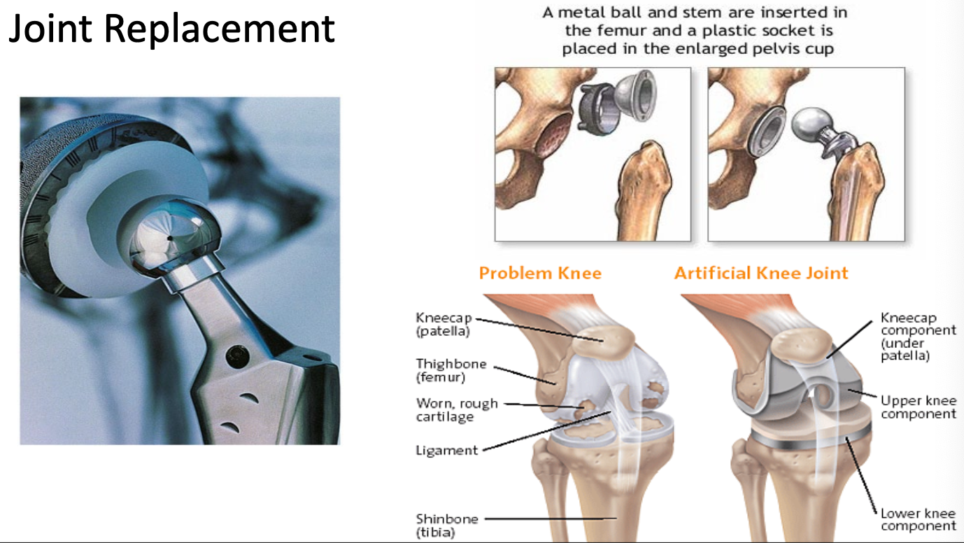
Joint Replacement
How is action created in a joint?
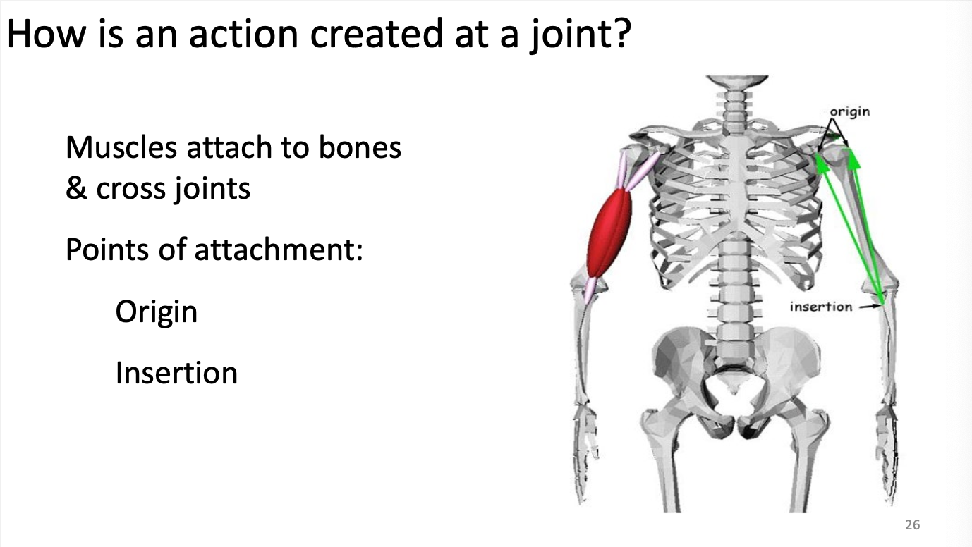
Muscles attach to bones and cross joints
Points of attachment
Origin = POA that doesn’t move
Insertion = POA that does move
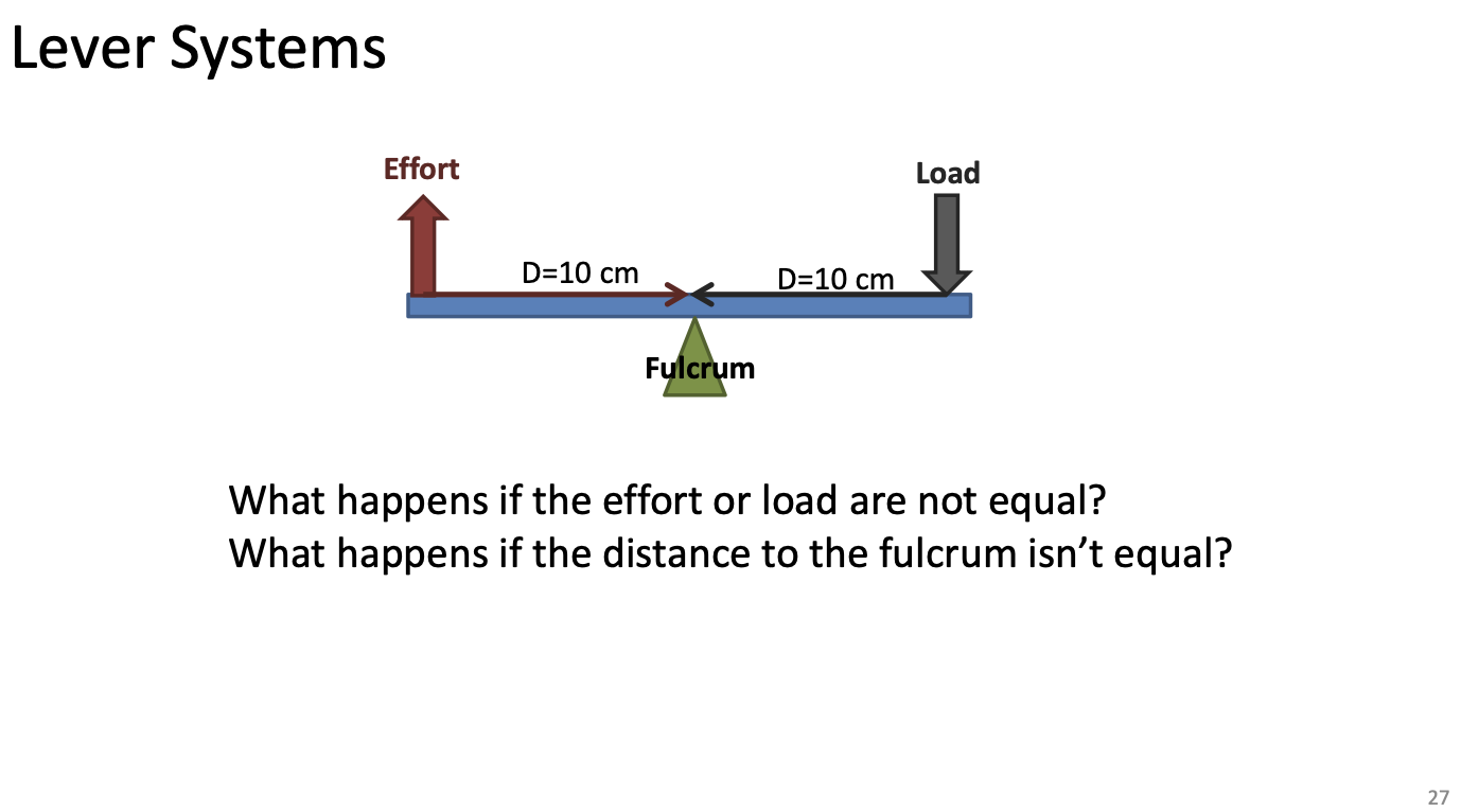
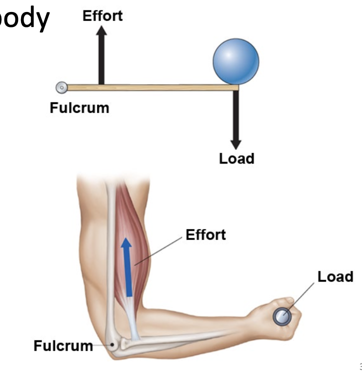
Lever systems
Longer effort arm = if the effort arm is longer than the load arm, the effort is smaller
Longer load arm = if the load is further from the fulcrum than the effort, you must apply greater force
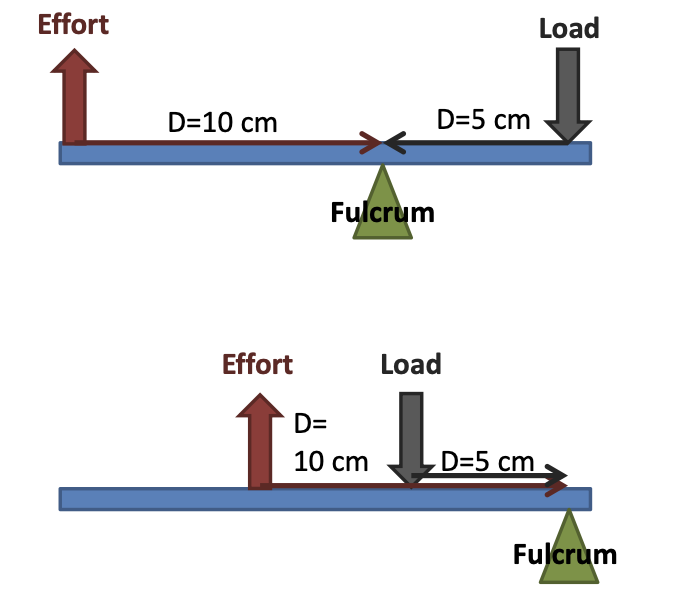
Lever Systems - Mechanical advantage
If the distange to the fulcrum is longer for the effort than compared to the load
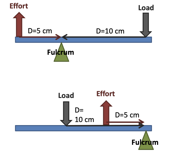
Lever system - Mechanical Disadvantage
If the distance to the fulcrum is shorter for the “effort” compared to the “load”, the lever is at a mechanical disadvantage
Lever systems in the body
Most joints work at a mechanical disadvantage
The insertion of the muscle is very close to the fulcrum compared to the weight of the load
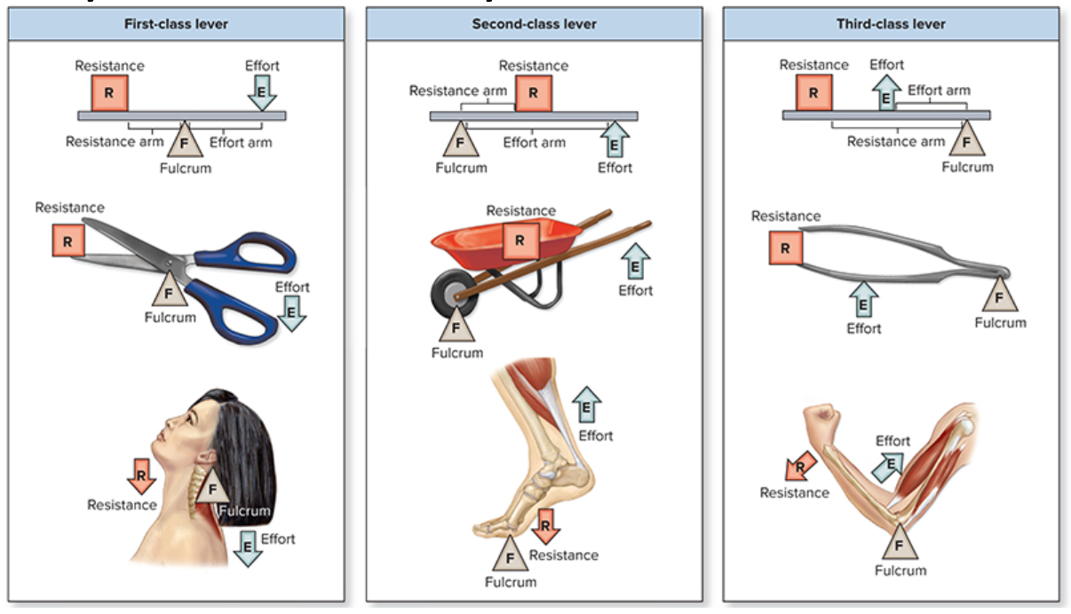
Lever systems - Classes
1 + 2 = mechanical advantage
3 = mechanical disadvantage
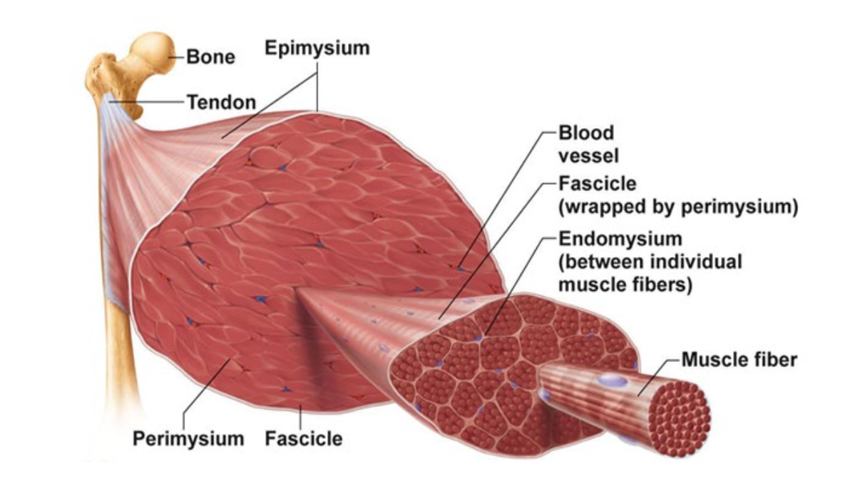
Organization of muscle tissue
Epimysium → Perimysium → Fascicle → Endomysium → Muscle Fiber
Muscle fiber = muscle cell
Miscroscopic Anatomy
Muscle cell = bundles of myofibrils
Muscle fiber → Sarcolemma → myofibril
Myofibril = intracellular proteins
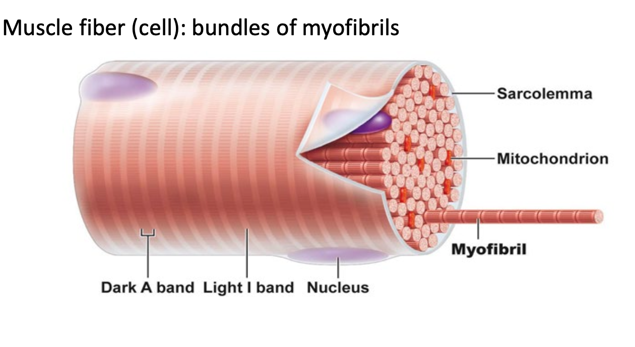
Microscopic Anatomy - Inside a muscle fiber (cell)
Between myofibrils = Sarcoplasmic reticulum, T-Tubules, mitochondria
A band = dark space (where there are myosin)
I band = light space (where there are only actin)
Sarcoplasmic reticulum = STORAGE FOR CALCIUM
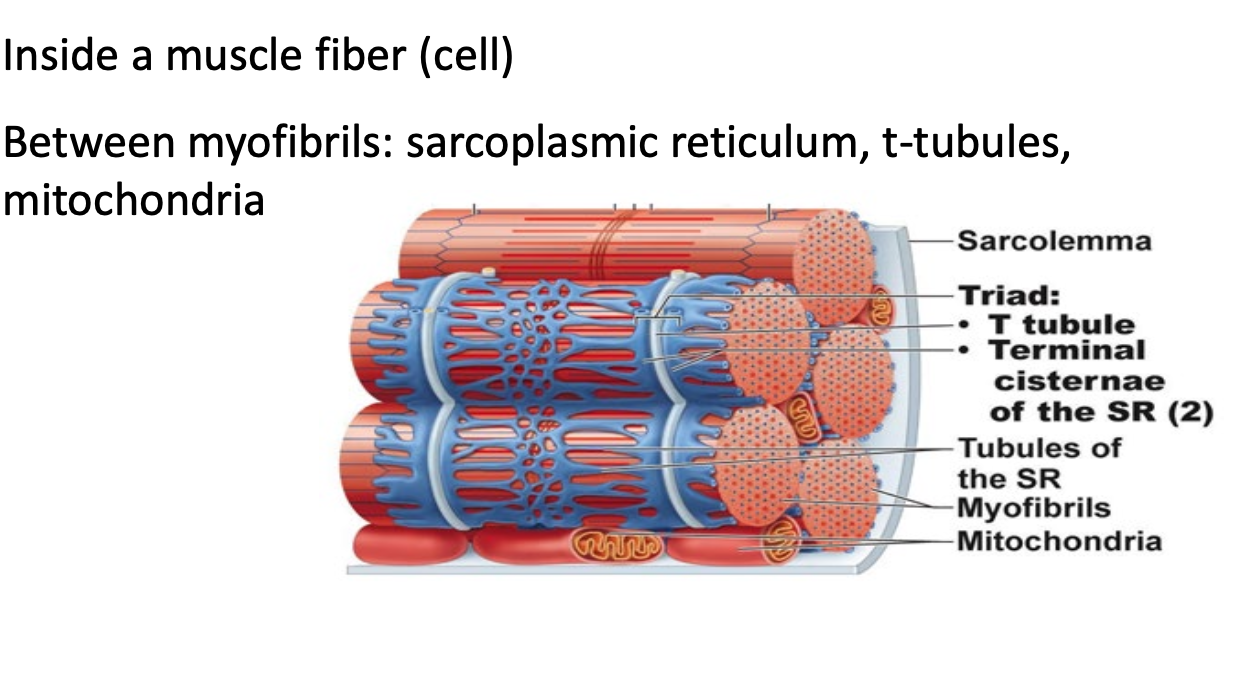
Microscopic Anatomy
Myofibrils are bundles of myofilaments
SARCOMERE = BETWEEN Z DISCS
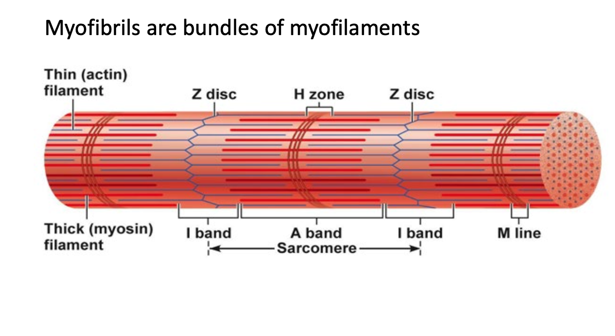
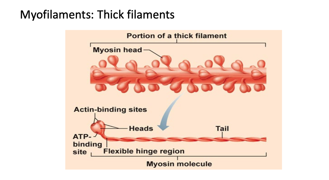
Microscopic Anatomy - Myofilaments: THICK
Thick filaments = Bundles of Myosin
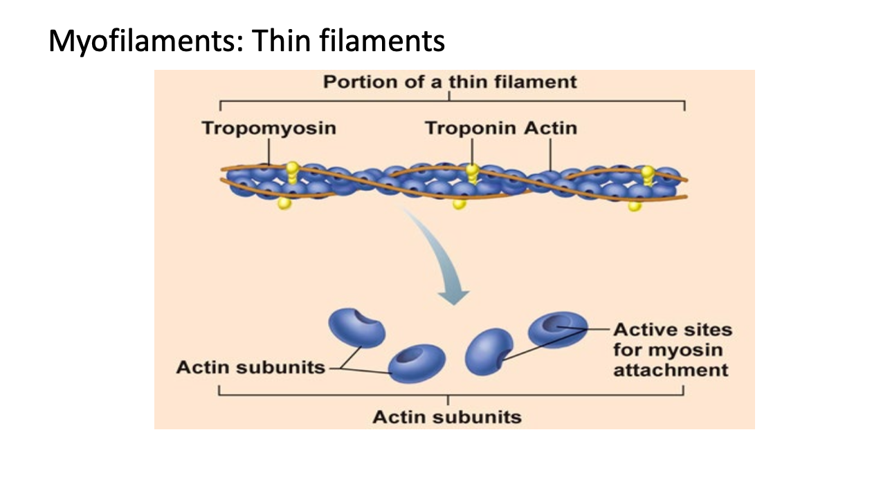
Microscopic Anatomy - Myofilaments: THIN
Thin filaments = Bundles of actin
Tropomyosin blocks myosin head from attaching to actin
Sliding Filament Theory
A band stays the same
H band disappears (Between ends of actin)
I band gets smaller (between Myosin)
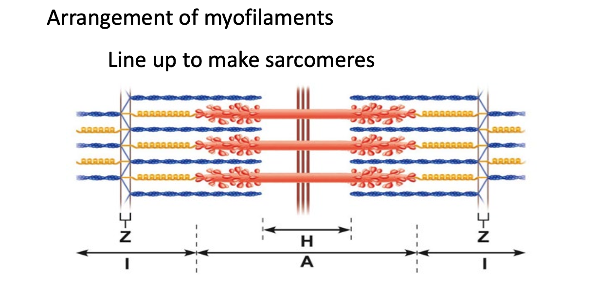
How does a muscle fiber generate force?
Interaction of myosin and actin generates force
Sarcomeres shorten
Filaments slide past each other (SLIDING FILAMENT THEORY)
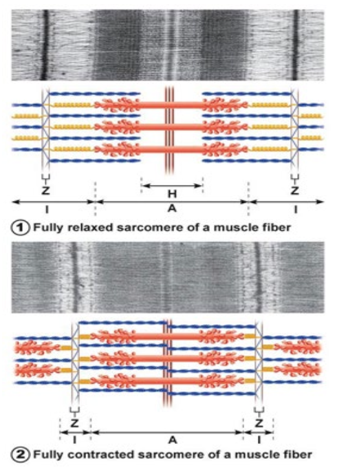
How does the muscle fiber generate force? - Cross bridge cycling
Bind
Calcium causes troponin to move tropomyosin so that myosin can attach
Powerstroke
ADP and P released, as head of myosin pushes/pulls
Release
Attachment of ATP causes myosin head to release
No release = rigor mortis = no ATP
Reset
Calcium go back to sarcoplasmic reticulum - ATP turns into ADP and P
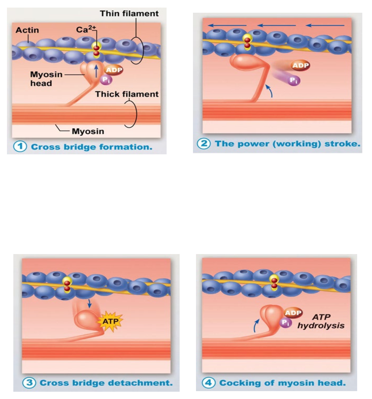
How does production stop?
calcium pumps move calcium to SR
Calcium no longer binds to troponin
Myosin cannot bind to actin - cross bridge cycling stops
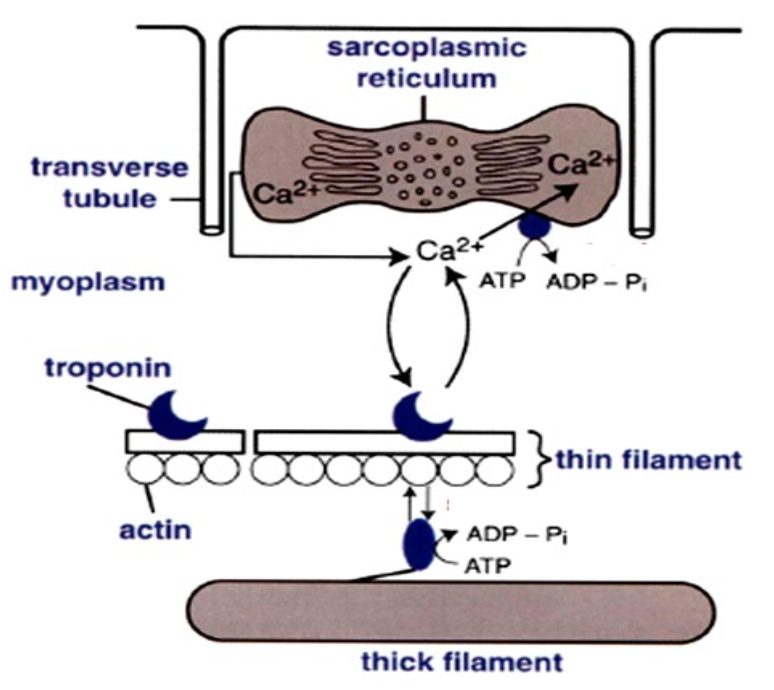
What stimulates force production in a muscle?
Stimulation of muscle fiber
Action potential arrives at axon terminal
ACh released, binds to receptos on sarcolemma
Ion permeability of sarcolemma change
Local change in membrane voltage (depolarization) occurs
Local depolarization (end plate potential) ignited AP in sarcolemma
Excitation-contraction coupling
AP travels across the entire sarcolemma
AP travels along t Tubules
SR releases Ca2+, Ca2+ binds to troponin; myosin-binding sites on actin exposed, contraction begins
Stimulation of muscle (excitation)
ACh effects are terminated by its enzymatic breakdown in the synaptic sleft by acetylcholinesterase
….
Smooth muscle
Innervation of Smooth Muscle
Autonomic nerve fibers have varicosities that act like axon terminals
This is so one neuron can stimulate multiple cells at one time
GAP JUNCTIONS ensure stimulation of smooth muscle cells all at once
Arrangement of Myofilaments
Smooth muscle cells “twist and scrunch”
don’t shorten in one direction