MCB 104
 Call Kai
 Learn
Learn Practice Test
 Spaced Repetition
 Match
Match Flashcards
 Knowt Play
1/104

There's no tags or description
Looks like no tags are added yet.
Name | Mastery | Learn | Test | Matching | Spaced | Call with Kai | Chat |
|---|
No analytics yet
Send a link to your students to track their progress
105 Terms
Nucleosomes
the basic relating structure unit of chromatin which consists of a segment of DNA wrapped around a core of histone proteins
How do nucleosomes associate with DNA?
DNA+histones is a nucleosome
Nucleosomes associate with DNA through electrostatic interactions, where the positively charged amino acids on histone proteins strongly attract to the negative sugar phosphate backbone of DNA.
Electrostatic interactions
Attraction- opposite charges pull toward each other
Repulsion- like charges pull away
Do histones differ in their functions (open/closed chromatin)? If so, how?
Histones directly regulate where chromatin is open or closed by controlling how tightly DNA is packaged. In open chromatin, histones weaken their electrostatic grip. This unwinds the chromatin, allowing transcription factors and cellular machinery to easily access and read genes.
Transcription factors
specialized proteins that bind to specific DNA sequences to control the rate of transcription (the process of copying DNA into RNA). They act as the primary "on/off switches" or "volume knobs" for gene expression.
nucleus
Compartment in which ribosomal RNASa re transcribed and ribosomes are assembled.
ribosomes
the macromolecular (big molecule) complexes responsible for protein synthesis (translation) (creation of proteins) in all living cells. They read the genetic code carried by messenger RNA (mRNA) and translate it into a specific sequence of amino acids to build a functional protein.
tiny cellular structure made of RNA and protein that builds proteins by reading genetic instructions
Nuclear envelope
two concentric membranes that surround the nucleus
Each chromosome has a sub structure

How is transcription set up in a eukaryotic cell?
Enhancer: A regulatory DNA sequence that acts as a specific docking site for activator proteins, speeding up transcription even when located far away from the gene.
Eukaryotic Activator Protein: A specific transcription factor that binds to the enhancer sequence. It triggers transcription by physically interacting with the Mediator complex to help recruit and stabilize the transcription machinery.
TATA Box: A conserved sequence within the core promoter region of DNA. It acts as a physical recognition landmark where general transcription factors bind to align the rest of the machinery.
Mediator (Purple Complex): A massive multiprotein co-activator complex that acts as a physical bridge. It connects the distant activator protein (via DNA looping) directly to the general transcription factors and RNA polymerase.
General Transcription Factors (Cluster of Colored Circles): A suite of essential proteins (like TFIID, TFIIB, etc.) that recognize the core promoter, position RNA polymerase correctly at the start site, and help unwind the DNA double helix.
RNA Polymerase (Large Light-Blue Enzyme): The core enzyme that carries out transcription itself. Once fully assembled and signaled, it moves along the DNA template strand to synthesize a complementary RNA transcript.
Start of Transcription (Yellow Sequence): The precise nucleotide location on the DNA strand where RNA polymerase begins copying the gene into RNA.

What does co-activation by the same enhancer tell us about the relationships between a set of gene products?
It means they are operating in the same protein complex, pathway, or time frame of the cell cycle.
TAD (Topologically Associated Domains)
Individual domains that contains enhancers works with the Cohesion complex acting like a molecular clamp or reel to physically organize DNA inside the nucleus.
It keeps enhancers closer to its repspective gne.

Disrupting genomic TADs would reduce overall gene transcription, True or False?
False because getting rid of the TADs would not disrupt gene transcription (turning DNA to RNA) it would cause the group of genes to not be grouped correctly/ organized but gene transcription will still happen.
Enhancers are always near some DNA, given the packing of DNA into the nucleus TAD keeps an enhancer near a specific subset of genes
Disrupted TAD= reduces specificity of activation, not reduced rate of activation
Transmission electron microscopy
measuring how many electrons come back at us
Eucchromotin vs Heterochromatin
Heterochromatin more dark under transmission electron microscopy. A lot of heterochromatin cell is silences meaning most of its genome is inactive it is so tightly packed that enzymes struggle to access genes
Euchromatin lighter in color and is less tightly packed because of loosely bound dan. It transcribes more genes because transcription factors can get through.
Cohesin depletion
larger, disorganized chromatin loops
histones
small proteins that DNA wraps around
2 cp[ies of the 4 core histones H2A, H2B, H3, and H4 come together to form a nuceosome
Positively charged amino acids, DNA has a negative charge
Linker DNA connects nucleosomes
Histones are highly conserved proteins (billions of years ago Histones looked the same/similar)
Levels of DNA packing

Histone core hypothesis
DNA wraps around histones → histone tails get modified → proteins recognize those modifications → gene activity/chromatin structure can change.
the concept that post-translational modifications (PTMs) on flexible histone tails act as biochemical signaling flags that cellular proteins read to regulate chromatin structure and gene expression.
The flexible N-terminal amino acid tails stick out from the dense core like tentacles, making them fully accessible to modifying enzymes in the nucleus.
The bottom half zooms into the H3 tail sequence to show specific chemical groups attached to targeted amino acids
These PTMs act as precise binding platforms ("docking sites") for specialized reader proteins and chromatin-remodeling complexes, which recognize these patterns to either open up chromatin (euchromatin) for transcription or close it down (heterochromatin) for silencing.

Chromatin remodeling complexes
Chromatin remodeling complexes dock onto both the histone octamer and the wrapped DNA (nucleosome) then physically move the DNA relative to the histones using energy from ATP hydrolysis.

H1
we know that a histone October is made with H2A, H2B, H3, and H4. H1 packages nucleosomes into dense fibers it sits on top of the assembled nucleosome and controls how tight DNA is wrapped.

Imagine that you’re a pathologist, a medical doctor that specializes in analysis of human tissue, cells, and fluids. One of your pediatric patients exhibits premature aging/ progeria. Design an assay that can distinguish cells from the progeria patient, from those of unaffected relatives.
Examine the patient nuclei by microscopy for bulges/ blebs. Compare to unaffected relatives. Should be more prominent in patient cell.
How does the nuclear envelope organize chromosomes in 3D space and regulates gene activity by physically anchoring specific chromatin regions to the inner periphery of the nucleus.
1. Nuclear Lamina & LADs (Lamin-Associated Domains)
Nuclear Lamina: The red crisscross network underlying the inner nuclear membrane composed of lamin filaments. The electron micrograph insert (top right) highlights its meshwork structure.
LADs: Large chromatin domains (shown anchored directly to the lamina) that interact with lamins. Because they are sequestered at the nuclear periphery, LADs are typically enriched for heterochromatin (silenced/inactive genes).
2. Transmembrane & LINC Complex Connections
Transmembrane Proteins: Green proteins embedded in the double membrane bridge the cytoskeleton (pink filaments outside) with the nuclear lamina and chromatin inside.
Mechanical Linkage: This allows physical forces from the outside environment to be transmitted directly into the nucleus to influence chromosome arrangement and gene expression.
3. Transcription Factor Regulation & Phosphorylation
Phosphorylation Signals (P): Dark blue transcription factors outside the nucleus become activated/modified via phosphorylation (P).
Translocation: Once signaled, they enter through nuclear pores (orange ring channels) into the nucleoplasm to bind target gene promoters and drive transcription.
4. Nuclear Pores & Signaling Molecules
Nuclear Pores: Regulated gateways (yellow star-like structures) spanning the inner and outer membranes that control transport between cytoplasm and nucleus.
Signaling Proteins (Purple): Signaling molecules enter through pores or dock near the lamina to interact directly with LADs or transcriptional machinery to alter chromatin positioning.
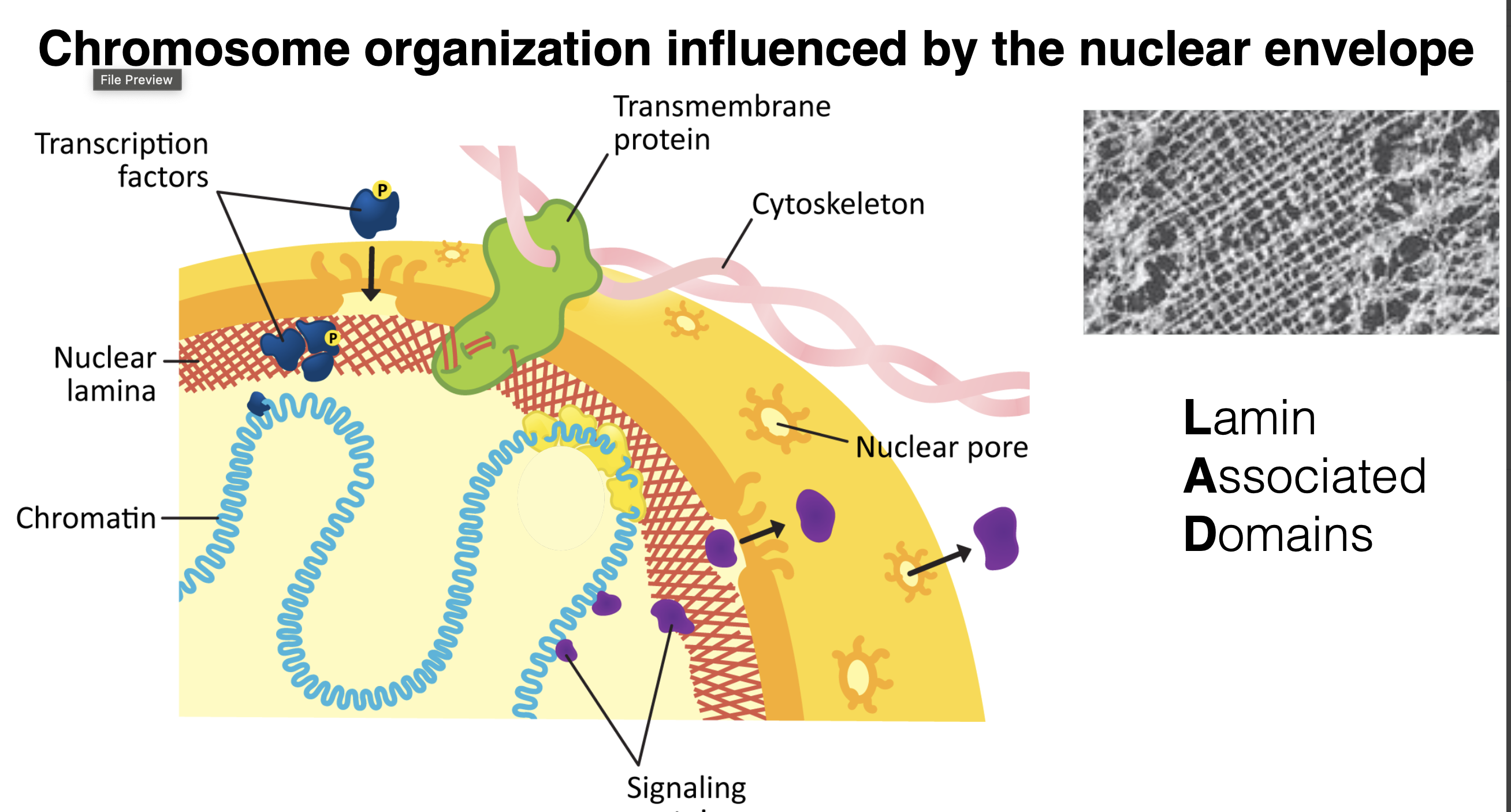
TAD vs LAD
LADs act as an "anchor system" that pulls inactive regions of chromosomes to the nuclear wall to keep them turned off. (silenced) turns into heterochromatin
TADs act as "insulated neighborhoods" within the interior of the nucleus that keep local genes and regulatory enhancers together so they can interact safely without interfering with neighboring genes.
phosphorylation—a post-translational modification (PTM)—controls the reversible assembly and disassembly of the nuclear envelope and nuclear lamina during the cell cycle.
1. Interphase (Intact Architecture)
Unphosphorylated State: During Interphase, phosphate groups are absent from the nuclear lamins.
Stable Structural Scaffold: The nuclear envelope (yellow) and underlying nuclear lamina (red mesh) remain fully assembled, providing a stable scaffold for chromatin (blue wavy lines) and maintaining nuclear integrity so normal transcription can occur.
2. Prophase (Disassembly Triggered)
Phosphorylation of Lamins: As the cell prepares to divide in Prophase, specific kinases attach phosphate groups (P) to lamin proteins.
Weakening Integrity: Adding these negatively charged phosphate groups causes electrostatic repulsion, disrupting the intermediate filaments that make up the lamina mesh.
Envelope Breakdown: The nuclear membrane fragments into small vesicles (yellow pieces), and the lamina disassembles (red fragments), freeing the condensed chromosomes (blue X-shapes) so spindle fibers can attach to them.
3. Anaphase to Telophase (Reassembly in Daughter Cells)
PTM Removal (Dephosphorylation): During Anaphase and into Telophase, phosphatases strip away the phosphate groups (P) from the lamins.
Nuclear Envelope Coalescence: Without the repelling phosphate groups, lamin proteins naturally reassociate around the segregated chromosomes.
Restoration: Membrane vesicles fuse back together around the chromatin, reforming two fully functional nuclei with intact nuclear envelopes as the cell returns to Interphase.

Nuclear Lamina anchors trans membrane proteins in Nuclear Envelope
describes how the cell physically connects its interior skeleton (the nuclear lamina) to the membranes surrounding the nucleus (the nuclear envelope).
Trans membrane proteins connected chromatin to cytoskeleton
Lamins organize nuclear envelope
Disrupted lamins= progeria disease
Kash domain proteins and Sun domain proteins prevents drift

Genome access is controlled by nuclear pore complexes
Nuclear localization signal- series of amino acids that is recognized by the receptor
Nuclear import receptor- they are checking for receptor not cargo if cargo is connected to the right receptor it goes through the pore
Nuclear pore complexes (NPC) control import and export through nuclear envelope
Control movement of both proteins and RNA

Import to the nucleus through the NPC
Importin aka Nuclear Import Receptor (NIR) Indicates import to the nucleus
Energy used for receptor recycling: taking NIR back to cytoplasm after cargo import is complete
NIR going down concentrating radiant uses no energy
But NIR going against concentration gradient required energy input
NIR moleculen in a cell are in high concentration within the cytoplasm

What is GEP and GEF, what is their job?
P is a inorganic phosphate group

RAN cycle
The Ran cycle is the chemical engine that powers nucleocytoplasmic transport through the Nuclear Pore Complex (NPC). It relies on a small protein called Ran—a monomeric GTPase—which acts as a directional switch depending on whether it is bound to GTP (active/high-energy) or GDP (inactive).
Ran- GTP binds the improtin, not to the cargo
The cargo and Ran-GTP never directly bind/interact
Ran molecules release GDP in favor of GTP in the nucleoplasm
Ran molecules are stimulated to hydrolyze GTP in cytoplasm
Ran GTPase = The physical light switch.
Ran-GTP = The switch turned "ON" (bound to high-energy GTP).
Ran-GDP = The switch turned "OFF" (bound to low-energy GDP).
What cell compartment is Ran GTPase inside when it hydrolyzes GTP?
cytoplasm
Ran GTPase regulated export by binding exporting proteins
Exportins- nuclear export receptors
Exportins are closely related to importing, but roles are not interchangeable
Nuclear export signals (NES)- an amino acid sequence found on the surface of the cargo
Ran facilitates export without directly binding cargo

exporting cycle
finds the protein after ran gets involved and protein sequence is NES instead of KKKRK
similar to import process RAN-GTP bind in the cytoplasm
Before step one in the nucleoplasm GEF stimulates RAN to exchange to release GDP and release GTP
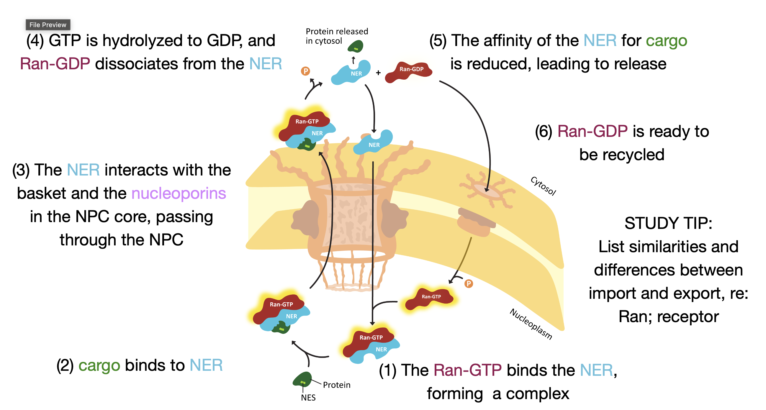
True or false. In both nuclear import and export pathways Ran will hydrolyze GTP while in the cytoplasm.
True
True or false? Import and Export receptor proteins always bind their cargo after binding Ran.
False
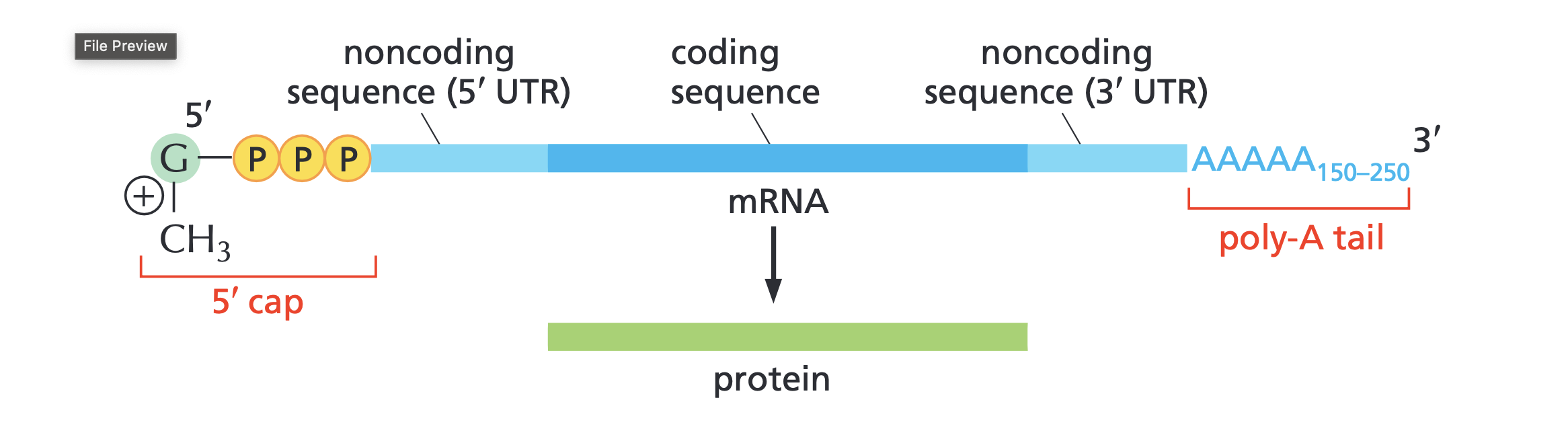
mRNA
Messenger RNA (mRNA) is the single-stranded intermediate transcript that carries genetic coding instructions from DNA to the ribosome for protein synthesis.
capped with a methyl at 5 prime end
mRNA cap protects from degradation; acts as “self” signal
Cap regulated by cell signaling pathways
How mRNA leaves the nucleus
In order to get a mature mRNA introns have to be removed introns is in the middle surrounded by 2 exons
Introns (Intervening sequences): Non-coding regions that are cut out and removed.
Exons (Expressed sequences): Coding regions that are stitched back together to form the final protein recipe.
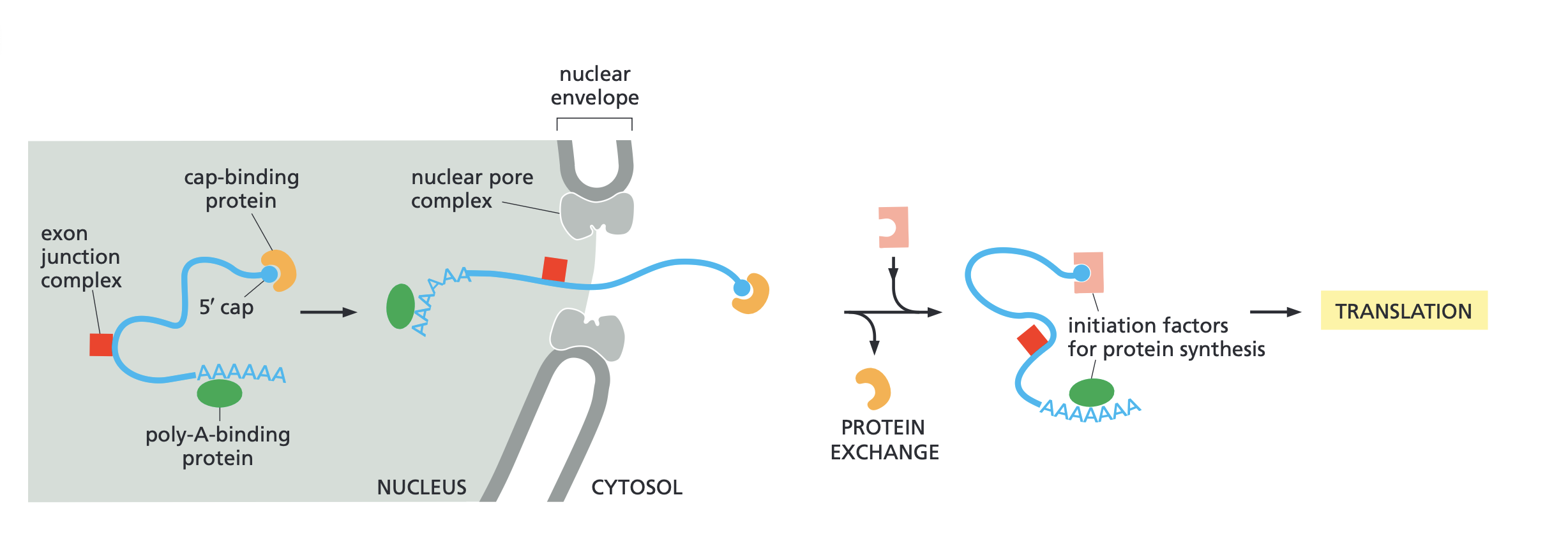
Poly H binding protein goes on poly A tail
CBP (exportin) goes on 5 prime cap
mRNAS caps are recognized by RNA export factors; CBP
The play A binding protein mediates stability of mRNA
Cap binding protein binds and recognized the 5 prime cap
We know mRNA is ready for export when we see this bind
CBP is removed before translation
translation- the biochemical process in which a ribosome reads the genetic code carried by a mature mRNA transcript to assemble a specific sequence of amino acids into a functional protein.
CBP acts as the mRNA exportin
EJC (electron junction complex)- a protein complex deposited onto mRNA during splicing. It acts as a positional memory tag, marking where introns were removed so the cell knows how to handle the transcript.
mRNAs get distributed among ribosome pools in cytoplasm
Free ribosome- orange part is ribosomal components, the blue string is the messenger RNA, and the green thing is the Nascent peptide nascent means beginning to exist or recently formed/ developed
Polyribosome bond and have the sane mRNA'
this is a temporary assembly that forms during protein synthesis
The cytosol and ER lumen represent two distinct, membrane-separated fluid compartments within a eukaryotic cell, each with its own chemical environment and specialized cellular functions.

Ribosomes cycle between cytoplasmic and rER pools
both path starts with a mana binding with a free ribosome
top path stays free in the cyctoplasm
they bth end up dissociating and recycling
membrane bound polyribosomes synthesize proteins that are targeted to the cell membrane, stored inside organelles, or secreted out of the cell.

SRP- Signal recognition particle
bring loaded ribosomes to ER surface
ER insertion sequence is recognized by SRP early in translation; translation pause
translation will stop once it binds this ensures they don’t translate in the cytoplasm but in the last step which is the membrane
SRP engaged the SRP receptor on ER membrane surface
Ribosome docks to translational channel
SRP is removed and translations resumes

Endoplasmic Reticulum.(ER)
It is a continuous membrane network within eukaryotic cells consisting of two distinct functional regions:
Rough ER: Studded with ribosomes; site of membrane and secretory protein synthesis.
Smooth ER: Lacks ribosomes; site of lipid synthesis, carbohydrate metabolism, and calcium storage.
Imagine that you are analyzing cells in which the CBP protein has been abruptly degraded. Which of the following is the most likely consequence?
CBP cap binding protein that tells the mRNA that it is ready for export. So without CBP mRNA will not go to export and it will be stuck in the nucleus.
cytosol vs cytoplasm
The cytosol is the liquid portion of the cell, while the cytoplasm encompasses that liquid plus all the organelles (except the nucleus in eukaryotes).
Cytosol: The semi-fluid, jelly-like aqueous matrix made of water, dissolved ions, small molecules, and soluble proteins. It excludes all membrane-bound organelles and structural components.
Cytoplasm: The entire cellular contents enclosed within the plasma membrane, excluding the nucleus. It includes both the liquid cytosol and all suspended organelles (such as mitochondria, endoplasmic reticulum, and ribosomes).
Cytoplasm = Cytosol + Organelles (minus the nucleus)
True or False- Both the SRP receptor and the translocon channel reside in the ER membrane.
True

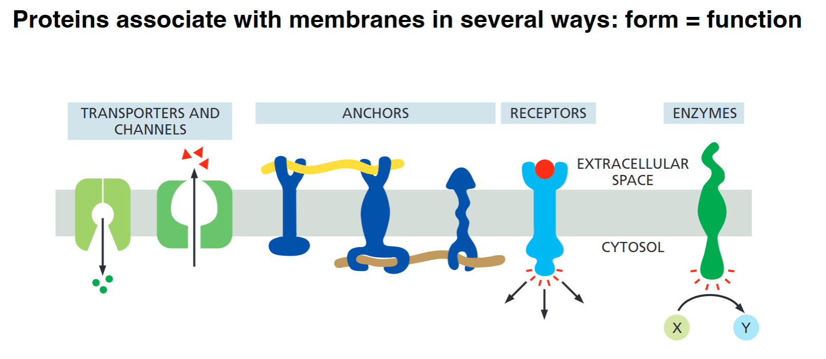
Proteins associate with membranes in several ways
hydrophobic domain allows them to be anchored in the membrane. The hydrophobic part they easily enter and sit inside the nonpolar interior of the lipid bilayer.
A transmembrane domain specifically refers to a region of a protein that spans all the way through the lipid bilayer—from one side to the other.
monolayer associated- not fully transversing the membrane bilayer
lipid-linked- protein linked to a lipid that is attached to the bilayer
protein attached- attached to a protein that is anchored in the bilayer

Integral and peripheral
Integral- permanently attached to biological membrane"; large fraction of animal genomes
Peripheral- temporarily attached to biological membrane
Proteins associate with membranes in several ways
Transporters and Channels (Green): Act as selective gates or pumps to move ions, nutrients, or waste across the hydrophobic lipid bilayer between the extracellular space and the cytosol.
Anchors (Dark Blue): Physical tethering points that attach to structural filaments—either extracellular matrix fibers outside or the cytoskeleton inside—holding the cell's shape or locking the protein in place.
Receptors (Light Blue): Bind specific extracellular signaling molecules (like hormones or growth factors) on the outside, triggering a conformational change that passes a signal into the cytosol.
Enzymes (Dark Green): Catalyze specific chemical reactions directly at the membrane interface (e.g., converting molecule X into molecule Y).
Soluble protein- Destined for ER lumen

Membrane protein- N terminus in cytosol, single pass
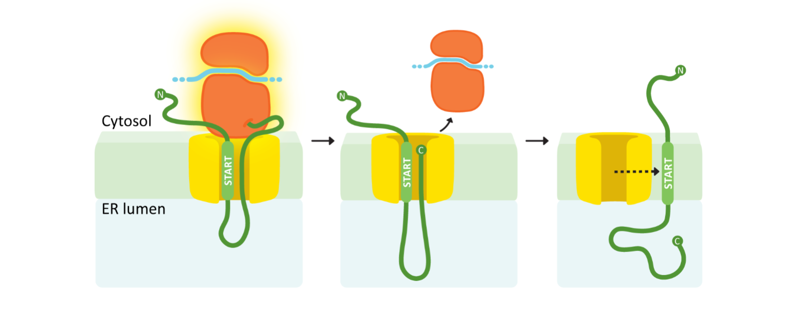
Membrane protein - C- terminus in cytosol, single pass

Membrane protein- double pass
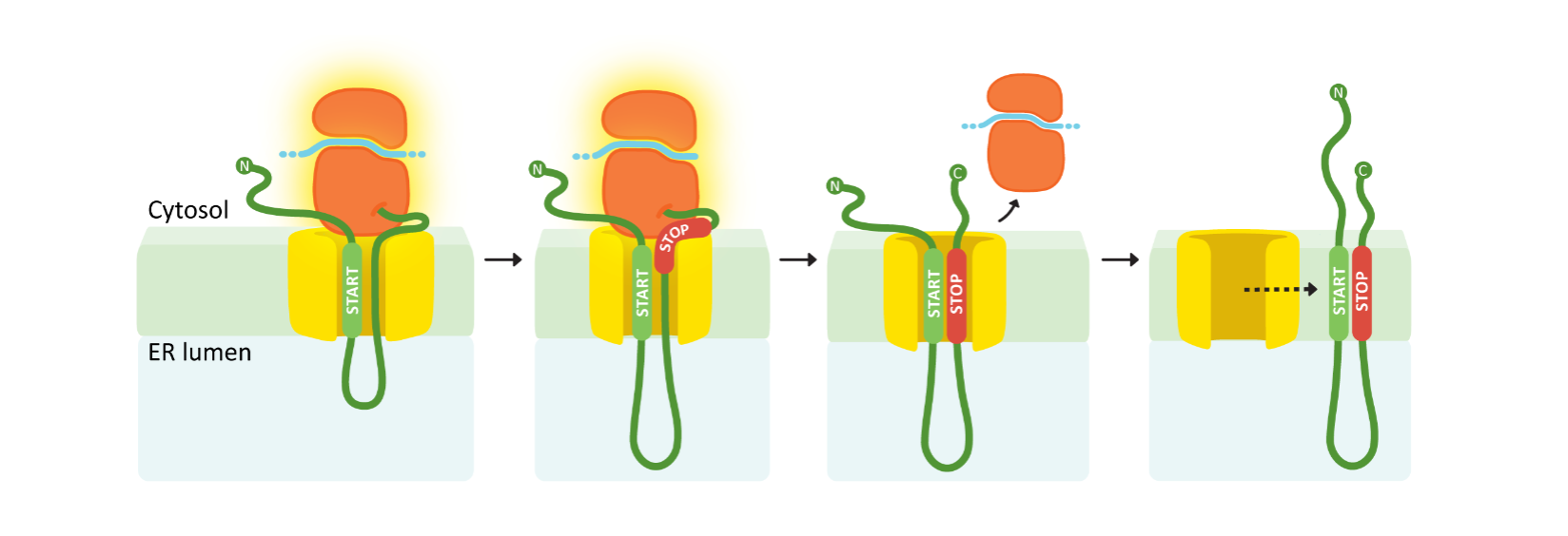
Signal Peptidase
a specialized enzyme located on the lumenal face of the Endoplasmic Reticulum (ER) membrane. Its main job is to recognize and chop off (cleave) the temporary N-terminal signal peptide from a newly synthesized protein as it enters the ER.
Vessicles are shuttles
"ER is point of entry to endomembrane system"
What it means: The Endoplasmic Reticulum (ER) acts as the main "front door." Newly synthesized proteins destined for membranes, lysosomes, or secretion outside the cell are imported directly into the ER during translation. Once inside, they enter the cell's internal shipping network (the endomembrane system).
"Membrane proteins do not return to cytosol once they enter"
What it means: Protein transport into the ER is a one-way trip. Because the hydrophobic transmembrane domains become permanently locked inside the lipid bilayer during insertion, these proteins can never slip backward out of the membrane and dissolve into the watery cytosol again.
"Cargo transfer from one organelle compartment to another must be achieved, without cargo 'escape' back to cytosol"
What it means: Vesicles act like sealed delivery pods. When moving proteins from the ER to the Golgi, or Golgi to the cell surface, the cargo is kept strictly enclosed inside the vesicle's membrane sphere or lumen. The membrane buds off from one compartment and fuses directly with the next, ensuring the contents are delivered safely without leaking out into the cytosol.

microtubules


Core Biological Principles Shown:
MTOC (Microtubule Organizing Center): The central hub (dark green circle) near the nucleus where all microtubule minus (-) ends are anchored.
Directional Orientation: The green microtubule tracks radiate outward, placing all their plus (+) ends toward the outer edges (periphery) of the cell.
A GPS System for Transport: Because the tracks have a strict direction (minus at the center, plus at the edges), motor proteins always know which way they are walking.
What the Text Means:
"Movement of vesicles and gene products from peri-nuclear to periphery": Newly made proteins (gene products) synthesized near the nucleus/rER are loaded into vesicles and shipped along these tracks outward to the cell membrane (periphery).
"MTOC associated with rER": The organizing center sits right next to the rough Endoplasmic Reticulum (rER) and nucleus, establishing the starting line for the endomembrane shipping route.
mitosis
process by which dividing cells partition their chromosomes to produce genetically identical daughters
During prophase the centrosome with the microtubules split and migrate until they are on opposite sides, the membrane also disappears
During pro metaphase the chromosomes become stabilized and will all wind up attached to a microtubule in the middle
during anaphase they separate
Assembly of microtubule spindle forms in the phosphate, attachment happens in the pro metaphase, and it is completely formed before the anaphase begins
Metaphase is when chromosomes reach the center of microtubles
The red dots are centromere, centromere are special chromosome regions that serve as sites for assembly of the kinetochore during mitosis.

kinetochore
Inner Plate (Red): The inner kinetochore layer that directly binds to the centromeric DNA.
Outer Plate (Green): The outer kinetochore layer housing microtubule-binding proteins that physically attach to the green spindle microtubules.
Pericentric Heterochromatin (Blue): The specialized, tightly packed DNA regions surrounding the centromere that provide structural support.
kinetochores contain many proteins that enable them to capture and move along the microtubules

Mitotic cell cycle
Interphase- G1, S(synthesis) -DNA replication, G2 not every cell has they cell growth gaps
Mitosis- spindle assembly, chromosome condensation, and division cytokinesis (cell division)
DNA replication (in S phase) and chromosome segregation (in mitosis) are highly regulated
Errors during the cell cycle can lead to aneuploidy, genomic instability, and cell death

MTOC (microtubule organizing centers)
Microtubules are nucleated(seed the initial growth) and organized by MTOCs which form the spindle poles during mitosis.
The position, number, and activity of MTOCs is regulated by the cell cycle and type of cell.
MTOCS often but not always have CENTRIOLES at their cores.
Centrioles are cylinder-like organelle structures composed primarily of protein fibers called microtubules. They have their own division cycle, which regulated the number of MTOCS
Cyclins
Tim hunt (Sea urchin dude) identified proteins that oscillated in abidance during each cell cycle and named them cyclins.
Cyclins - a family of regulatory proteins that control the progression of a cell through the cell cycle
MPF
MPF is literally the specific protein complex formed when Cyclin B binds to CDK1.
Cyclin B regulatory subunit
catalytic subunit: cyclin- dependent kinase (Cdk1)
doesn’t work without cyclin B
Kinase- an enzyme that phosphorylates target proteins (adds a phosphate group from ATP) to change their shape and turn them "on" or "off."
Cyclin B binds to Cdk1 to form the MPF complex, and then active Cdk1 transfers phosphate groups to other cellular proteins to trigger mitosis.

Ubiquitin chains
Anaphase transition is triggered by destruction of cyclin through ubiquitin mediated proteolysis. Adds ubiquitin chain to cyclin B.
These chains are recognized by proteasome, a large protein destroying machine, which degraded cyclin, leading inactivation of Cdk
This causes the cell cycle to be irreversible

Cohesins
ring-shaped multi-protein complexes that hold sister chromatids together from the moment DNA is replicated until they are ready to be separated during mitosis or meiosis.
Cohesin is released at the metaphase to anaphase transition when one of its subunits is cleaved by a protease called separate
Protease- an enzyme that breaks down proteins by cleaving the peptide bonds that join amino acids together—a chemical process called proteolysis.
as we can see ubiliquene chain attaches and securing leaves making separase become activated
Securing- a target of the ubiquitin ligase APC/C, which triggers it destruction through ubiquitin mediated proteolysis
the destruction of cyclin b and securing though ubiquitin mediated proteolysis triggers the metaphase- anaphase transition

Checkpoints
Dan damage S- phase entry
Is the DNA damaged?
Is the cell big enough, is there enough energy?
DNA damage M-phase entry
Is the DNA damaged?
Did all the chromosomes replicated correctly
M to A transion
Are all the chromosomes aligned at the center?
Mitotic exit
Have the spindles moved correctly to divide the sister chromatid

Cohesin vs Condensins
Cohesin = holds chromatids together
Condensin = condenses/comapcacts chromosomes
not yet fully discovered but from what we know
Condensin I = higher impact on width
Condensin II = higher impact on length
How chromosomes become compacted and organized during prophase/prometaphase, especially through cohesin and condensin?
In G₂/interphase, chromatin is relatively loose and cohesin helps organize/hold DNA regions.
In prophase, condensin I and II start reorganizing the chromosome into loops and compacting it.
By prometaphase, the chromosome is much more condensed and organized.

The 2 cell cycles
even though meiosis is 1 way and it doesn’t go backwards it is still called a cycle

meiosis
Pairing & synapsis → homologous chromosomes line up with each other.
The synaptonemal complex holds the homologs closely together. Spo11 is the enzyme that causes these breaks.
Double-strand DNA breaks are intentionally made.
Recombination/crossing over happens, where homologous chromosomes exchange DNA. A lot of breaks happen but only a few become crossovers usually 1 per homolog pair
Then in meiosis I (MI), the homologous chromosomes separate. homologous mean matching maternal + paternal chromosome pair
In meiosis II (MII), the sister chromatids separate.
cool fact: early meiotic prophase the nuclear envelope still present and the chromosomes can attach to it and cluster up due to moving this is called a meiotic bouquet

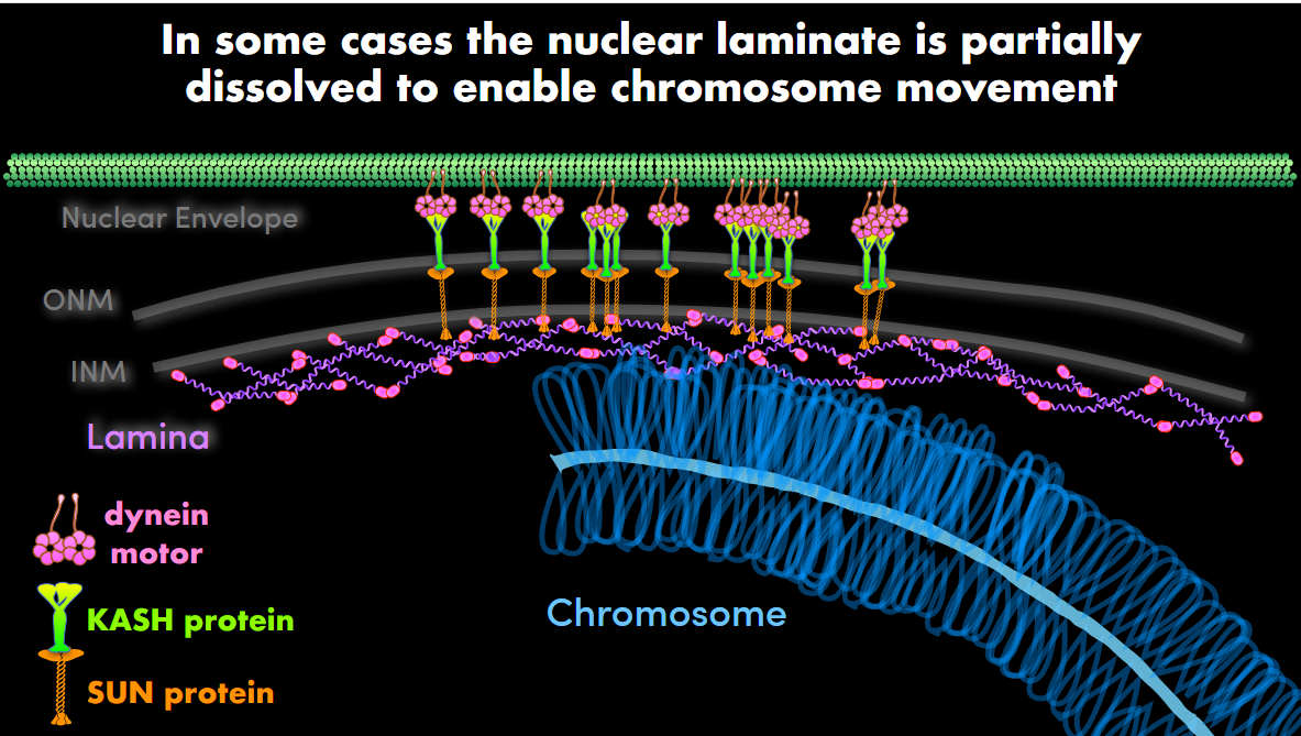
LINC
SUN + KASH = LINC complex
LINC complexes are bridges that span the nuclear envelope and connect chromosomes/nuclear structures inside the nucleus to the cytoskeleton outside the nucleus.
Dynein motors interact with cytoplasmic microtubules and generate movement.
Connects lamina to dynein motor
Kinetochroes in meiosis 1 vs meiosis 2
In meiosis I, the kinetochores of the two sister chromatids act together as one unit and attach toward the same spindle pole. That way, the sister chromatids stay together while the homologous chromosomes separate.
In meiosis II, the sister kinetochores act separately and attach to opposite poles, so the sister chromatids separate.
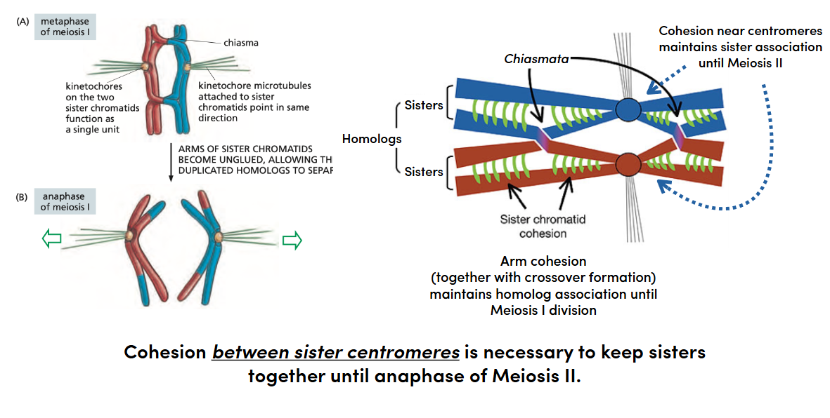
Meiotic cohesion is released in 2 steps to allow 2 rounds of chromosome segregation
Homologs pair and crossing over occurs during prophase I.
SC disassembles, but chiasmata + arm cohesin keep homologs connected.
In metaphase I, homologs attach to opposite spindle poles (spindle microtubules are pulling the two homologs in opposite directions) while sister kinetochores act together.
In anaphase I, arm cohesin is cleaved → homologs separate.
Centromeric cohesin remains protected.
In anaphase II, centromeric cohesin is removed → sister chromatids separate.

Chiasma
crossover +cohesin
meiotic cells use a special version of cohesin
Think of cohesin like a ring-shaped clamp around sister chromatids. The kleisin acts like the “latch” that connects the SMC1 and SMC3 parts of the ring.

Shugoshin
Sister chromatid cohesion at the centromere is protected during the first division by recruit of a special “guardian spirit” protein called Shugoshin (SGO)
Shugoshin → recruits phosphatase → dephosphorylation → cohesin protected → centromeric cohesion maintained
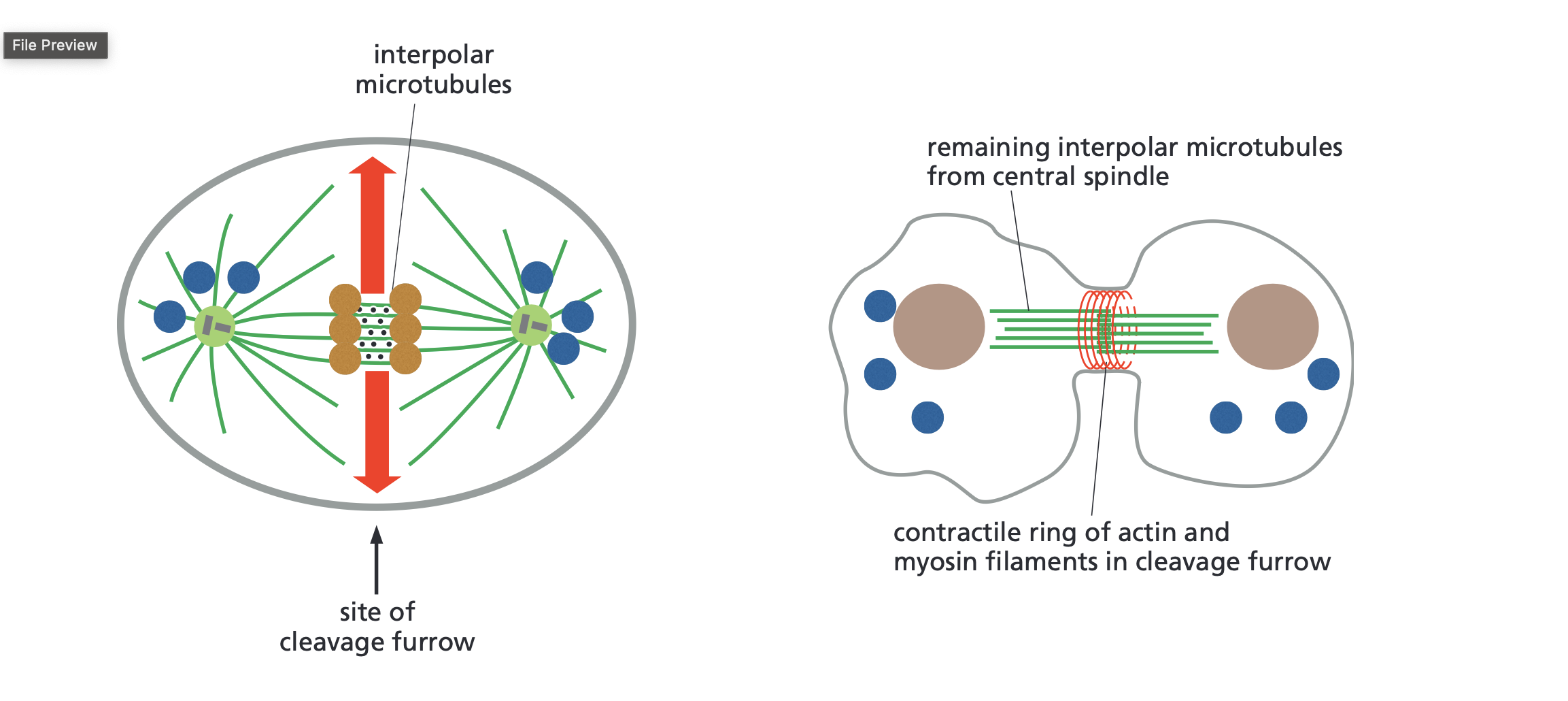
cytokinesis
Process of when one cell splits into 2, very similar to mitosis but mitosis is 1 nucleus into 2 nucleus happens at the end of mitosis.
Orange dots are proteins that help organize/crosslink the microtubles
Site of furrow is a pinch that forms in the cell membrane during cytokinesis
contractile ring of actin and myosin filaments tightens and creates the cleavage furrow until the cell separates into 2
The brown is the chromosomes/nucleus of future daughter cells
Motor proteins
Kinesis move toward the plus end of microtubules
Dyneins move toward the minus end of microtubules
Transport on microtubule relies on motor proteins
Motors position cargo for daughter cell inheritance, it makes sure each daughter cell has what it needs

Cytoskeleton
keep its shape
move things around inside the cell
move the cell itself
separate chromosomes during cell division
position organelles and vesicles
Transport sequence
The rough ER puts out small transport vesicles that contain proteins and lipids made in the ER.
This travels to the cis side of the Golgi apparatus cisternae is one flattened membrane sac of the Golgi
The Golgi apparatus then sorts, modifies (by adding or modifying sugar/lipids), and packages the proteins.
Then the tran cistern sends out secretory vesicles
There are 3 main places that they could go
the plasma membrane / outside the cell for secretion
endosomes, which act as sorting/recycling compartments
lysosomes, where material is broken down
Something to know is that Golgi mass is dynamic it is constantly being changed, removed by budding vesicles on one side and added by fusing vesicles on the other

4 main steps link donor to acceptor compartments
Budding
A transport vesicle forms from the donor compartment and pinches off carrying cargo.Transport
The vesicle moves through the cell along the cytoskeleton, often with help from motor proteins. The snare is the purple wiggly line it has no job right now.Docking
The vesicle reaches the correct acceptor compartment and attaches to it. Tethering protein is the blue thing and raw is the pink thing it gets recognized by the protein. Key thing is that it is longer than the snare so it can check if it is the right vesicle. Snare then attaches to the t snare on the membrane.Fusion
The vesicle membrane fuses with the acceptor membrane, and the cargo is delivered inside the target compartment.

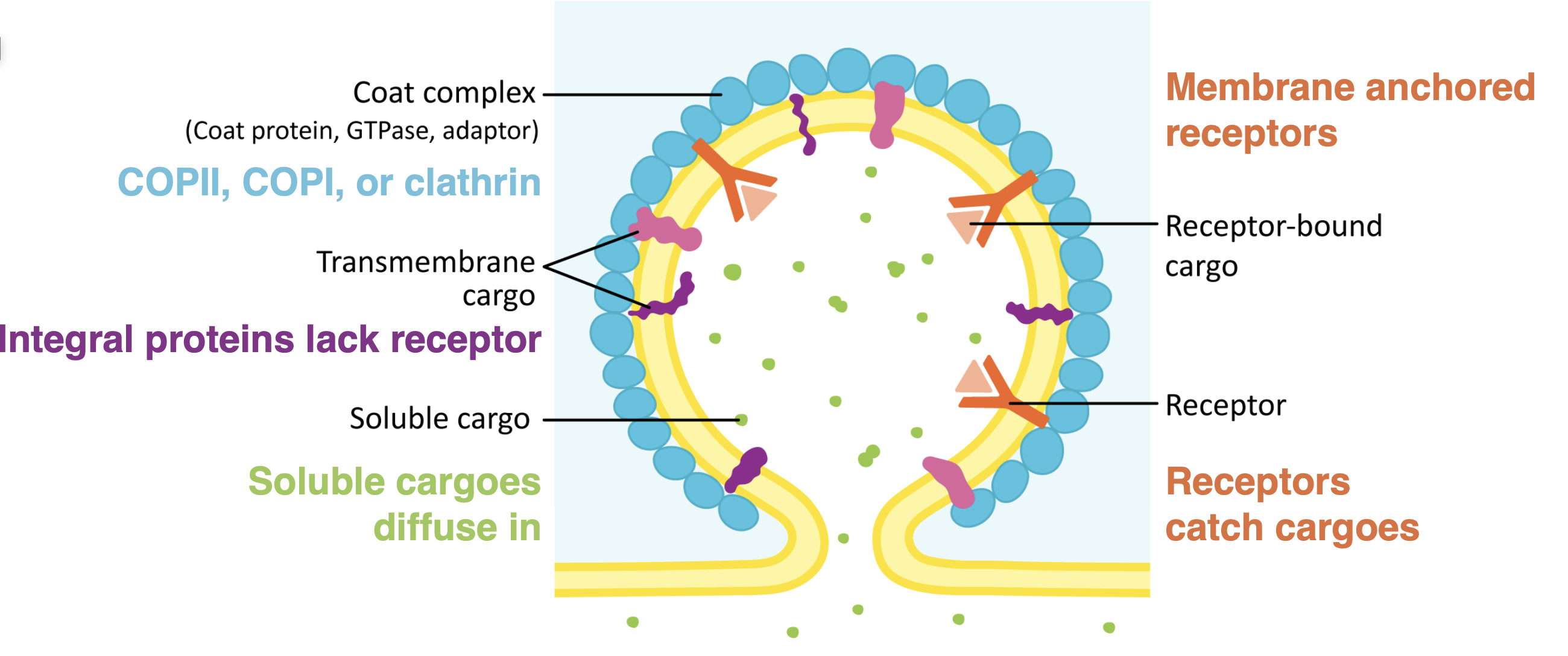
Budding
Coat complex are proteins that stabilizes the membrane curve forms on the cytosolic side
the curve is not favorable and has to have energy or supporting structure
adaptor protein is part of the coat complex- it acts as a bridge between the cargo receptor and the coat protein
Vesicle relies may be conducted by the coat protein, or by a a protein such as dynamic gtpase
when vesicle has budded off the coat is removed, the vesicle can be transported to the organelle
the coat protein determines the size and curve Clathrin and COP1 have very similarly sizes but COP11 is smaller
microbial transport
Microtubules = tracks/highways
Kinesin usually moves toward the + end → away from nucleus → anterograde
Dynein usually moves toward the − end → toward nucleus → retrograde
Adaptor proteins connect cargo to motor proteins
Different motor + adaptor combinations send different cargo to specific places
Cargo can be vesicles, proteins, RNA, or whole organelles like mitochondria
Adaptors can help control when cargo attaches to or releases from the motor
Fusion extra notes
Key ideas
without tethering protein the chance of v snare finding t snare is super low we can say that it is basically impossible
vesicle get so close to the membrane that they fuse and everything in the vesicle rapidly enters the acceptor membrane
there is usually more than 1 set of snares working together

Budding: Cathrin assembly
Coat assembly + cargo selection
Cargo binds to a cargo receptor in the membrane. Adaptin binds the receptor on the cytosolic side, and clathrinbinds adaptin.Bud formation
More clathrin assembles and bends the membrane into a bud.Vesicle formation
Dynamin forms a ring around the neck of the bud and pinches it off from the membrane.Uncoating
The clathrin coat and adaptin are removed, leaving a naked transport vesicle that can travel to its destination. The naked transport vesicle can then recruit rabs.
There are many dynamin molecules Dynamin is a GTPase that assembles into oligomers/helical rings around the neck of a budding vesicle. Once enough dynamin molecules are assembled, they hydrolyze GTP. That causes a conformational change that tightens/constricts the dynamin helix, which helps pinch the vesicle off from the membrane. Dynamic is used in many membrane remodeling processes

GTPase
a protein/enzyme that hydrolyzes GTP
What does it mean when people say mitochondria is the exception to vesicular trafficking rules?
Mitochondria are an exception to the normal rules of vesicular trafficking because they do not usually receive proteins through vesicles from the ER and Golgi. Instead, most mitochondrial proteins are made by free ribosomes in the cytosol and are imported directly across mitochondrial membranes through protein translocators. So, unlike organelles in the endomembrane system, mitochondria mainly use direct protein import rather than vesicle transport.

Mitochondria import most of their proteome (all proteins) from the cytoplasm
Notes
Translocator of outer membrane (TOM) the signal sequence we see is a tom specific signal sequence the TIM signal sequence has not been discovered
until that signal is cleaved off the protein will not function
the inner membrane is very different compared to the outer membrane in temp, ph, and the things residing there
All proteins destined for mitochondria matrix have the TOM-specific START sequence right at N terminus
Signal peptidase enzymes cleave all soluble mitochondria proteins

Mitochondrial fusion and fission are balanced by Dynamin GTPases
Fusion = two mitochondria join together
Fission = one mitochondrion splits into two
For fission, a dynamin-related GTPase called Drp1 wraps around the mitochondrion like a ring. When it hydrolyzes GTP, it tightens and helps pinch the mitochondrion apart.
For fusion, other dynamin-family GTPases, mainly mitofusins and OPA1, use GTP hydrolysis to help mitochondrial membranes come together and fuse.
Three main types of endocytosis
Endocytosis is the process by which a cell takes substances in from its surroundings by engulfing them with the cell membrane and forming a vesicle inside the cell.
Phagocytosis (to eat)
ingestion of large, solid particles by cells
particles are taken into phagosomes/food vacuoles
stimulated by activation of cell surface receptors and Calcium channels
Transmembrane signaling remodels cytoplasmic cytoskeleton, initiating membrane deformation.
• Occurs primarily in specialized immune cells, in humans.
• Aquatic single-celled protists, such as amoeba or paramecium, are also capable.
Pinocytosis (to drink)
Consititutive and non-selective (water, small ions, etc). Used to counteract volume loss due to continuous flow of vesicles to the plasma membrane via secretion.
May or may not use clathrin-coat. Alternative structure, caveola is used.
Caveolae are marked by caveolin protein complex/Cav1 (adaptin)
Characteristic stable bulb shape at the plasma membrane.
Receptor-mediated endocytosis
Much more selective than pinocytosis.
Triggered by binding of macro molecules to receptors.
Cargo concentrated up to a thousandfold at cell surface by before vesicle budding.
Cargoes bind dedicated receptors that enrich via the adaptor protein (adaptin).

Example of receptor mediated endocytosis
LDL= Low density lipoprotein, carries cholesterol inside because cholesterol is not very soluble in water/ blood so the outside of the LDL has hydrophilic parts.
Cargo delivery and receptor recycling
A subset of endosomes will mature into lysosomes
Lysosomes become acidified
lysosomes are sited of intracellular digestion
ATP- dependent hydrogen pumps in lysosomal membrane
Lysosome is the rate limiting step endocytosis can happen but if its bring it in faster than the lysosome can sort or break it down a bottle neck forms and material will start to accumulate

endosome
An endosome is a membrane-bound compartment involved in endocytosis. It receives material that the cell brings in from outside and helps sort it.
M6P influence of receptors cycle between plasma membrane and endosomes
M6P- It is a chemical tag placed on certain proteins, especially lysosomal enzymes, so the cell knows where to send them.
Cargo M6P will attach on a Receptor- M6PR on the vesicle and the vesicle will go to the endosome, There are 2 options now either the endosome becomes a lysosome and degrades or sends out the cargo inside the vesicle or they are recycled tot the plasma membrane with the help of retromer.

Cells convert extracellular signals into intracellular ones
4 general phases of signaling cascades
The signal is sent. Molecular cues, environmental signals, temperature, gases (oxygen availability)
The signal is received. Receptor recognizes signal, usually at plasma membrane, but sometimes internally if signal can diffuse. Internally is usually a gas.
The signal is interpreted. Transfer of signal across, membrane, signal amplification, internal responses, “transduction”(converting a signal received by a cell into a response inside the cell).
The cell responds May be fast or slow
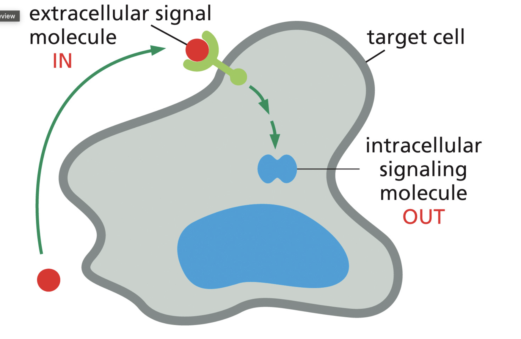
Signaling may be fast or slow
Singaling factors
Biomolecules, such as peptides carried in blood stream
Electricl signals
Odernants
photons
nuerotransmitters
mechanical forces
4 main signaling tipes
Endocrine signaling: hormones sent throughout the organism via the bloodstream
pacarine signaling: local signaling mechanism ligand released into extracellular space, and it diffuses to be picked up by nearby receptors
Neuronal signaling: electrochemical signals sent long distances yet through one cell
contact dependent signaling: direct cell to cell contact very rare

what happens after a signal binds to a receptor?
Signal/ligand binds receptor → receptor changes shape → intracellular signaling starts → cell produces a response
The terms on the slide mean:
Relay = pass the signal from one signaling protein to the next.
Transduce = convert the outside signal into an intracellular signal.
Amplify = one signal activates many molecules, making the response much bigger.
Integrate = the cell combines information from multiple signaling pathways into one response.
Feedback = the response feeds back to either increase or decrease the pathway.
Distribute = one signaling pathway can trigger several different cellular responses at once.
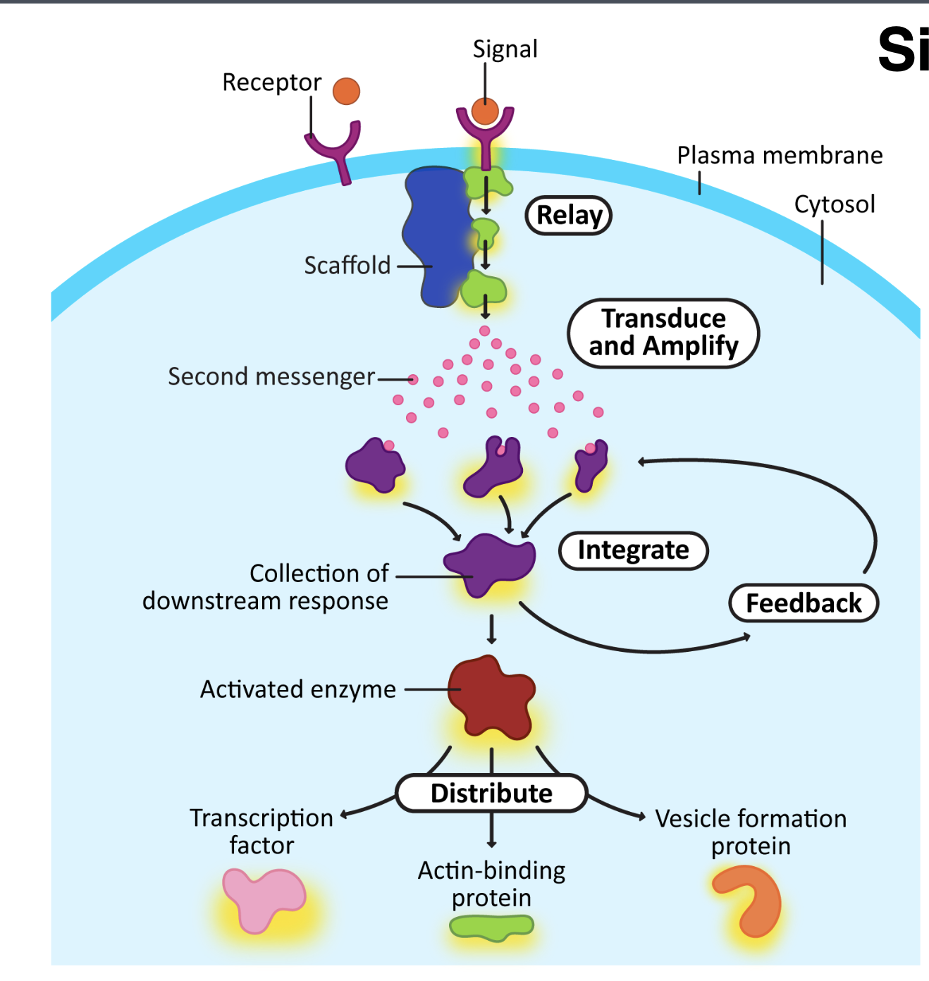
How are signals turned off?
3 main ways
Receptor sequestration = receptor is temporarily removed from the plasma membrane and brought into the cell, often into an endosome. It can later be recycled back to the membrane.
Receptor down-regulation = the cell reduces the total number of receptors, often by sending receptors to lysosomes for degradation or making fewer new receptors. This is usually more long-lasting.
Receptor inactivation = the receptor is still there, but it is turned off and can’t signal properly. For example, a receptor may be phosphorylated so downstream proteins can no longer interact with it.

G protein activation by GPCR
Background
GPCR are most common type in animals
7 transmembrane domains; internal loop transducer signal to nearby G protein
GTP required to trigger cascade leading to enzyme activity
G protein composed of trimeric complex
Function
Receptor acts as a GEF for a G- protein change GDP to GTP
An activated GPCR activated a G protein by inducing the GTPASE(alpha subunit) to release GDP and bind to GTP.
When the alpha subunit binds to GTP it releases the other 2 subunits (beta and gamma), thereby activating them as well

NLS
Cargo that gets recognized by NIR and NER have a Nuclear localization sequence.
Difference between RNA polymerase 1 and RNA polymerase 2
RNA polymerase I → ribosomal RNA
Works mainly in the nucleolus
Produces most of the rRNA that becomes part of ribosomes
Think: Pol I = ribosome production
RNA polymerase II → messenger RNA
Transcribes protein-coding genes from DNA into pre-mRNA
That pre-mRNA then gets processed with a 5′ cap, poly-A tail, and splicing
The mature mRNA can then leave the nucleus and be translated
Think: Pol II = protein-coding genes
Protofilament
one linear chain of tubulin dimers. 13 protofilaments associate to make one microtubule.
Nucleation
the process of starting a new microtubule, usually at an MTOC.