Histotechnology HTL Exam - Comprehensive Review
1/1524
There's no tags or description
Looks like no tags are added yet.
Name | Mastery | Learn | Test | Matching | Spaced | Call with Kai |
|---|
No analytics yet
Send a link to your students to track their progress
1525 Terms
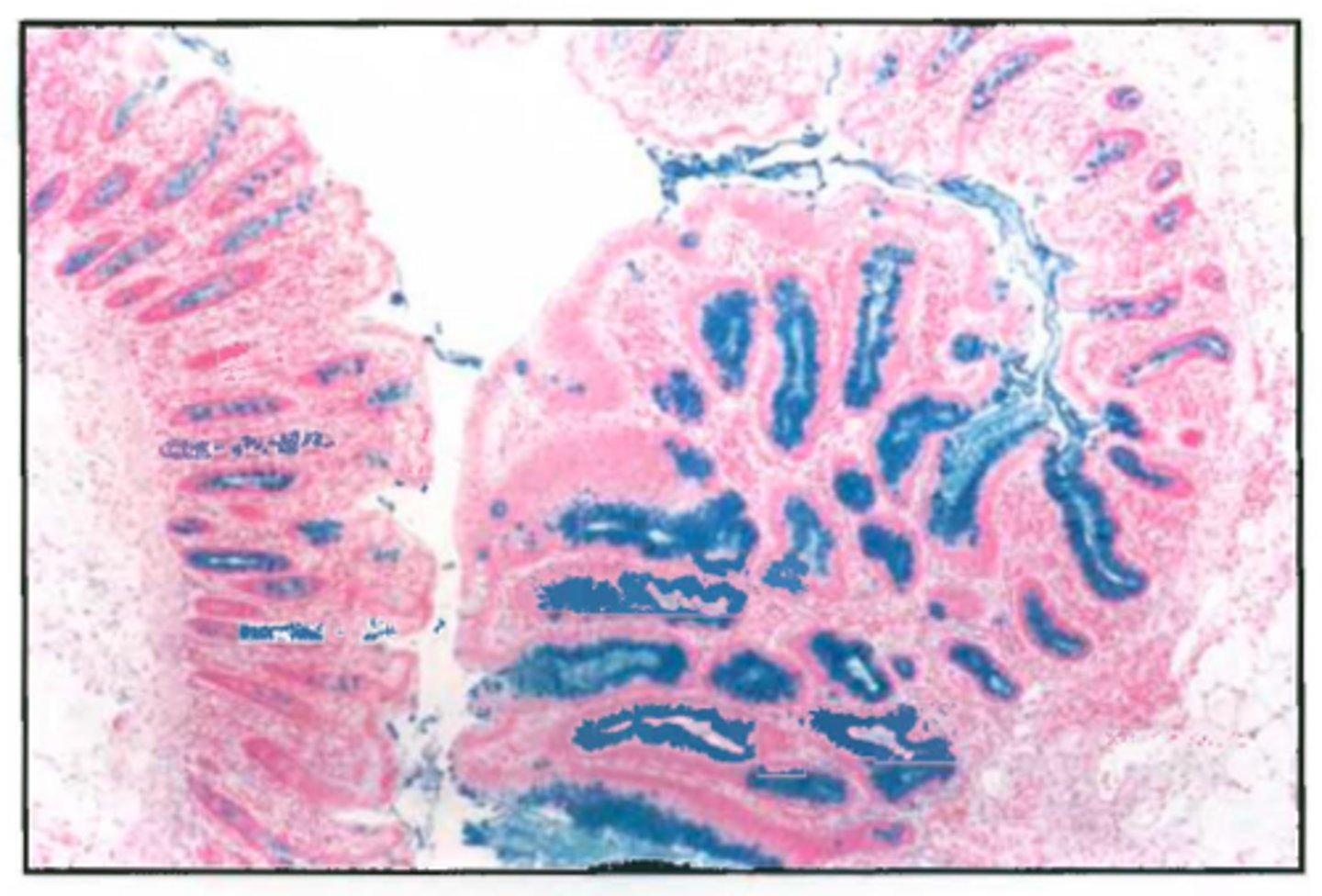
Alcian Blue demonstrates
Acid mucins
ATPase Enyme Histochemistry demonstrates
ATPase in muscles
Bielschowsky's Silver Technique demonatrates
Dendrites, axons, neurofibrils
Brown & Brenn gram stain demonstrates
bacteria
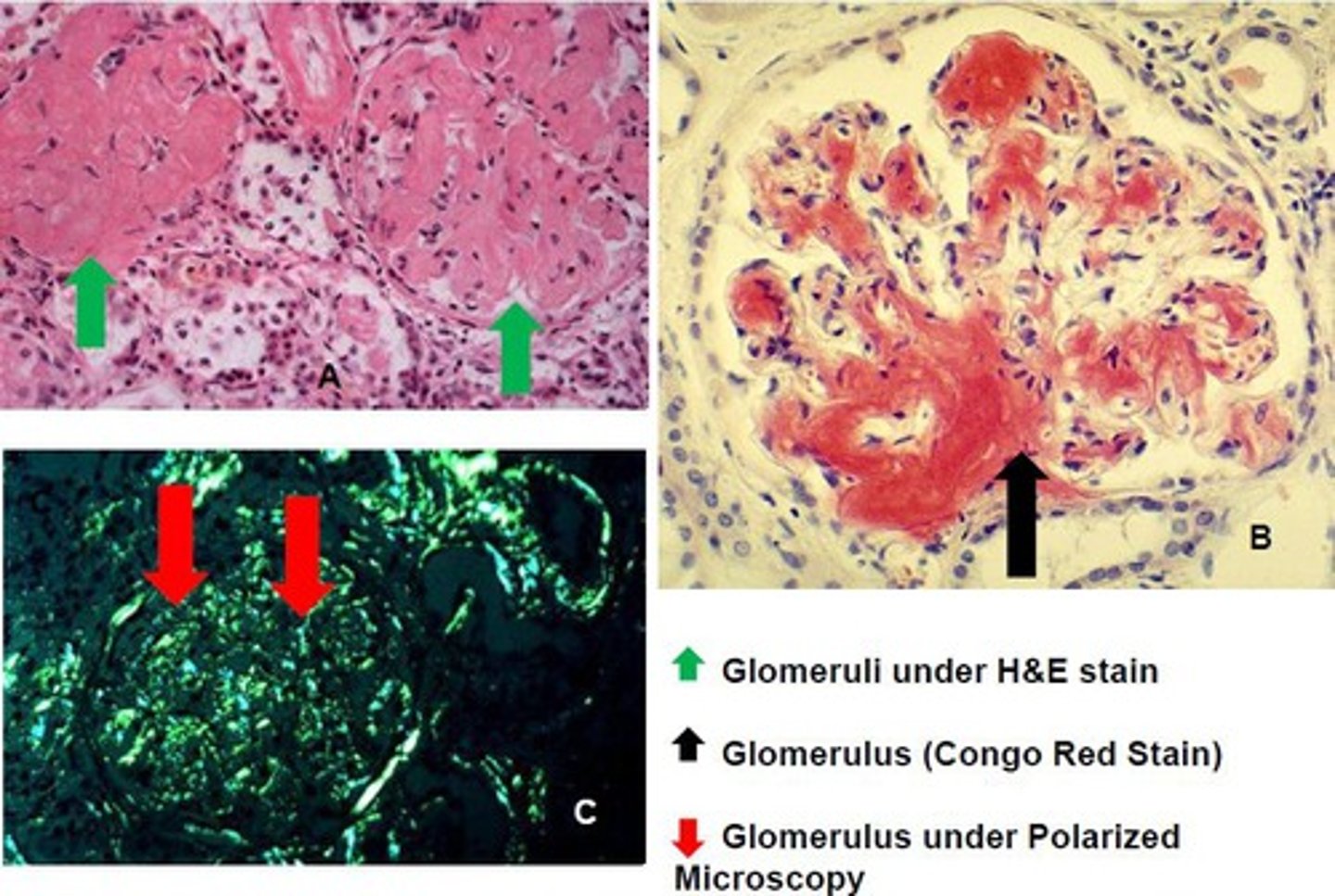
Congo red demonstrates
amyloid
Cresyl Echt Violet demonstrates
Nissl substance
Geimsa stain demonstrates
mast cells or H. pylori
Gomori Burtner demonstrates
melanin
Gomori's Aldehyde Fuchsin
demonstrates
elastic and mast cells
Gordon & Sweet's demonstrates
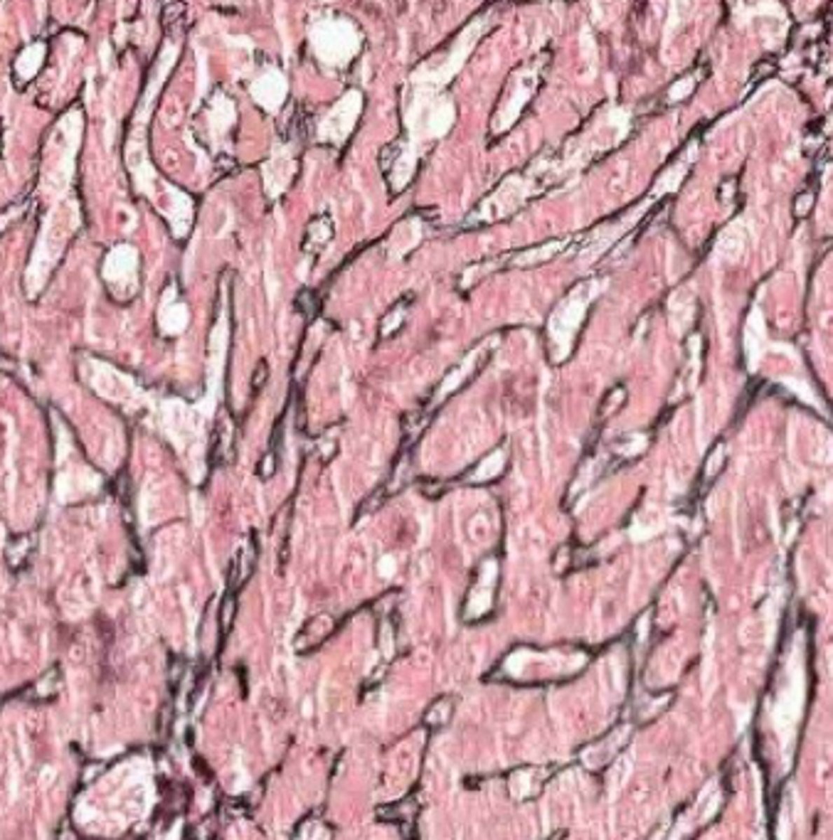
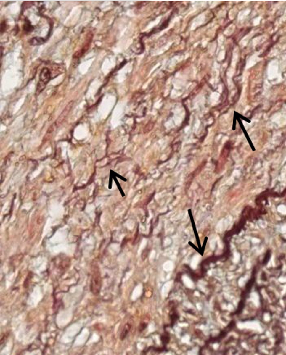
reticulin
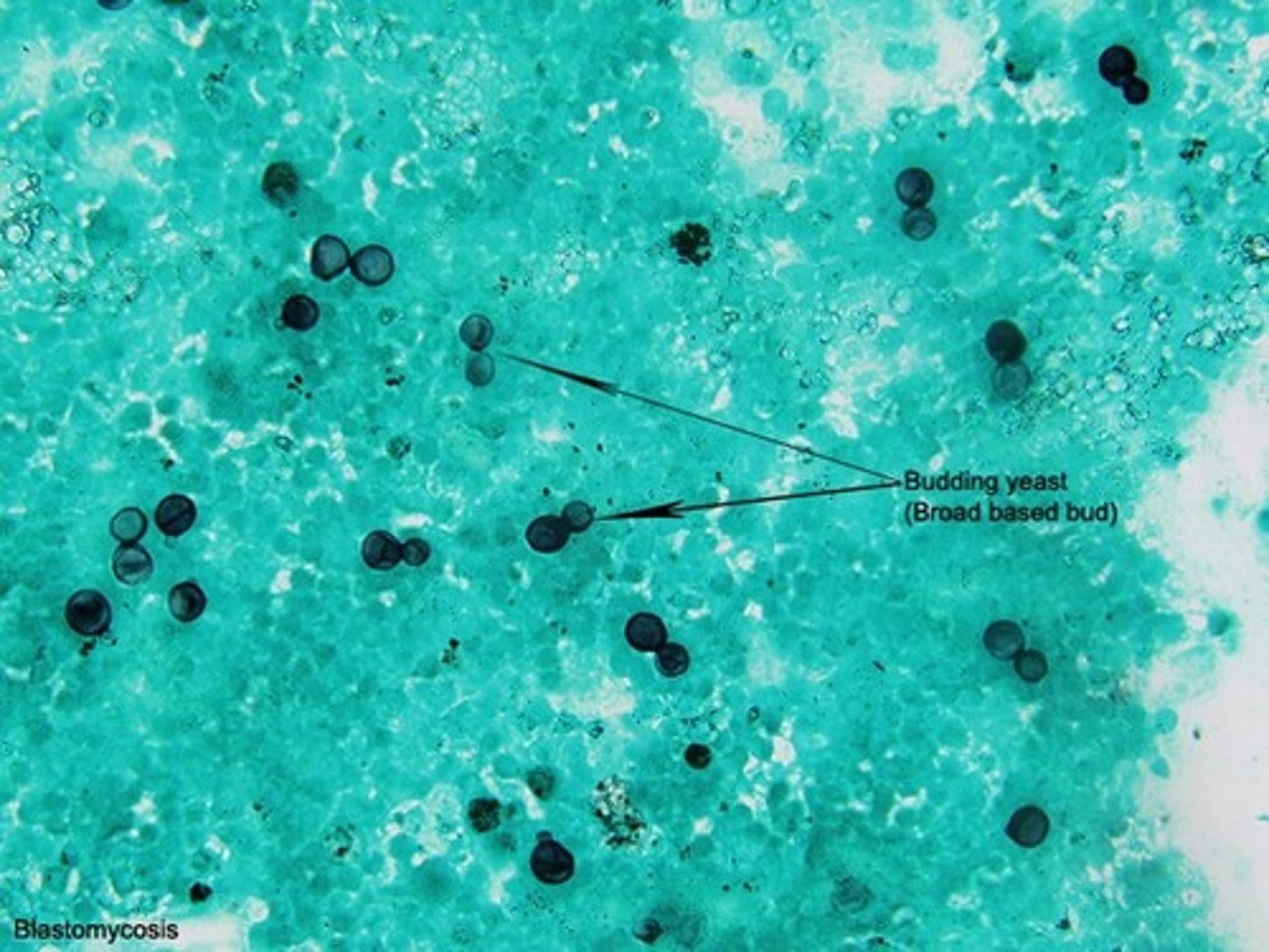
Grocott's Methenamine Silver demonstrates
fungi (eg. Pneumocystis)
Hematoxylin & Eosin shows
general stain
Immunohistotochemistry
Anything an antibody can be made for
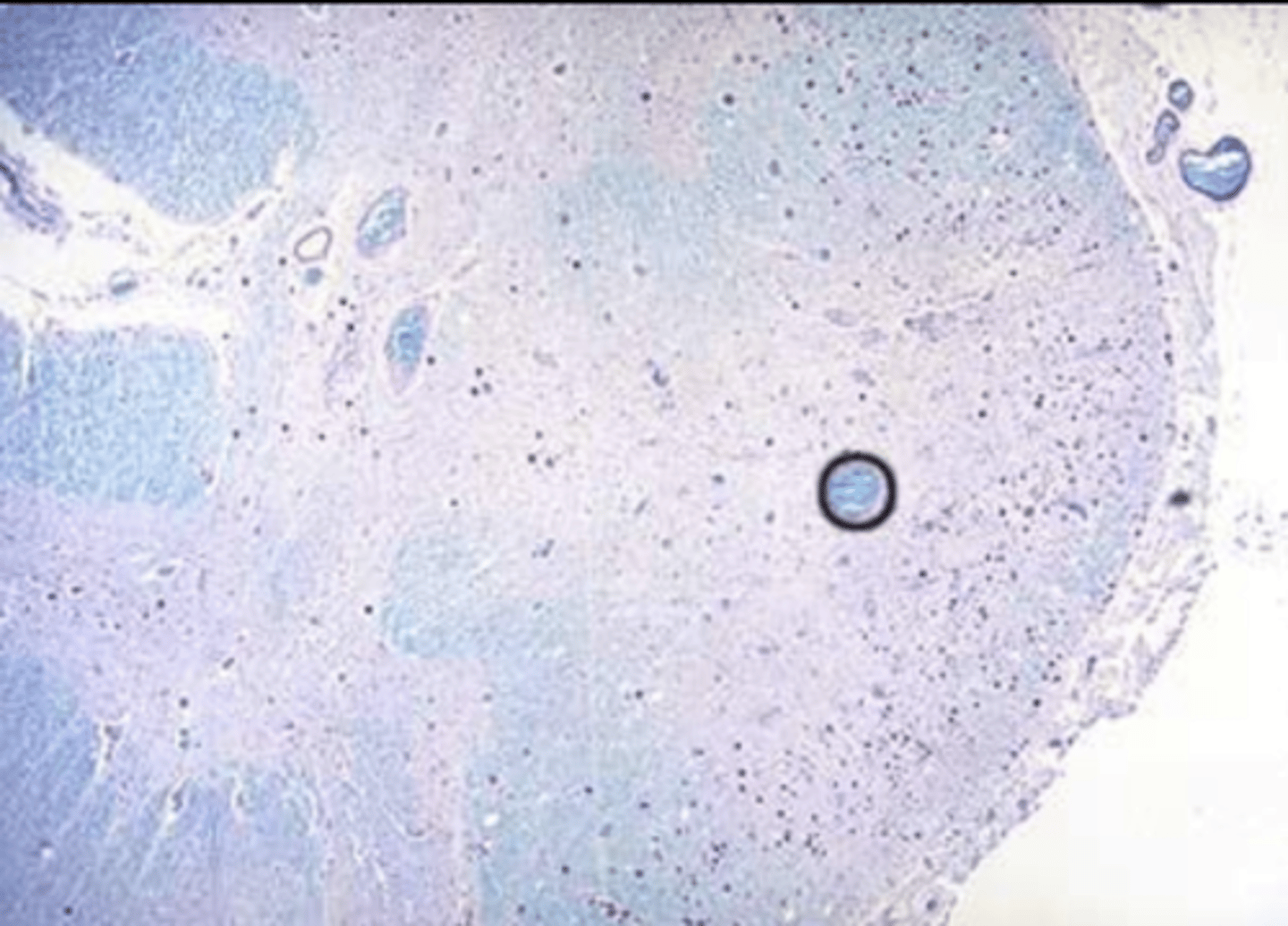
Luxol fast blue stain shows
myelin
Toluidine blue stain shows
mast cells or H. pylory
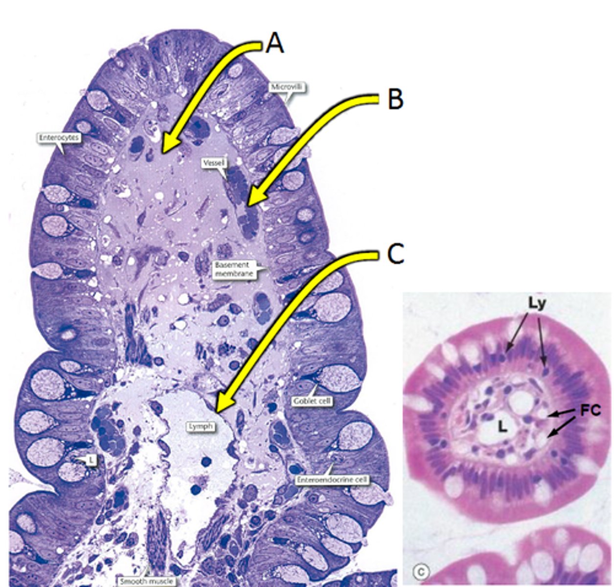
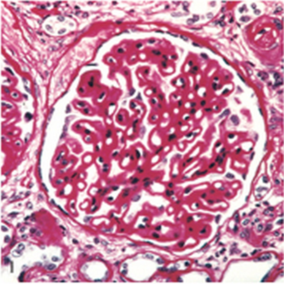
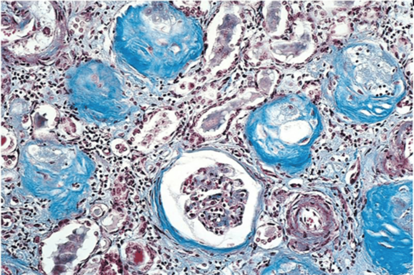
Masson's Trichrome shows
collagen
Oil Red O technique shows
neutral fats (simple fats)
Papanicolaou technique shows
cytology smears
Periodic Acid Schiff's shows
neutral mucin, glycogen
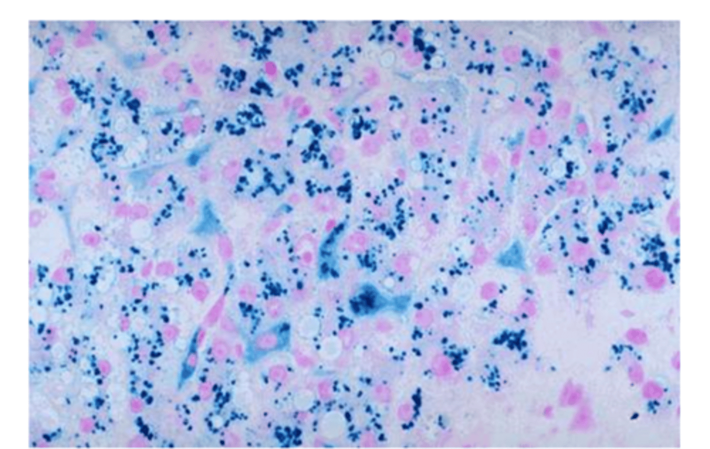
Perl's Prussian Blue shows
Hemosiderin
Southgate's mucicarmine
epithelial acid mucins
Verhoeff's Van Gieson
elastic
Van Kossa shows
calcium
Ziehl Neelsen shows
Acid fast bacilli (T.B.)
Nuclei in Gomori's Aldehyde Fuchsin technique
yellow - no specific stain for nuclei
Mast cells in a Gomori's Aldehyde Fuchsin technique
purple - GAF not specific for elastic
Gram positive bacteria if over-differentiated after crystal violet
red
Nuclei in the Brown and Brenn Gram stain if over differentiated with picric acid/acetone
yellow
Fungi in the Periodic Acid Schiff (PAS) technique
magenta - chitin binds with Schiffs
Fungi in the Grocott's Methenamine Silver technique
black - silver impregnation
Neutral mucins in a PAS technique if potassium permanganate is used as the oxidizing agent
light pink - colour of background - CHO overoxidized from aldehydes to acid state
Cytoplasm in an H&E if over differentiated with acid alcohol
pink - only affects nuclei
Epithelial acid mucins in the Southgate's mucicarmine technique
red
Cytoplasm in an H&E if the blueing agent is not properly removed
very pale pink
Nuclei in the Congo Red technique
blue - stained with aluminum hematoxylin
Calcium in the Von Kossa technique
black - silver impregnation
Glycogen, after diastase in the Periodic Acid Schiff technique
pale pink - background colour
Mycobacterium after Ziehl Neelsen technique
red - AFB stain with carbol fuchsin
Mycobacterium after Ziehl Neelsen technique
red - AFB stain with carbol fuchsin
Streptococcus after Ziehl Neelsen technique
blue - colour of background
Neutral lipids for Oil Red O cut on a cryostat
red to orange
Neutral lipids for Oil Red O cut from wax block
not demonstrated - removed
Hemosiderin after a Perl's Prussian Blue
blue
Nuclei after Masson's trichrome using an aluminum Hematoxylin
red - same as cytoplasm
Fine elastic fibres in Verhoeff's Van Gieson (VVG) after leaving in Van Gieson reagent too long
not black/not demonstrated - iron hematoxylin removed by the acid
Cytoplasm if underdifferentiated in the VVG technique
grey to black
Reticulin fibres if iron alum is skipped in the Gordon and Sweet's
not demonstrated
Melanin if potassium permanganate was used as an oxidizing agent in the Gomori Burtner technique
not demonstrated - removed
Melanin in the Luxol fast blue if overdifferentiated with lithium carbonate
pink
Nissl substance using the Cresyl echt violet technique
violet
Carboxylated mucins after Alcian Blue technique ph 1.0
no demonstration - not blue
Mast cells using Geimsa or Toluidine Blue
violet - both metachromatic dyes
Control slide for Alcian Blue
small intestine
Control slide for Bielschowsky's Silver Technique
grey matter
Control slide for Brown & Brenn gram stain
infected tissue
Control slide for Congo Red
known amyloid slide
Control slide for Cresyl Echt Violet
grey matter
Control slide for Gomori Burtner
skin
Control slide for Gomori's Aldehyde Fuchsin
aorta or skin
Control slide for Gordon & Sweet's
spleen or liver
Control slide for Grocott's Methenamine Silver
known fungi slide
Control slide for Masson's Trichrome
kidney
Control slide for Hematoxylin & Eosin
small intestine
Control slide for Luxol Fast Blue
cerebellum
Control slide for Masson's Trichrome
kidney
Control slide for Oil Red O
fatty liver
Control slide for Periodic Acid Schiff's(PAS) glycogen
liver
Control slide for PAS for neutral mucins
small intestine
Control slide for Perl's Prussian Blue
known hemosiderosis slide
Control slide for Verhoeff's Van Gieson
aorta or skin
Control slide for Ziehl Neelsen
known TB lung
A pathologist is reading the gross description report and it indicates that the patient sample they are examining should be uterus but under the microscope they see tissue from a breast on the slide. What could be the reason for the pathologist not receiving the correct specimen with the gross description report? (3)
The Pathologist Assistant opened two specimen containers at a time and grabbed the wrong specimen while grossing.
-The Pathologist Assistant gave the wrong requisition to the wrong specimen after grossing.
-The Technologist while embedding opened two cassettes at one time and put the wrong tissue in the wax mold with the wrong cassette on top.
_______counteracts the shrinking of the cells that occurs from picric acid in Bouins fixative
Acetic acid
What information would you find on a tissue cassette?
Block number
Year and surgical number
Number of tissue pieces
Before processing decalcified tissue, what is the next step?
Neutralize acid
The grossing area receives a liver biopsy that appears to be fatty. The pathologist wants to preserve the lipids in the tissue. What is the best fixative to use for lipid studies?
Osmium Tetroxide
A technologist completed an H +E stain on breast tissue and decided to check the slide under the microscope to verify if the stain worked correctly. The technologist noticed that all the lipids were destroyed in the tissue. What fixative was used in the grossing area that could of destroyed all the lipids on the slide?
100% Ethanol
Which fixative is the best in providing maximum preservation of glycogen in the tissue?
Picric Acid 95-100% Ethyl Alcohol
Problems associated with Picric Acid
Yellow discolouration of tissue
Hemolyzes RBCs
Dissolves Iron
Explosive when dry
Problems associated with 95-100% Ethyl Alcohol
Flammable Poisonous Excessive hardness & shrinkage
Government regulated
Describe the choice of fixative to use and three reasons why you chose that fixative for immunological studies.
B5 (or Zinc Formalin)
- Enhances immunoreactivity
- Coagulant fixative
- gives greater permeability
- better antibody penetration
- more intense staining
- Enhances nuclear detail, and good morphology
- Enhances staining of both nuclei & cytoplasm
Upon microscopic examination, an H & E stained section of routinely processed spleen shows small brown to black granules evenly distributed throughout the tissue.
Suggest a possible causative agent and describe how this artifact can be removed.
Formalin pigment - Acid Formaldehyde Hematin (AFH)
Formaldehyde / formalin, acid pH, hemoglobin (blood rich tissues)
1) Bring section to absolute alcohol
2) Place section in saturated picric acid for 5 min to 2 hours depending on the amount of pigment present
3) Wash for 15 to 20 min in running tap water
4) Proceed to desired stain
surface decalcification
Rough in to expose the tissue
- Surface of the tissue is placed in 1% HCl for 15-60 minutes
- This allows only several sections to be cut
a. Disposal of used 10% NBF
Wear appropriate PPE - mask, gloves and safety glasses. Work in a chemical fume hood. Mix with formalex in a ratio of 1:5 with formalin. Leave for a minimum of 1 hour (when a precipitate has formed, the fixative has been "neutralized"). This solution can now safely be poured down the sink with running tap water.
The 3 ingredients in B5 fixative are:
Mercuric chloride, Sodium acetate, Formaldehyde
Which fixative is classified as a non-additive
95% Ethyl alcohol
Which fixative is considered a substitute for B5?
zinc formalin
Commercial stock formaldehyde solution contains:
40% formaldehyde
Formalin pigment (AFH), may be removed from tissue by: (acidic ph and blood cause formalin pigment)
Alcoholic picric acid
Non-coagulant fixatives
Creates a gel that makes penetration by subsequent solutions difficult. Example:
10% NB Formalin
Formaldehyde, Acetic acid
additive fixatives
(non "A" words)
Formalin, Mercury, Osmium, Glutaraldehyde
Non-additive fixatives
("a" words)
Alcohol, acetic acid, Acetone
Goagulants (ZAPAM)
Zinc, Alcohol, Picric acid, Acetone, Mercury
Tissue is fixed in order to:
Stop autolysis
Which fixative is considered the "routine fixative"?
10% NB Formalin
Best secondary fixative for cytoplasmic (trichrome) staining
Bouin's
After fixing tissue in Bouin's solution the excess picric acid is removed by washing in:
70% Ethanol
Which fixative makes lipids insoluble?
Osmium tetroxide
A dye appears colorless because its chromophore was reduced. The reaction is reversible.
Leuco compound
Basophilic tissue has a ___________ charge.
Negative