ANHB2212 - Labelling Practice
1/55
Earn XP
Description and Tags
Name | Mastery | Learn | Test | Matching | Spaced | Call with Kai |
|---|
No analytics yet
Send a link to your students to track their progress
56 Terms
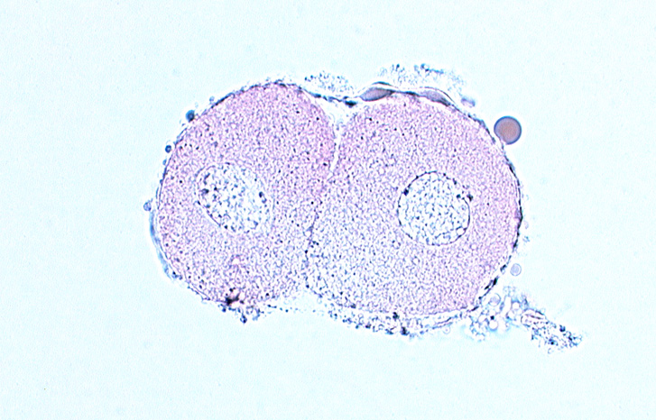
Label the following:
zona pellucida
nuclei
cytoplasm
cleavage plane
Day 2-3 Embryo

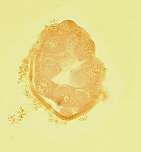
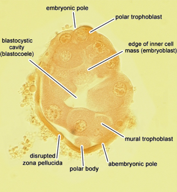
Label the following:
trophoblasts
embryoblasts
polar body
zona pellucida
blastocoele
Day 4 Embryo
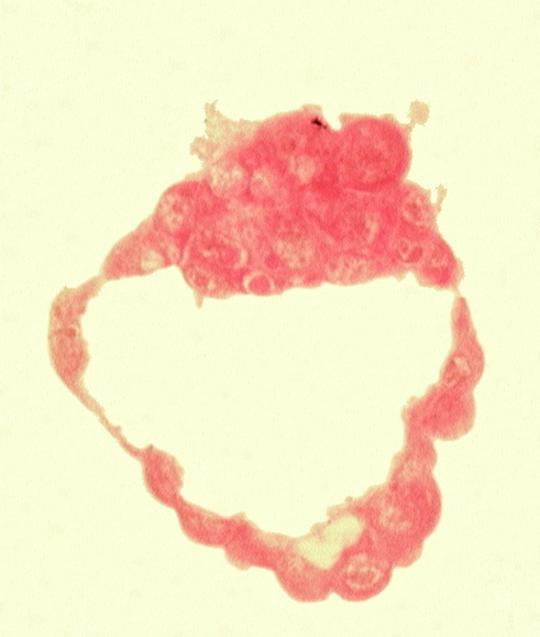
Label the following:
trophoblasts
epiblast
hypoblast
blastocoele
Day 4-5 Embryo
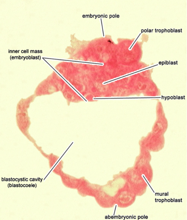
Label the following:
syncytiotrophoblast
cytotrophoblast
hypoblast
epiblast
blastocoele
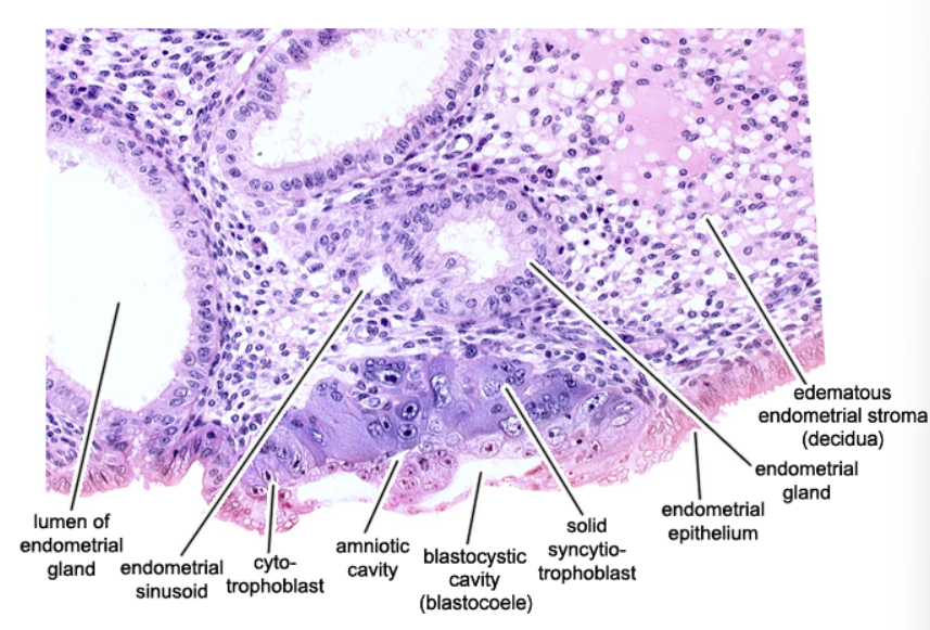
Day 6 Embryo
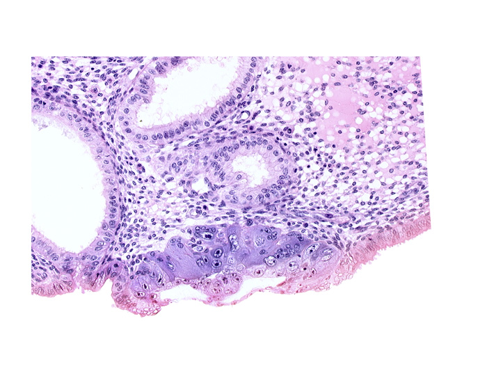
Label the following:
endometrial epithelium
endometrial gland
amniotic cavity
lumen of endometrial gland
blastocoele
syncytiotrophoblast
cytotrophoblast
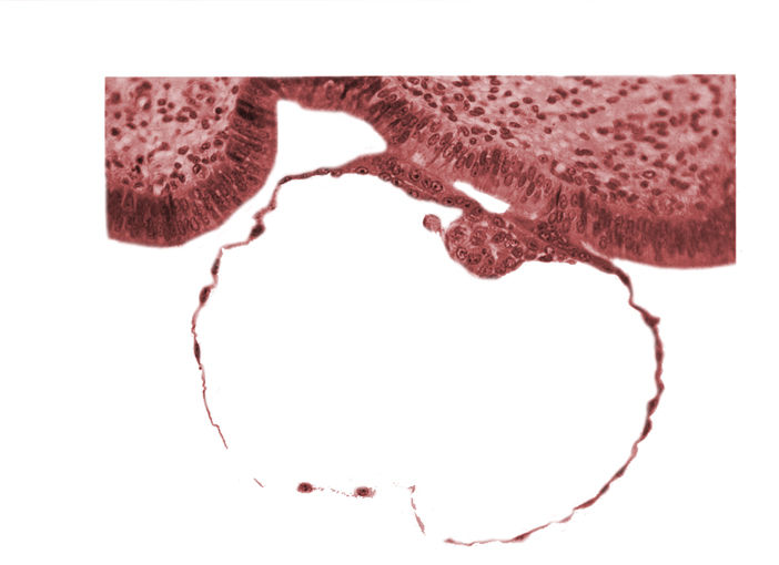
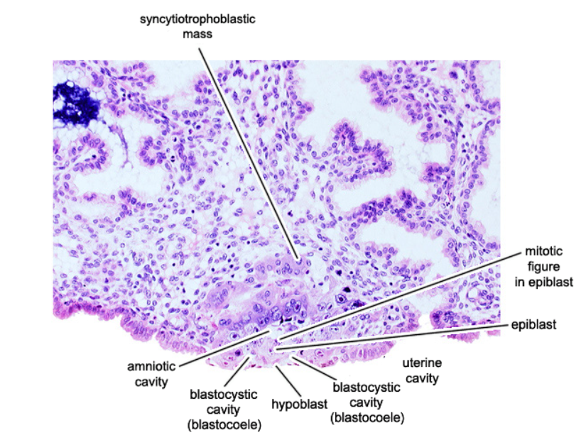
Day 7 Embryo
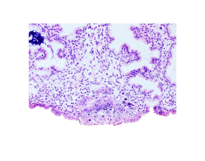
Label the following:
syncytiotrophoblast
epiblast
blastocoele
hypoblast
amniotic cavity
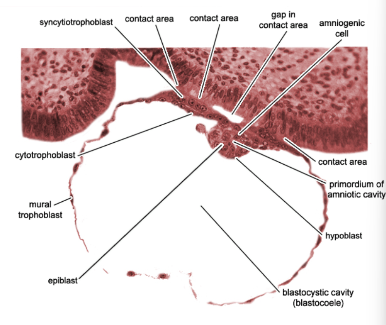
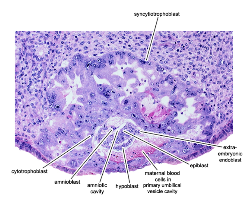
Day 8 Embryo
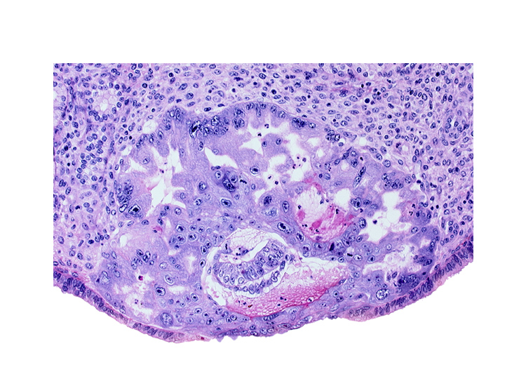
Label the following:
syncytiotrophoblast
epiblast
hypoblast
amniotic cavity
amnioblast
cytotrophoblast
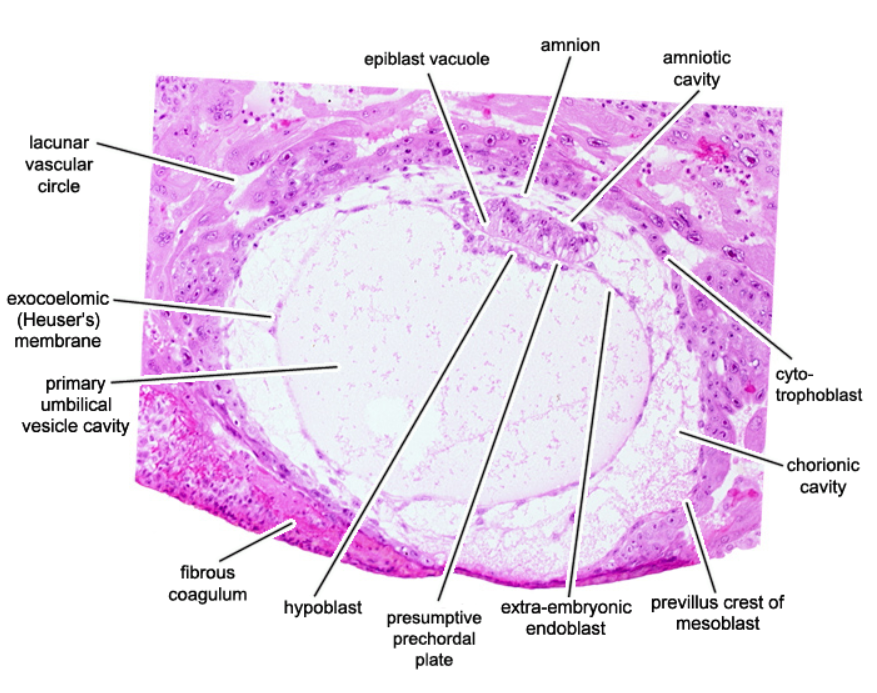
Day 9 Embryo
Label the following:
amniotic cavity
amnion
lacunar vascular circle
Heuser’s membrane
chorionic cavity
hypoblast
cytotrophoblast
Day 12 Embryo
Label the following:
amnion
amniotic cavity
epiblast
embryonic disc
hypoblast
yolk sac
endoderm
mesoderm'
connecting stalk
primitive streak
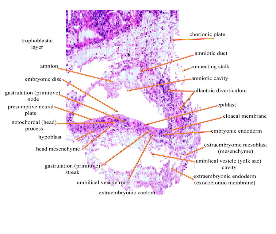
Day 13-17 Embryo
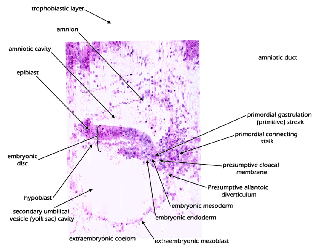
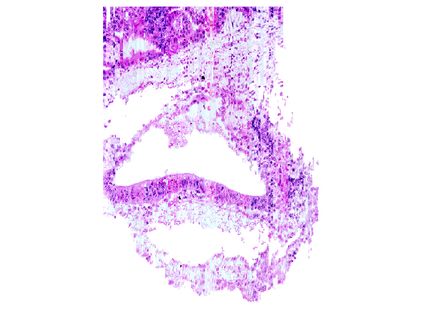
Label the following:
connecting stalk
amniotic cavity
epiblast
endoderm
yolk sac
primitive streak
hypoblast
amnion
Day 16-19 Embryo
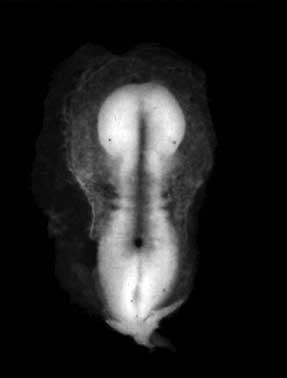
Label the following:
neural plate
neural groove
primitive streak
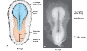
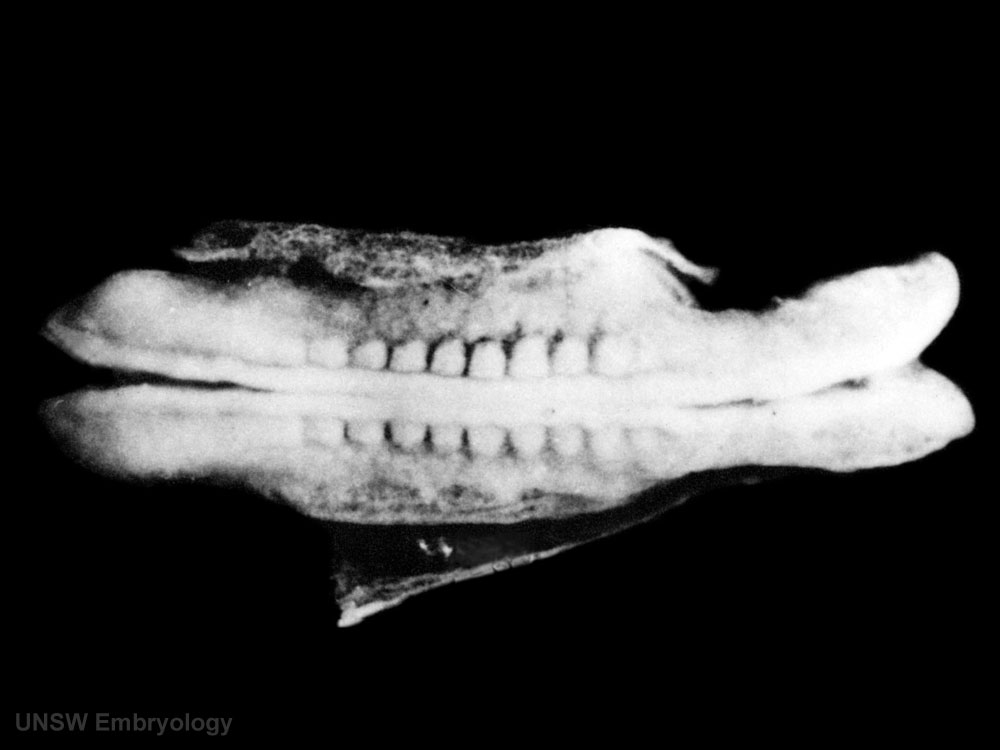
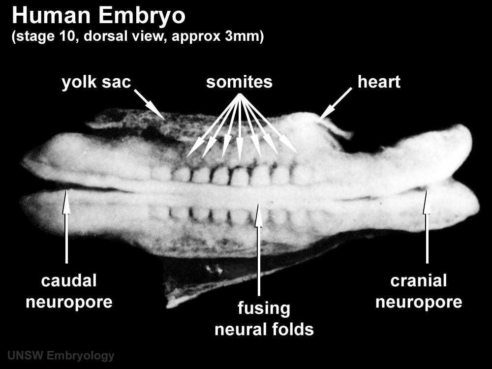
Label the following:
yolk sac
somites
caudal neuropore
cranial neuropore
fusing neural folds
heart
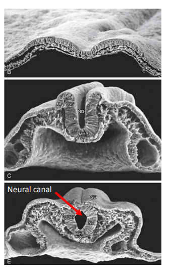
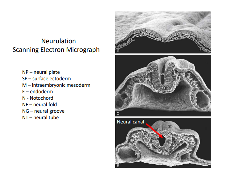
Label the following:
Neural Plate
Surface Ectoderm
Intraembryonic Mesoderm
Endoderm
Notochord
Neural Fold
Neural Groove
Neural Tube
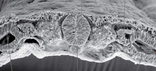
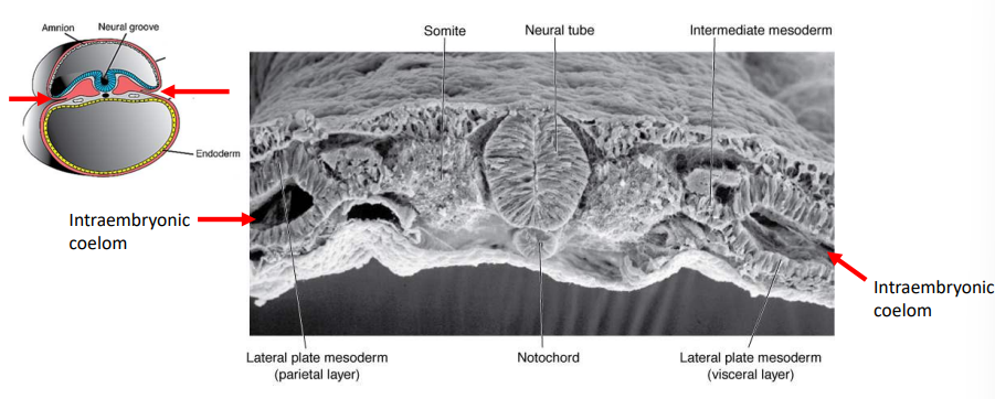
Label the following:
somite
neural tube
intermediate mesoderm
intraembryonic coelom
lateral plate mesoderm
notochord
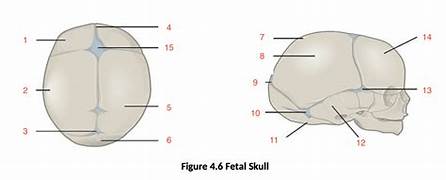
Label the following:
anterior, anterolateral, and posterior fontanelles
coronal and lamboid sutures

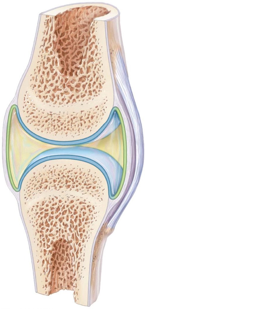
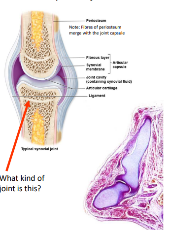
Label the following:
periosteum
articular capsule (fibrous layer and synovial membrane)
joint cavity
articular cartilage
ligament
Synovial Joint
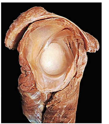
Label the following:
capsule
articular cartilage
tendons
femoral head and neck
labrum
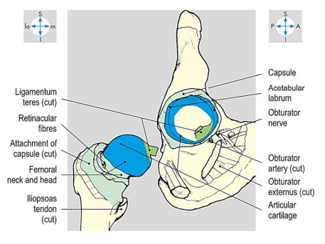
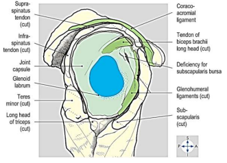
Label the following:
tendons
ligaments
joint capsule
labrum
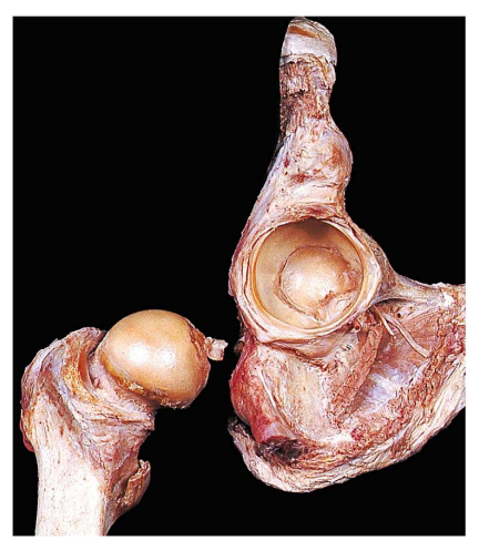
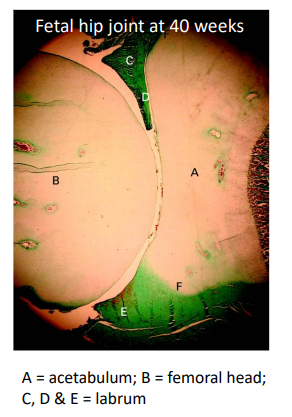
Label the following:
acetabulum
femoral head
labrum
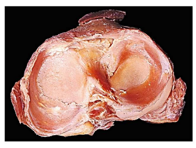
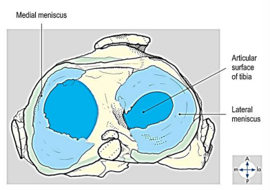
Label the following:
articular surface of tibia
meniscus
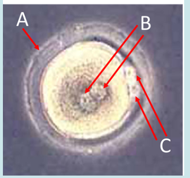
Label A, B and C
Where is this structure normally located in the female body?
What triggers the second structure C to develop?
How many chromosomes are contained in B?
How many chromosomes are contained in C?
What process immediately follows the joining of structures B?
A = zona pellucida, B = pronuclei, C = polar bodies
ampulla of uterine tube
fertilization
23
23
cleavage
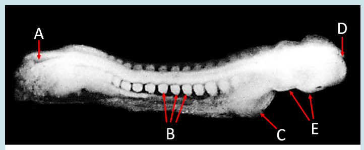
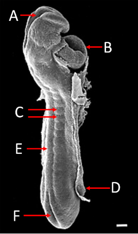
Label A, B, C, D, and E
What view is this?
What is the next developmental process?
A = caudal neuropore, B = somites, C = heart tube, D = cranial neuropore, E = pharyngeal arches
right dorsolateral
folding
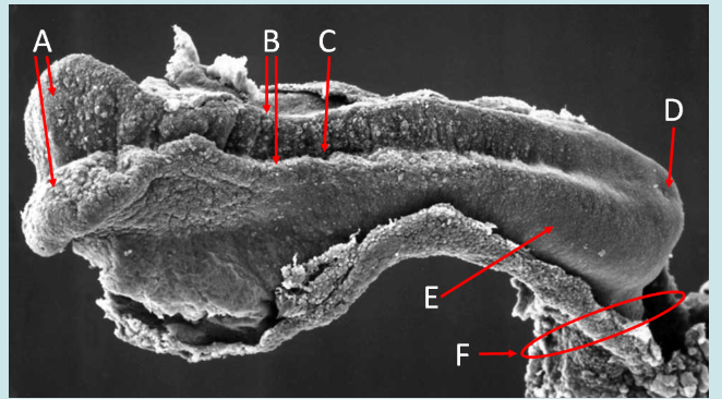
Label A, B, C, D, E and F
What view is this?
A = neural plate ectoderm, B = neural folds, C = neural groove, D = primitive streak, E = surface ectoderm, F = connecting stalk
left dorsolateral
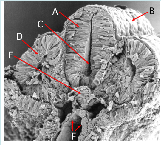
Label A, B, C, D and E
Name the part of the primitive streak that gives rise to E
Name the primary germ layer the cells lining tube F are derived from
At this stage, tube F opens into the
What view is this?
A = neural tube, B = surface ectoderm, C = neural canal, D = paraxial mesoderm, E = notochord
primitive pit
endoderm
yolk sac
transverse
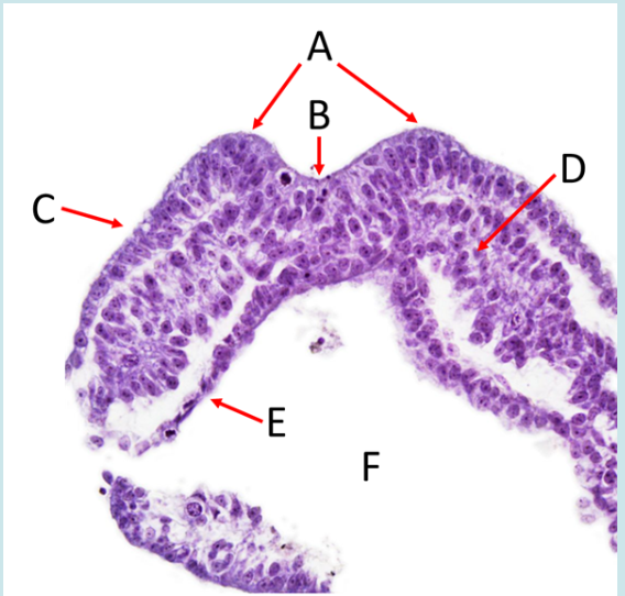
Label A, B. C, D, E and F
What embryonic structure does F open into?
What view is this?
A = neural fold, B = neural groove, C = ectoderm, D = paraxial mesoderm, E = endoderm, F = vitelline duct
yolk sac
transverse
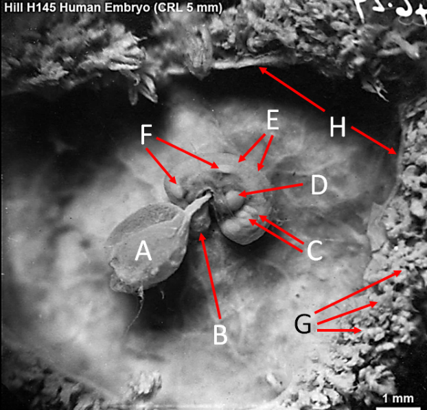
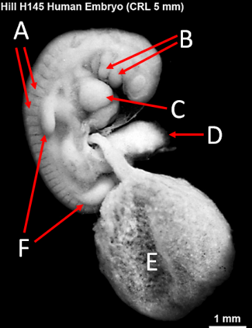
Label A, B, C, D, E, F, G and H
What view is this?
A = yolk sac, B = umbilical cord, C = pharyngeal arches, D = heart tube, E = somites, F = limb buds, G = tertiary villi, H = chorionic cavity
right lateral
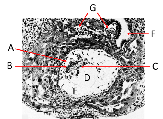
Label A, B, C, D, E, F and G
A = amniotic cavity
B = epiblast
C = hypoblast
D = yolk sac
E = chorionic cavity
F = maternal gland
G = lacunae
Label A, B, C, D, E and F
What view is this?
A = cranial neuropore, B = heart tube, C = somites, D = amnion, E = neural tube, F = caudal neuropore
right dorsolateral
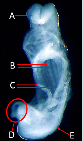
Label A, B, C, D and E
What view is this?
A = neural plate ectoderm, B = somites, C = yolk sac, D = connecting stalk, E = ectoderm
ventral
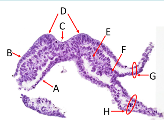
Label A, B, C, D, E, F, G and H
What are the upper and lower layers of G?
What are the upper and lower layers of H?
What view is this?
A = endoderm, B = ectoderm, C = neural groove, D = neural fold, E = paraxial mesoderm, F = intermediate mesoderm, G = somatopleure, H = splanchnopleure
upper = ectoderm, lower = lateral plate mesoderm
upper = lateral plate mesoderm, lower = endoderm
transverse
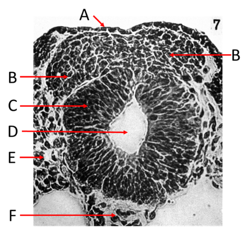
Label A, B, C, D, E and F
What view is this?
A = ectoderm, B = neural crest cells, C = neural tube, D = neural canal, E = paraxial mesoderm, F = notochord
transverse
Label A, B, C, D, E and F
A = somites
B = pharyngeal arches
C = heart tube
D = connecting stalk
E = yolk sac
F = limb buds
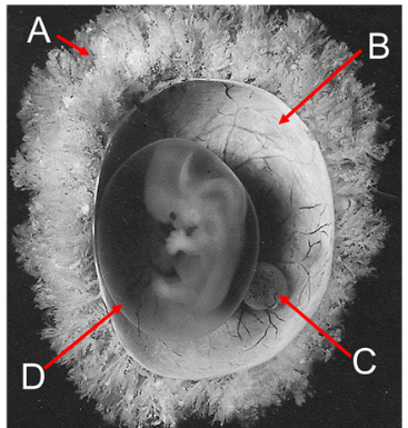
Label A, B, C and D
A = tertiary villi
B = chorion
C = yolk sac
D = amnion
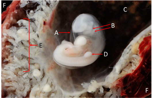
Label A, B, C, D, E and F
A = amnion
B = pharyngeal arches
C = chorionic cavity
D = left lower limb bud
E = tertiary villi
F = uterine cavity
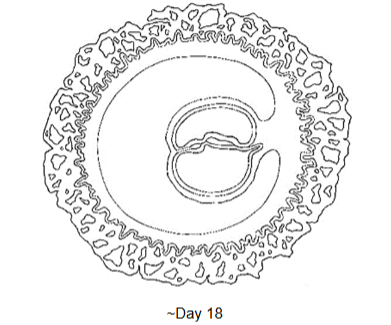
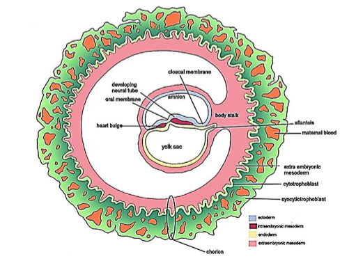
LABEL where possible: amniotic cavity, chorionic cavity, yolk sac, ectoderm,
intraembryonic mesoderm, endoderm, extraembryonic mesoderm, connecting stalk, cytotrophoblast, syncytiotrophoblast, lacunae, oropharyngeal membrane, cloacal membrane, allantois, developing heart, foregut, midgut, hindgut.
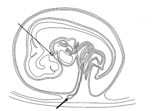
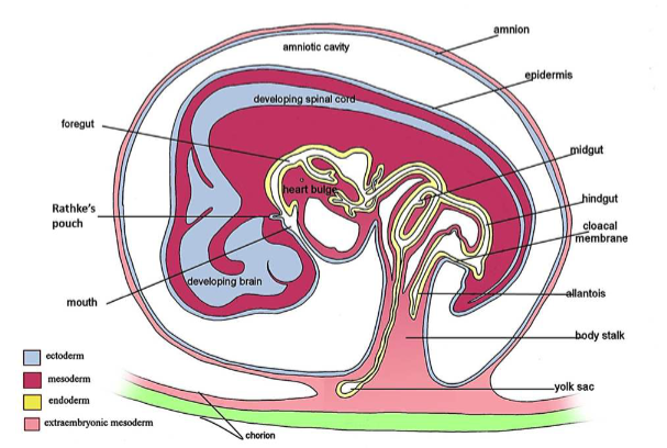
LABEL where possible: Rathke’s pouch, amniotic cavity, chorionic cavity, yolk sac, ectoderm, intraembryonic mesoderm, endoderm, extraembryonic mesoderm, connecting stalk, cytotrophoblast, syncytiotrophoblast, lacunae, oropharyngeal membrane, cloacal membrane, allantois, developing heart, foregut, midgut, hindgut.
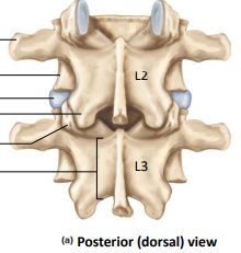
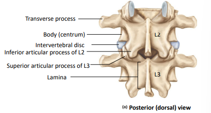
Label the following:
transverse process
body (centrum)
intervertebral disc
inferior articular process (L2 and L3)
Lamina
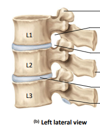
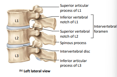
Label the following:
superior and inferior articular process of L1
superior vertebral notch of L2
spinous process
intervertebral disc
inferior articular process of L3
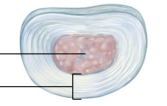
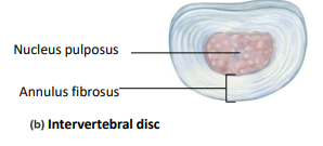
Label the following:
nucleus pulposus
annulus fibrosus
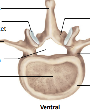
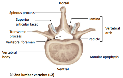
Label the following:
spinous process
lamina
pedicle
vertebral arch
annular apophysis
vertebral body
vertebral foramen
transverse process
superior articular facet
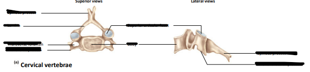
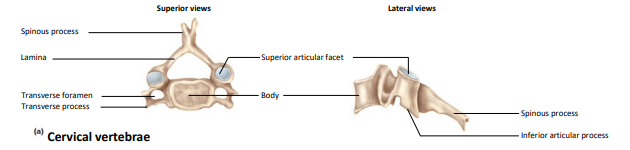
Label the following:
spinous process
lamina
transverse foramen
transverse process
vertebral body
superior articular facet
inferior articular process
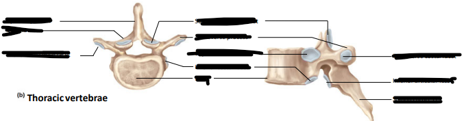
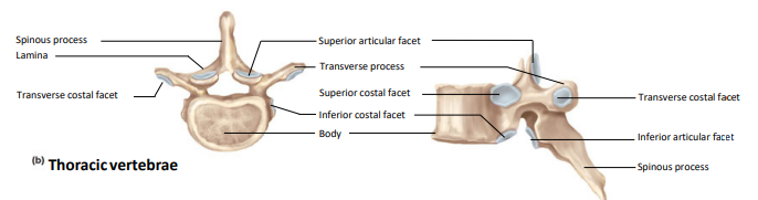
Label the following:
spinous process
lamina
transverse costal facet
body
superior articular facet
transverse process
superior costal facet
inferior costal facet
inferior articular facet
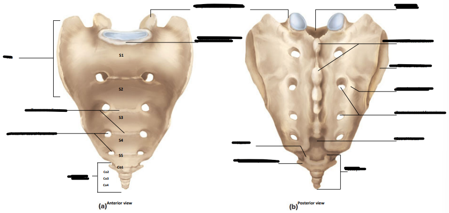
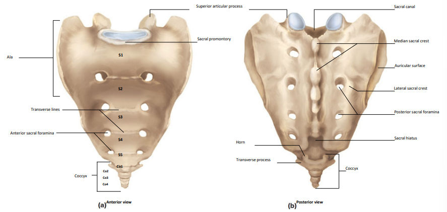
Label the following;
ala
transverse lines
anterior sacral foramina
coccyx
sacral promontory
superior articular process
sacral canal
median sacral crest
articular surface
lateral sacral crest
posterior sacral foramina
sacral hiatus
horn
transverse process
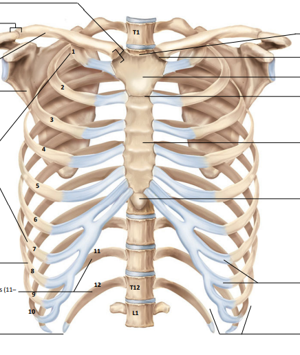
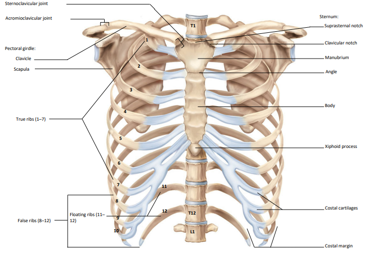
Label the following:
sternoclavicular joint
acromioclavicular joint
clavicle
scapula
true ribs
false ribs
floating ribs
sternum
suprasternal notch
clavicular notch
manubrium
angle
body
xiphoid process
costal cartilages
costal margin
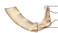
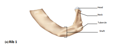
Label the following:
head
neck
tubercle
shaft
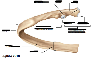
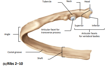
Label the following:
head
neck
tubercle
inferior and superior articular facets for vertebral bodies
articular facet for transverse process
angke
costal groove
shaft
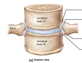
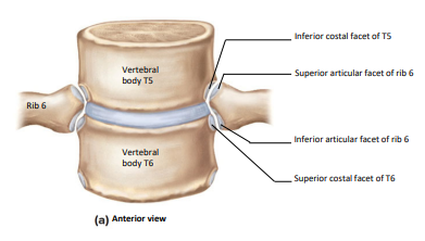
Label the following:
inferior costal facet of T5
superior articular facet of rib 6
superior costal facet of T6
inferior articular facet of rib 6
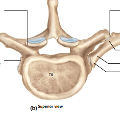
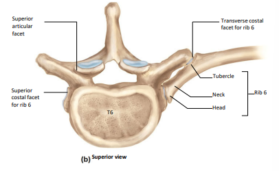
Label the following:
superior articular facet
superior costal facet of rib 6
transverse costal facet of rib 6
tubercle
neck
head
rib 6
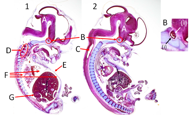
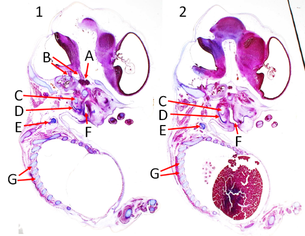
Which image is more parasagittal?
Label A, B, C, D, E, F and G
What part of the pituitary gland is i and ii?
Image 1
A = brain, B = pituitary gland, C = central canal, D = dorsal root ganglia, E = epidermis of the abdomen, F = peritoneal cavities, G = adrenal gland
i = anterior, ii = posterior
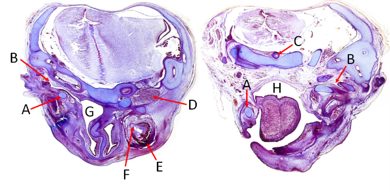
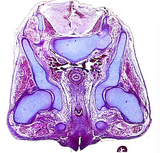
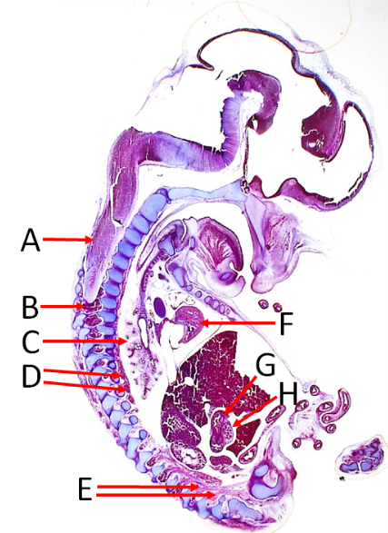
Label A, B, C, D, E, F, G and H
A = meckel’s cartilage
B = auditory tube
C = notochord
D = cranial ganglion
E = eye
F = optic nerve
G = nasopharynx
H = oropharynx
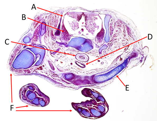
Label A, B, C, D and E
A = dorsal root
B = dorsal root ganglion
C = esophagus
D = trachea
E = clavicle
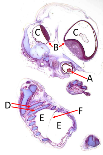
Label A, B, C, D, E and F
A = lens
B = brain
C = ventricles
D = ribs
E = peritoneal cavities
F = diaphragm
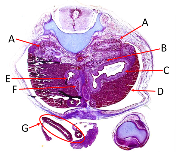
Label A, B, C, D, E, F and G
A = central canal
B = right adrenal gland
C = diaphragm
D = liver
E = epaxial muscles
F = hypaxial muscles
G = stomach
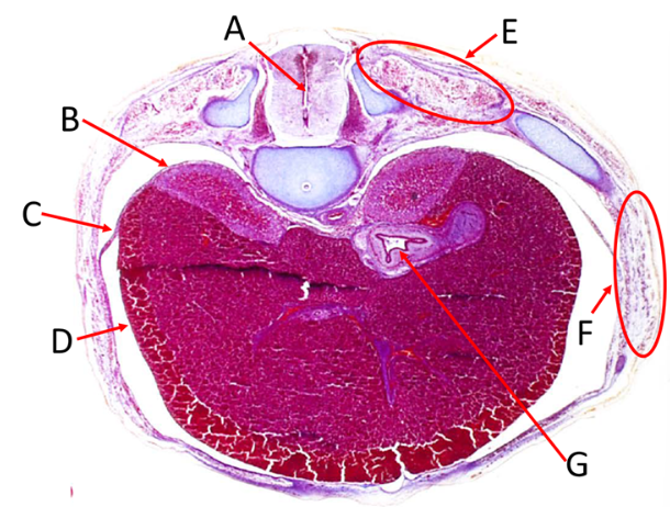
Label A, B, C, D, E, F, G and H
A = spinal cord
B = dorsal root ganglion
C = right lung
D = sympathetic trunk
E = mixed spinal nerves
F = ventricle
G = duodenum
H = pancreas
Label A, B, C, D, E, F and G
A = nerve ganglion
B = cranial nerves
C = meckel’s cartilage
D = mandible
E = clavicle
F = oral cavity
G = ribs
Label A, B, C, D, E, F and G
A = kidney
B = adrenal gland
C = stomach
D = liver
E = duodenum
F = pancreas
G = umbilical artery