Chapter 7: Nervous System & Neuron Excitability
1/53
Earn XP
Description and Tags
Final Exam
Name | Mastery | Learn | Test | Matching | Spaced | Call with Kai |
|---|
No study sessions yet.
54 Terms
What are the functions of the Nervous System?
integration: processes sensory information and controls all body responses and actions and stores information
sensory input: detecting and monitoring internal stimuli and and external stimuli
motor output: causes a response in effectors like muscles and glands
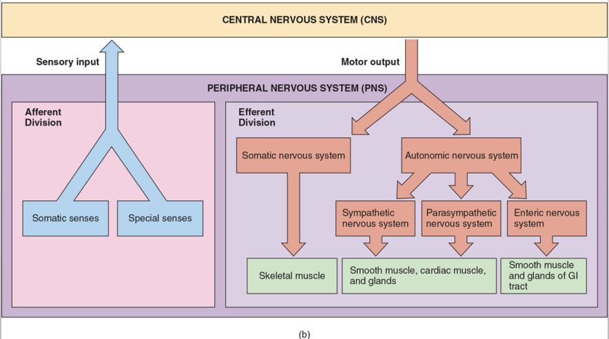
What are the nervous system divisions?
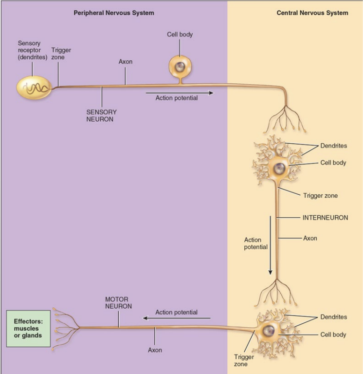
central nervous system: brain & spinal cord
peripheral nervous system: cranial & spinal nerves as well as sensory receptors
How do we refer to the peripheral nervous system (directional)?
the peripheral nervous system is bi directional
this means it is divided into two subdivisions based on the direction the impulse travels
sensory afferent: sensors that detect stimuli and send information toward the CNS (think arriving)
motor efferent: neurons that carry impulses from the central nervous system to various targets like muscles and glands
What are some of the cells in the nervous system?
cells in the nervous system are called neurons
Neurons: (primary signaling cells) are a functional unit of the nervous system that have a limit on repair and cannot go through mitosis once formed
Excitable cells: these are cells that respond to physical and chemical stimuli then produce and conduct electrical signals
These cells release chemicals for regulation and communication
aka neurotransmitters
What are some of the cells in the nervous system? cont.
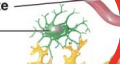
Neuroglia: (supporting cells) these are glial cells
there are 4 types in the CNS
there are 2 types in the PNS
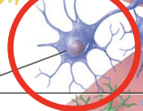
Describe the neuron structure
cell body: the big circle at the beginning contains a single nucleus
axon hillock: just below the cell body is the thick base of the cells body
(processes) axon: the long tube after the axon hillock
(processes) dendrites: located on the cell body the little wires on it
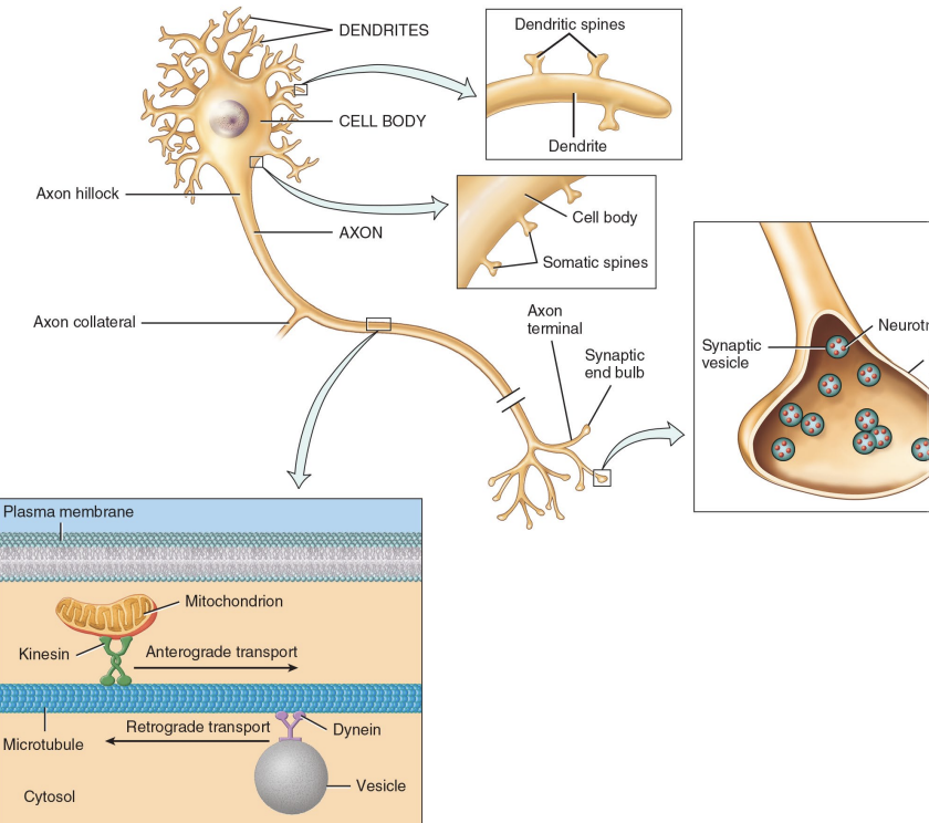
What do the dendrites do?
they carry signals TO the cell body
made for communication/contact with other neurons
often branched and contain dendritic spines for increase in points of contact
What does the axon do?
they carry impulses away from cell body (think axon away)
it is a single long process
it starts at the axon hillock
it branches at the end to create axon terminals
the axon terminals that contains vesicles that are filled with neurotransmitter
What is the basic pattern of information flow?
sensory neurons
transmit info from pns to cns
interneurons
interpret sensory info which may elicit a response
motor neurons
transmit info from cns to pns
What are different groups of neurons called?
clusters of cell bodies in the PNS are called: ganglia/ganglion
clusters of cell bodies in the CNS are called: nucleus/nuclei
a bundle of axons in the CNS is called: tract
a bundle of axons in the PNS is called: nerve
Describe neuroglia/glial cells
much smaller than neurons
they are 10-50x more numerous
can replicate & divide
four types in CNS: ependymal, oligodendrocytes, astrocytes, & microglia
two types in PNS: Schwann & satellite cells
Describe astrocytes and their function
they are the most common glial cells
they provide structural support
they form the blood brain barrier by covering capillaries in blood
they direct and guide new neural connections during development when they are being made
star shaped
Describe oligodendrocytes and their function
they are fairly common
they form myelin sheaths around axons (myelination)
similar to Schwann cells in PNS
Describe microglia and their function
they are found near blood vessels
phagocytic cells - engulf away dead or damaged cells, debris, and pathogens
Describe ependymal cells and their function
they form an epithelial layer lining canals and chambers in the central nervous system
produce cerebrospinal fluid

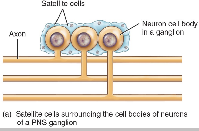
(PNS) Describe satellite cells and their function
they are flat cells that surround the cell bodies in ganglia
provide support and structure for neurons that make up ganglia
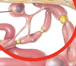
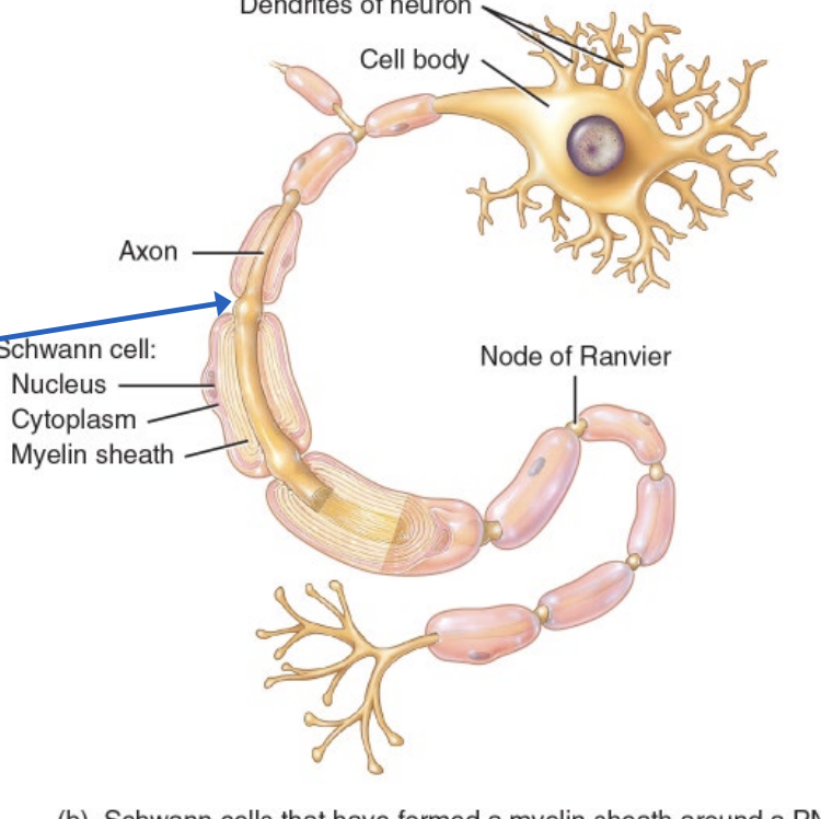
(PNS) Describe schwann cells and their function
they wrap around axon of PNS neurons
produce myelin sheath (myelination) similar to oligodendrocytes in CNS
What is myelination?
most axons in the PNS are myelinated
only some in the CNS are myelinated
the sheaths are formed by cells wrapping tightly around axons
acts as an insulator to speed up conduction of electrical impulses
What are the nodes of ranvier?
they are gaps between axons that are unmyelinated
these nodes are in contact with extracellular fluid
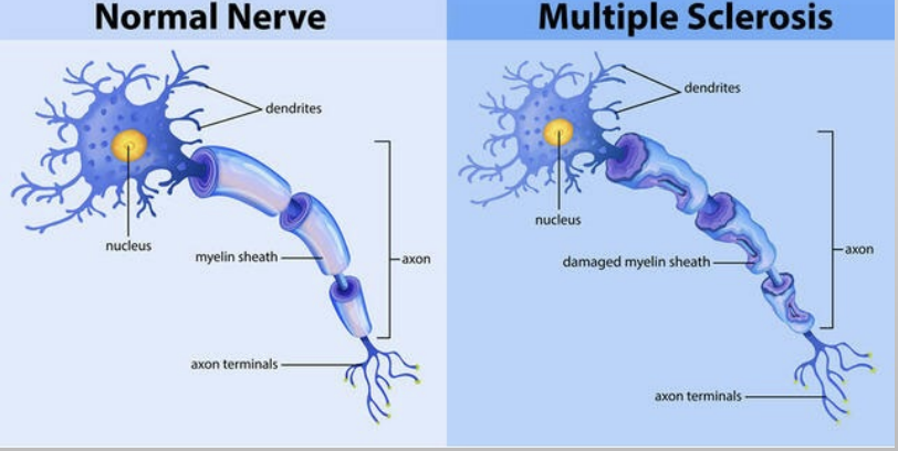
What are some of the results of loss of myelination?
the loss of myelin sheaths in the CNS neurons: multiple sclerosis
the loss of myelin sheaths in the PNS neurons: guillian-barre syndrome
autoimmune diseases: the immune system attacks itself and destroys myelin sheaths
neuron function is impaired (sensory & motor)
What is the physiology of excitable cells?
excitable cells change membrane potential when adequately stimulated
it creates an electrical signal at one small end of the cell membrane that spreads along the entire membrane (similar to dominos)
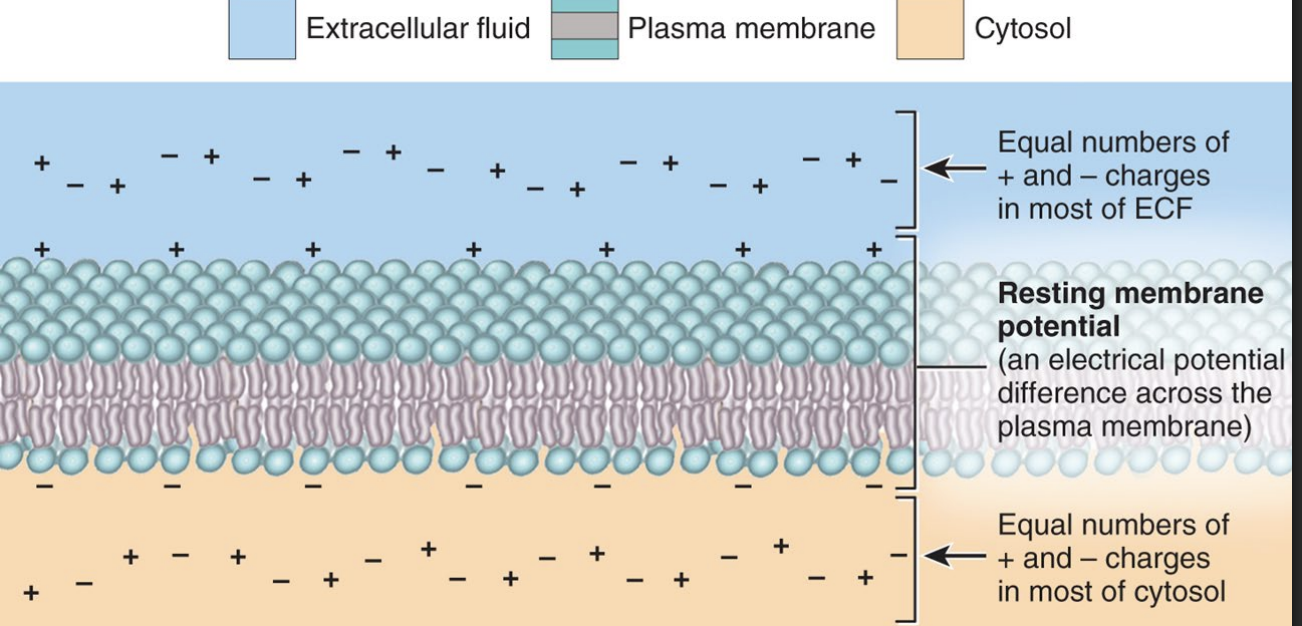
Describe electricity within membranes
charged particles are in solution outside and inside of our cells
opposite attract, like repels - electrical gradient
electrical charge can be different on either side if these are separated by a boundary → this difference is called a potential
a “potential” contains potential energy
within the cells of the nervous system, the boundary is the cell membrane and the difference in charge inside/outside of cell = membrane potential
Describe membrane potential cont.
a cell at rest = resting membrane potential (RMP)
the range of rmp is from -50 to -100 the negative tells us the inside is negatively charged relative to the outside
excitable cells can rapidly alter rmp when stimulated (neurons, muscles)
What is rmp in neurons? & how is it created?
resting membrane potential is -70mv
the charge inside vs outside is different because…
1. the difference in ionic makeup of the extracellular fluid and cytosol
Na+ is higher on the outside, K+ is higher on the inside
2. the membrane has selective permeability to various ions due to the number of leakage channels
barely permeable to Na+ (little trickles in), while very permeable to K+ (more flows OUT)
THIS IMBALANCE MAKES THE INSIDE MORE NEGATIVE
How is the RMP created in neurons cont.
there are Na+/K+ pumps that return the ions back to where they came from (pump Na+ out & K+ in) so gradient and RMP is maintained
ONE PUMP CYCLE MOVES 3 NA+ OUT 2 K+ IN
Describe the function of ion channels in excitable cells
leakage channels: always open to it creates a continuous slow leak that is non gated
gated channels: must be stimulated to open or close
there are 3 types of these in neurons
What are the three types of gated channels in neurons?
Ligand binding channels will open chemically gated channels which then close when the ligand is no longer present
Changes in membrane potential will open and close voltage gated channels
Mechanical forces (pressure, touch, vibration, stretch) will open and close mechanically gated channels
What is the key to excitable cell activity?
stimuli applied to neurons will open gated channels that would normally be closed
this changes the normal RMP as distribution of ions as they diffuse along their concentration gradient through newly opened channels
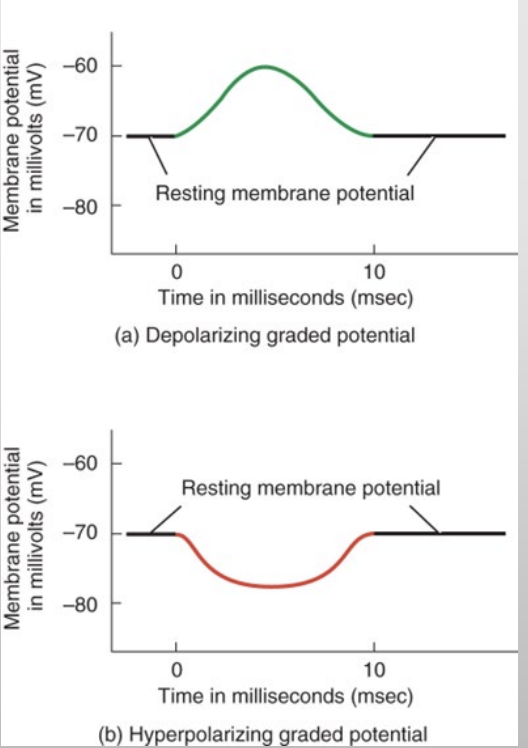
changes in membrane potential away from -70 as a result of a stimuli is called a GRADED POTENTIAL
can be cause of 2 changes
Describe graded potentials
depolarization - makes the inside of the cell less negative than RMP
hyperpolarization - makes the inside of the cell become more negative than RMP
“graded” potential means the change can be large or small depending on the strength of the stimulus
graded potentials usually occur at dendrites or cell bodies of neurons (site of stimulation)
Explain some details about graded potentials
the stimuli that creates these potentials can be strong or weak
multiple graded potentials can be added together (summation)
the strength of a graded potential deteriorates as it moves along the membrane (decremental speed)
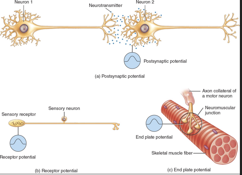
What are some names for graded potentials based on the stimulus that caused them?
postsynaptic potential:
first neuron is causing a graded potential in a second neuron
receptor potential:
neuron is acting as a receptor for sensory information in PNS
endplate potential:
potential is found on skeletal muscle (also excitable)
Details about stimulus in graded potentials
a stimulus can be designed to inhibit neural activity - which prevents it from being activated
a stimulus can also excite neural activity - which causes the generation of an action potential
What are action potentials?
this is a rapid set of neural events starting from the axon hillock spreading down the axon to the axon terminals
ultimately results in the release of a neurotransmitter
neuron “fires”
What does trigger zone activation mean?
it is located in the axon hillock
activated (triggered) if graded potential is strong enough
depolarize to threshold which is -55 mv
if the graded potentials is less than -55mV there is no response
if the graded potential is greater than or equal to -55 mV then an action potential is generated in the axon
ACTION POTENTIALS ARE A ALL OR NOTHIN
What is present in the trigger zone?
there is a high concentration of voltage gated Na+ channels
if threshold is reached:
these channels will open
Na+ will rush in
trigger zone depolarizes
generate an action potential in axon
What are action potentials?
they are an all or nothing phenomena - must reach threshold to activate one
all are identical in strength and duration
they are driven by voltage gated na+ channels and k+ channels
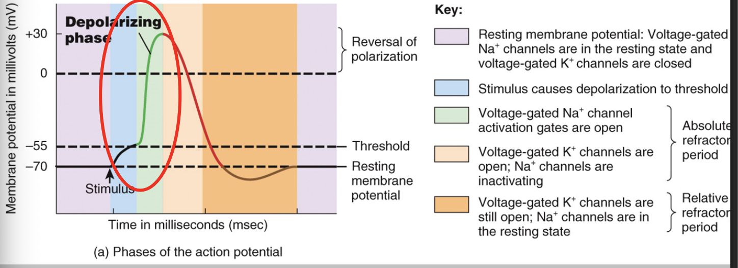
Describing the four stages of an action potential
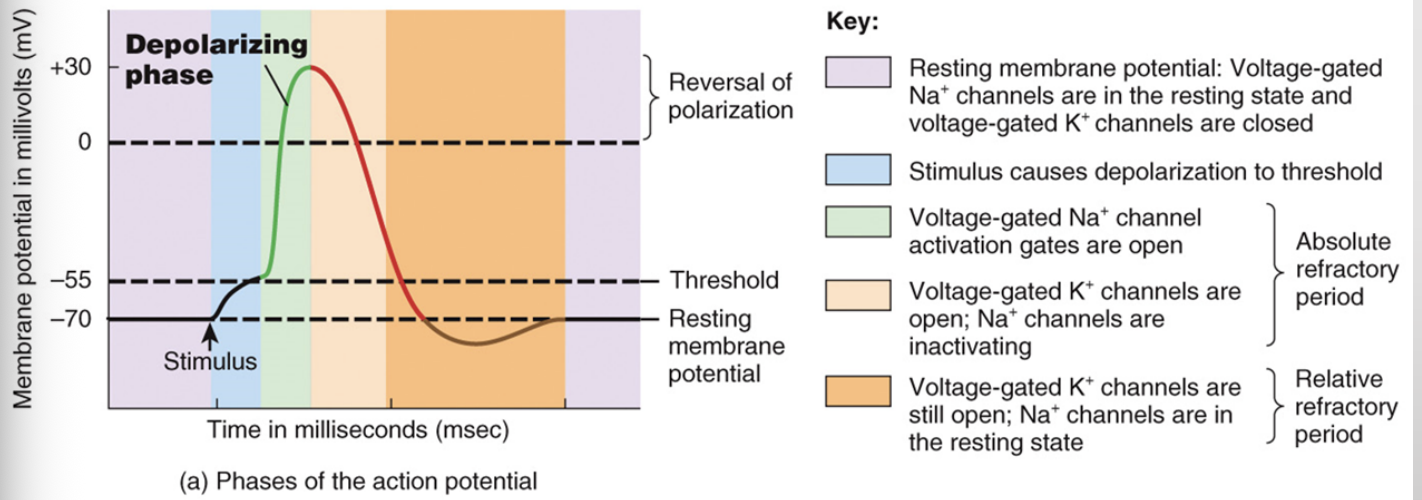
Describing the four stages of an action potential: Stage 1 - resting stage
the neurons are at rest during this stage both Na+ & K+ channels are closed
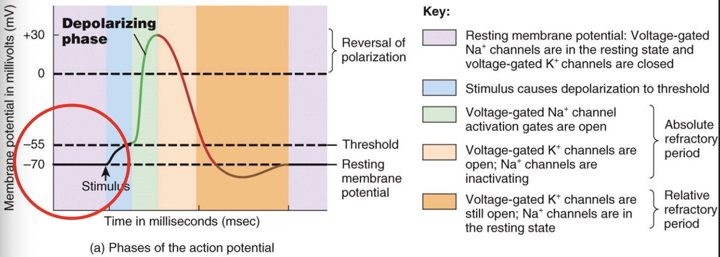
Describing the four stages of an action potential: Stage 2 - depolarizing phase
a graded potential depolarizes neuron to threshold - ap starts
many gated Na+ channels open and it rushes in
only a few K+ channels open - it slowly trickles out
the rapid entry of Na+ causes depolarization to +30mV
at the peak of depolarization a second gate in Na+ channel closes - this causes depolarization to stop
Describing the four stages of an action potential: Stage 3 - repolarization phase
this new +30mV causes gated K+ channels to open now, K+ leaves cell and membrane repolarizes
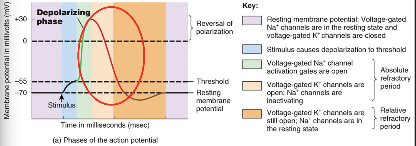
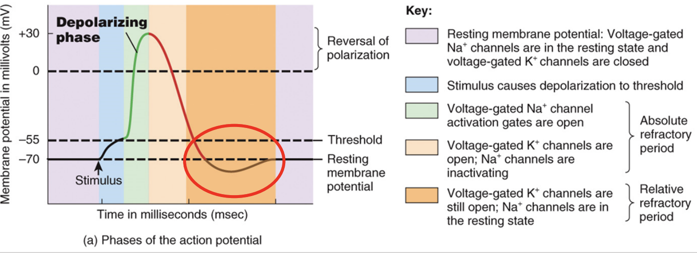
Describing the four stages of an action potential: hyperpolarization phase
after repolarization, some K+ channels are still open and membrane overshoot RMP & hyperpolarizes a bit
eventually all k+ are closed and Na+/K+ pumps restore to original position
membrane returns to RMP ready for next AP
What is energy used for in action potentials?
ATP is required to fuel Na+/K+ pumps that restore equilibrium after an AP is generated
No ATP is needed to initiate an AP, it is all done by diffusion - only needed to restore
Describe conduction of action potentials
one action potentials only occurs along a small distance (<1mm)
one action potential stimulates the next in the next region of the axon membrane and they propagate (conduct) along the axon to its end
Describe conduction in myelinated axons and its effects
where myelin exists - no channels open or close
action potentials jump from node of ranvier to next node of ranvier
this is called saltatory conduction it is 10-50x faster
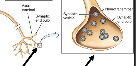
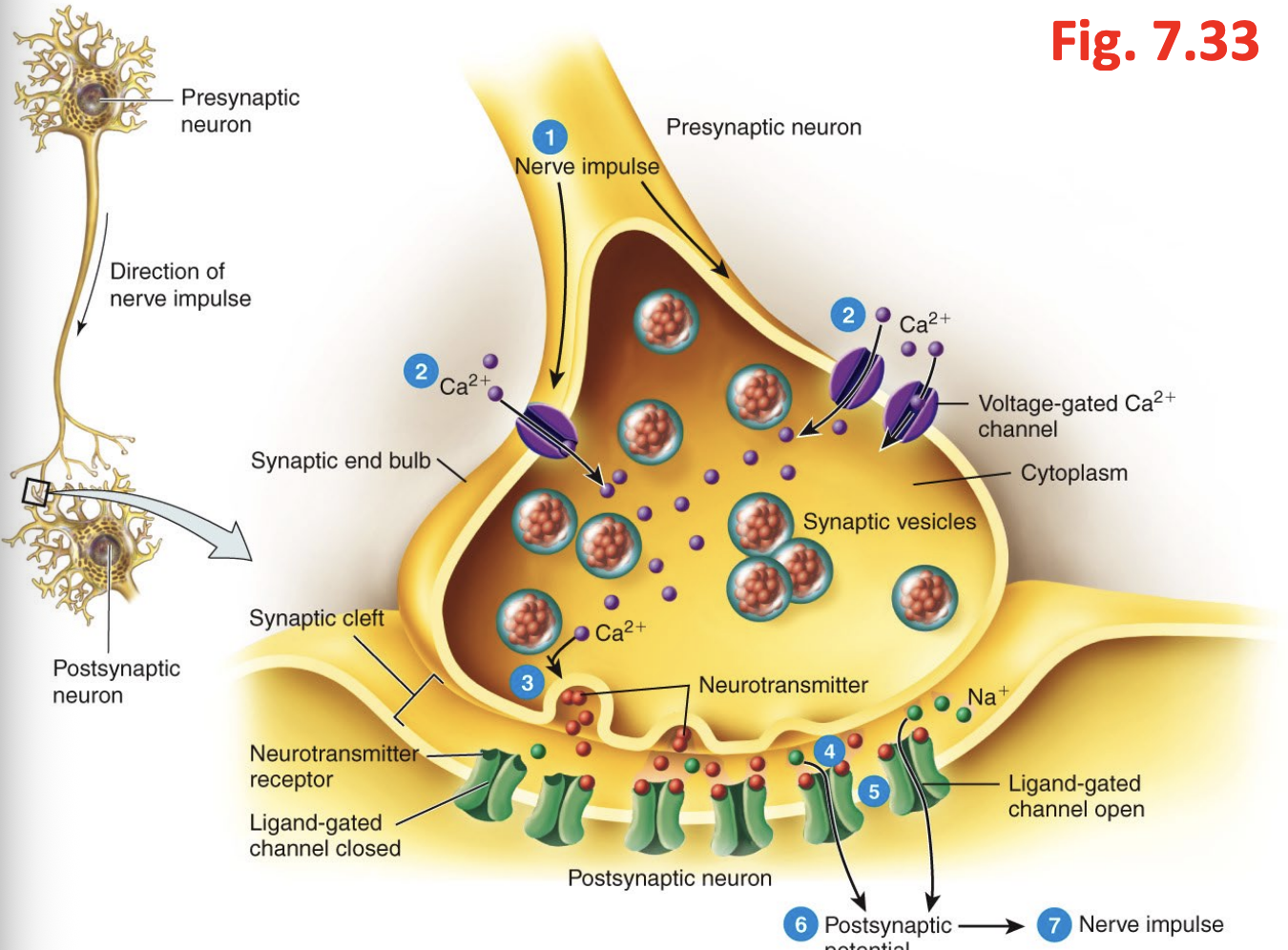
What happens at the end of the axon?
once the action potential reaches the end of an axon → a synapse occurs
What is a synapse?
a junction that allows for communication between a neuron and another cell
neuron to neuron
neuron to effector cell
the transmission of information is always in one direction from presynaptic to postsynaptic
What is the process of the release of a neurotransmitter?
an action potential arrives at the axon terminal
voltage gates ca channels open and allow ca to diffuse into axon terminal
ca stimulates vesicles to release neurotransmitter through exocytosis
the neurotransmitter then crosses the synaptic cleft and diffuses and binds to a receptor on postsynaptic cell
the neurotransmitter binding causes receptors that are chemical gated ion channels to open which allows ions to move across membrane of post synaptic cell
then from here ions can either depolarize (excite) or hyperpolarize (inhibit) it
the neurotransmitter activity usually ends when it is removed by an enzyme or moves away through diffusion
Describe neurotransmitters and name some common ones
they are used for regulation of normal nervous system function
over or underproduction of these leads to deficiencies that can be fixed through medications
common neurotransmitters:
acetylcholine (ach)
used in cns for neuron neuron communication
used in pns for neuron muscle communication
can be both excitatory or inhibitory
deactivated through achE then recycled
What are some common neurotransmitters?
monoamine family (mostly cns)
dopamine
serotonin
histamine
epinephrine
norepinephrine
broken down and deactivated by enzyme monoamine oxidase
What is a neural circuit?
these neural circuits can enhance or inhibit neural activity in the CNS or out in the body
it is common for multiple neurons to interact with each other forming complex circuits
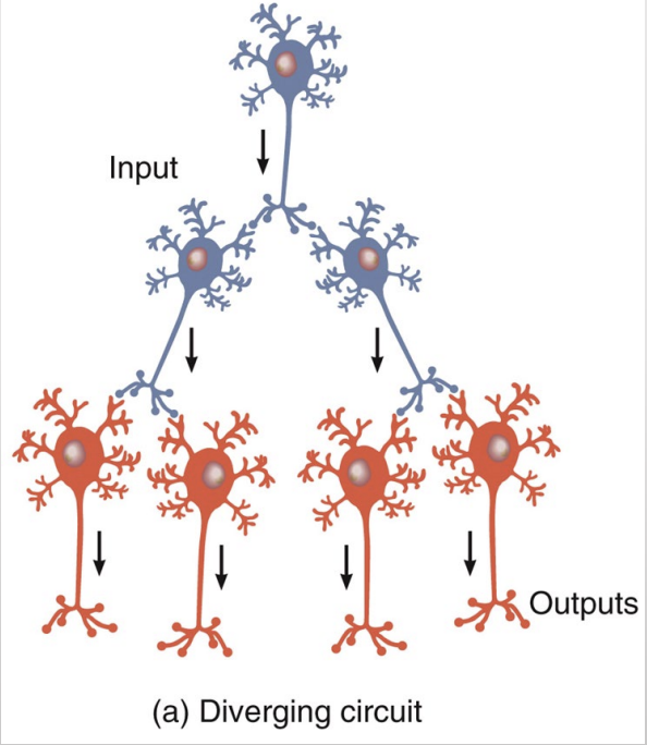
What is a diverging circuit?
diverging circuits: a single presynaptic neuron synapses with several post synaptic neurons
stimulate multiple muscle groups simultaneously
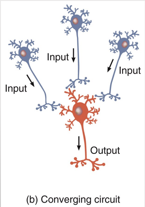
What is a converging circuit?
converging circuit: the postsynaptic neuron receives action potentials from several different presynaptic neurons
makes summation more effective
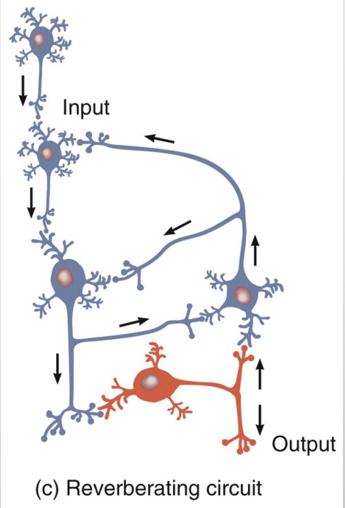
What is a reverberating circuit?
an action potential stimulates a neuron which stimulates a second then stimulates a third and another and so on
branches from downstream neurons synapse with earlier ones, sending action potentials back through the circuit again and again
takes place in epileptic seizures in millions of neurons
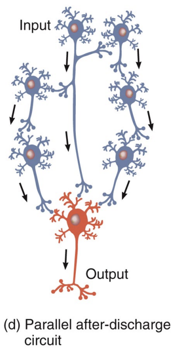
What is a parallel after discharge circuit?
a single presynaptic cell stimulates a group of neurons each of which synapses with a common post synaptic cell but at different times creating a continuous input to the target