Physical signs of retinal disease
 Call Kai
 Learn
Learn Practice Test
 Spaced Repetition
 Match
Match Flashcards
 Knowt Play
1/103

There's no tags or description
Looks like no tags are added yet.
Name | Mastery | Learn | Test | Matching | Spaced | Call with Kai | Chat |
|---|
No analytics yet
Send a link to your students to track their progress
104 Terms
a complete retinal description answers what 6 questions?
what, color, morphology, where, depth, extent/context
cotton wool spots are...
superficial ischemic lesions
what are two words used to describe CWS?
fluffy white
the fluffy white lesions associated with CWS are lesions from....
microinfarction
CWS are caused by what???
precapillary arteriole occlusion
CWS are associated with what 4 systemic conditions?
HTN, diabetes, CT disease and HIV
CWS are known as indicators of what disease?
systemic microvascular
unexplained, multiple, recurrent or bilateral CWS warrant????
targeted systemic evaluation
most CWS absorb over __-__ weeks
6 8
CWS can resolve with control of what two things?
BP BS
why are CWS often next to flame hemorrhages?
both are superficial
HARD exudates are caused by what two things?
chronic vascular leakage or damage from retinal vessels
hard exudates are what color?
yellow/white
hard exudates are yellow/white lipid/protein deposits in what two layers of the retina?
OPL (major) and NFL (minor)
what pattern are hard exudates normally found in?
circinate or macular star
hard exudates are linked with what retinal conditions? (2)
DR and hypertensive retinopathy
is macular edema fluid or a fundus color?
fluid
macular edema is macular thickening from....
fluid leakage
macular edema is common in what four findings?
DR, vein occlusion, inflammation, post-surgical
macular edema is a major cause of ____ ____ in retinal vascular disease
vision loss
what is essential for diagnosis and monitoring of macular edema?
OCT
drusen are deposits beneath the ___
RPE
what color are drusen?
yellow
if drusen are small and hard, what does that indicate?
benign finding
if drusen are large and soft, what does that indicate?
AMD risk
drusen are a waste buildup between what two layers?
RPE and Bruchs
if you find drusen, monitor the patient for what two things?
pigmentary changes and SRF
RPE atrophy and hypertrophy are _____ pigment responses
opposite
atrophy is a sign of cell loss from what two things?
retinal thinning and AMD
hypertrophy is a sign of cell enlargement from what two things?
injury and inflammation
retinal scars/pigment signify CHRONIC or PRIOR retinal _____
insult
what finding is known to obscure retinal detail?
vitreous hemorrhage
a ____ hemorrhage is superficial, feathery, and follows the NFL
flame
a _____ hemorrhage is round or oval, deeper intraretinal blood
dot blot
a ______ hemorrhage is boat or D shaped with a horizontal fluid level
preretinal
a ________ hemorrhage is a diffuse haze, clumps, strands or inferior layering that obscures detail
vitreous
a _______ hemorrhage is a dark, deep-appearing blood beneath retinal vessels
subretinal
vitreous blood may track _______ across the _____ _____
inferiorly posterior pole
flame hemorrhages are associated with what 4 blood disorders?
HTN, RVO, anemia, leukemia
disc-margin flame hemorrhages demand ____-_____ context
optic nerve
dot-blot hemorrhages are present in what two layers of the retina?
INL and OPL
dot blod hemorrhages are leakages from ___ retinal capillaries
deep
dot blot hemorrhages are findings associated with what two things?
diabetic retinopathy and RVO
mixed hemorrhage patterns suggest _____ vascular injury
diffuse
a subretinal hemorrhage lies beneath the ________ retina
neurosensory
a subretinal hemorrhage is blood between what two structures?
retina and RPE
what are the four causes for subretinal hemorrhages?
CNV, AMD, truma and high myopia
what color and shape is associated with subretinal hemorrhages
dark red/black and dome
subretinal hemorrhage can cause ____ _____ vision loss, as blood is toxic to RPE
severe fast
active _____ _______ cna produce expanding subretinal hemorrhage
choroidal neovascularization
_____ _____ are white centered hemorrhages (NOT A DIAGNOSIS)
roth spots
roth spots are found in what layer of the retina?
NFL
what constitutes the white center of a roth spot?
fibrin/WBCs
roth spots are seen in what three blood disorders?
endocarditis, leukemia or anemia
___ ____ filter/imaging improves hemorrhage detection
red free
an ____ distorts the inner retinal surface
ERM
ERM is a _______ proliferation on the ___
fibrocellular ILM
what are the three main causes for ERM?
idiopathic, post-surgical or inflammatory
ERM may cause what two things?
macular pucker or metamorphopsia
what are the two management techniques used for ERMs?
observation or membrane peel
retinal vessel straightening reveals what???
tangential traction
retinal folds reflect...
mechanical deformation
what are the three main causes for retinal folds?
hypotony, tumors or post-surgical changes
retinal folds can cause what visually?
metamorphopsia
choroidal folds create parallel ________ bands
alternating
what can you use to separate retinal folds from choroidal folds?
OCT
in a retinal fold, look for ____ ____ of the retinal tissue
focal buckling
in a choroidal fold, follow the hyperreflexive ____-____ line
RPE bruch
neovascularization is fragile vascular growth driven by _____
ischemia
what are the three causes for neovascularization?
PDR, ischemic RVO or sickle cell
what are the two risks associated with neo?
vitreous hemorrhage or tractional RD
what are two ways we can treat neo?
anti-VEGF and PRP
CWS

CWS and flame hemes
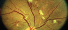
exudates

hard exudates and flame heme

macular star
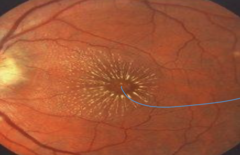
mac edema

mac edema

drusen
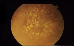
hard drusen

soft drusen

hypertrophy and atrophy

chRPE

vitreous hemorrhage
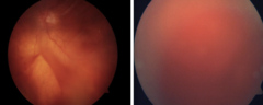
vitreous hemorrhage

flame heme

disc margin flame heme
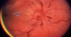
dot blot heme

subretinal heme

subretinal heme

roth spots
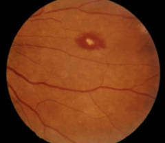
roth spots
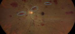
ERM

ERM

vessel straigntening (tangential traction)

retinal folds
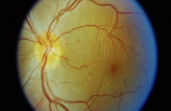
choroidal folds

neovascularization

NVD
