Chapter 9 Neurons
 Call Kai
 Learn
Learn Practice Test
 Spaced Repetition
 Match
Match Flashcards
 Knowt Play
1/118

Earn XP
Description and Tags
Starting 9.3
Name | Mastery | Learn | Test | Matching | Spaced | Call with Kai | Chat |
|---|
No analytics yet
Send a link to your students to track their progress
119 Terms
excitable tissue
nerve and muscle cells are described as excitable tissues bc of their ability to propagate electrical signals rapidly in response to a stimulus.
Two factors influence the membrane potential
1. The uneven distribution of ions across the cell membrane. Na+, Cl- and Ca2+ are more concentrated in the extracellular fluid than in the cytosol. K+ is more concentrated in the cytosol than the EC fluid.
2. Differing membrane permeability to those ions. cell membrane is more permeable to K+.
Nernst equation description
describes membrane potential that would result if the membrane were permeable to only ONE ion. Also called equilibrium potential of the ion (E subscript ion).
Eion (in mV)= 61/z log [ion]out/ [ion]in
z is the electrical charge on the ion (+1 for K+)
Average value for the resting membrane potential of neurons
is -70 mV because, although K+ would make it -90 mV, the cell is permeable to leaking Na+ entering the cell, making it slightly more positive. Thanks to the GHK equation
Goldman-Hodgekin-Kratz equation (GHK)
Calculates the membrane potential that results from the contribution of ALL ions that can cross the membrane. Na+, K+, and Cl-.
Vm =61 log Pk[k+]out + Pna[Na+]out + Pcl[Cl-]in/Pk[K+]in + Pna [Na]in + Pcl[Cl-]out
p is the relative permeability of the membrane to the ion shown in the subscript.
Ions with higher permeability have more influence on the membrane potential.
If membrane is not permeable to an ion, the permeability value for that ion is zero.
Nernst vs GHK equations
Nernst is for only one ion, GHK is for ALL ions that are permeable to the membrane
GHK calculates overall membrane voltage
By combining the concentration gradients and membrane permeability of all ions that can cross the membrane. The concentration gradient tells you what an ion wants to do; permeability tells you how much opportunity it has to do it.
Depolarization
Brings closer to zero
Hyperpolarize
Away from zero
Changing membrane permeability
Opening/closing channels in the membrane changes ion permeability in the cell.
Slower method for changing membrane permeability is for the cell to insert new channels into the membrane or remove some existing channels.
4 major types of selective ion channels in the neuron
Na + channels
K+ channels
Ca2+ channels
Cl- channels
other channels are less selective
Conductance
{meaning to escort}, the ease with which ions flow through a channel is called the channels conductance.
Gated channels fall into 3 categories
Mechanically gated ion channels
Chemically gated ion channels
Voltage-gated ion channels
There are many different subtypes in each major channel. Within each subtype, there may be multiple isoforms or associated proteins that modify channel properties.
Closely related channels may express different opening and closing kinetics.
Mechanically gated ion channels
Are found in sensory neurons and open in response to physical forces such as pressure or stretch.
Chemically gated ion channels
Most neurons respond to a variety of ligands, such as extracellular neurotransmitters and neuromodulators or intracellular signals.
Voltage-gated ion channels
Respond to changes in the cell’s membrane potential.
Voltage-gated Na+ and K+ channels are very important in the initiation and conduction of electrical signals along the axon,
Isoforms
any of two or more functionally similar proteins that have a similar, but not identical, amino acid sequence.
Kinetics
{moving} the speed with which the channel opens and closes.
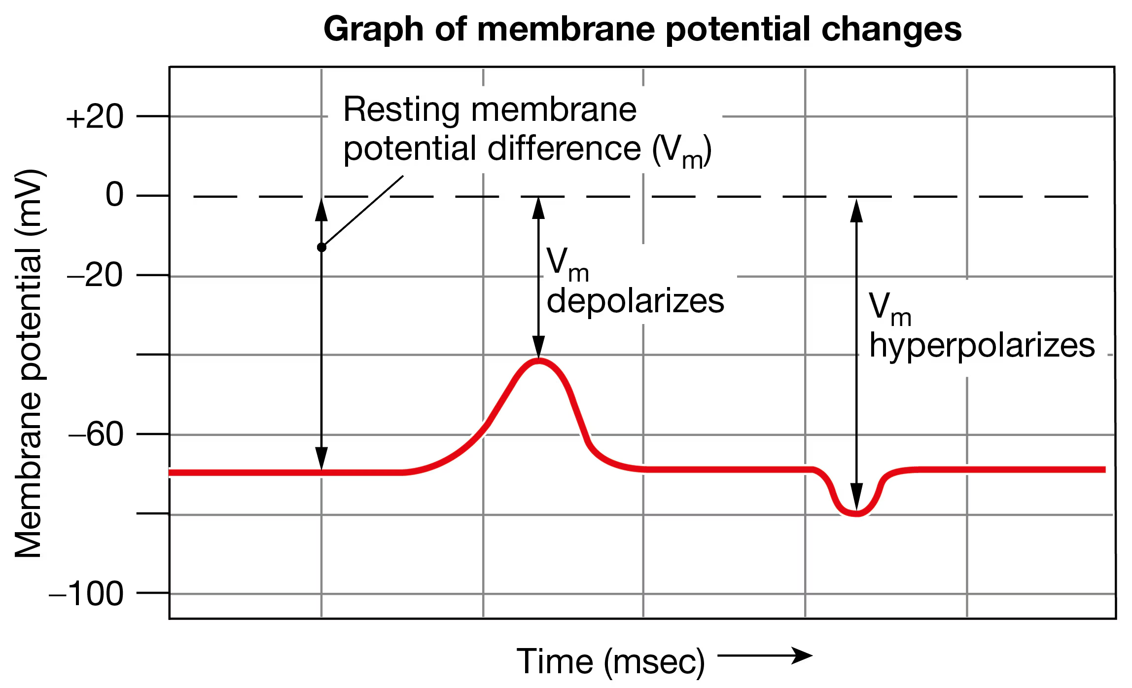
What is depolarization and hyperpolarization?
Depolarization is going closer to zero. Hyperpolarization is going away from zero.
Activation
Channel opening to allow ion flow is called activation.
Ex: voltage-gated Na+ channels and voltage-gated K+ channels of axons are both activated by cell depolarization.
Speed of Na+ and K+ channels
Na+ channels open very rapidly, K+ channels are slower to open..
Channels closing
Many channels that open in response to depolarization close only when the cell repolarizes. The gating portion of the channel protein has an electrical charge that moves the gate open/close as membrane potential changes.
What is channel inactivation?
Some ion channels spontaneously inactivate. Even though the activating stimulus that opened them continues, the channel “times out” and closes.
Ex: automatic door after 10 secs closes even if you stand in the doorway.
An inactivated channel returns to its usual closed state shortly after the membrane repolarizes.
Ion’s current (I subscript ion)
The flow of electrical charge carried by an ion is called the ion’s current. The direction of ion movement depends on the electrochemical (combined electrical and concentration) gradient of the ion.
Ion’s net flow
K+ usually leaves the cell. Na+, Cl-, and Ca2+ usually enter the cell.
Net flow of ions across the membrane depolarizes or hyperpolarizes the cell, creating an electrical signal.
Ohm’s law
I= v/r , as resistance (R) increases, current flow (I) decreases.
2 types of resistance to current flow
(Rₘ): the resistance of the cell membrane
(Rᵢ): the internal resistance of the cytoplasm
Extracellular fluid creates resistance, but it’s very small so it's ignored (R₀)
Internal resistance is determined by?
In most neurons, it’s determined by the composition of the cytoplasm and the diameter of the cell. The cytoplasmic composition is relatively constant.
Rᵢ decreases as cell diameter increases. larger diameter neurons have lower resistance.
What determines how far current will flow
Rᵢ and Rₘ together determine how far the current will flow through a cell before energy is dissipated and the current dies.
Length constant/space constant
Rᵢ, Rₘ, and R₀ create a length constant for a given neuron. Length constant is sometimes called space constant.
Voltage changes across the membrane can be classified into 2 types of electrical signals
Graded potentials and action potentials
Graded potentials
Variable-strength signals that travel over short distances and lose strength as they travel through the cell. Short-distance communication.
Graded potential initiates an action potential.
Action potentials
Very brief, large depolarizations that travel for long distances through a neuron without losing strength. Function: rapid signaling over long distances. Ex: from toe to brain.
Graded potential vs Action potential

What do graded potentials do?
Graded potentials in neurons are depolarizations or hyperpolarizations that occur in the dendrites and cell body, or less frequently, near axon terminals. They are called “graded bc their size, amplitude {large} is directly proportional to the strength of the triggering event.
They are graded depending on size of stimulus, so a large stimulus=strong graded potential, small stimulus=weak graded potential.
Graded potentials can also occur when an open channel closes (K+): few K+ leave the cell, K+ depolarizes the cell.
local current flow
The wave of depolarization that moves through the cell. By convention, current in biological systems is the net movement of positive electrical charge.
Explain how the strength of the graded potential affects how far it spreads.
The strength of the potential is determined by how much charge enters the cell. More channels open →more ions enter → stronger depolarization → further cascade. Ex: how the size of waves caused by a stone tossed in water.
Why do graded potentials lose strength as they move through the cytoplasm? hint: 2 factors
Current leak. As the electrical signal moves through the cytoplasm, some of the positive charge leaks out of the neuron through open leak channels.
Cytoplasmic resistance. The cytoplasm creates resistance to the flow of electricity, just like water creates resistance that diminishes the waves from a stone.
Trigger zone
Graded potentials that are strong enough eventually reach the region of the neuron known as the trigger zone. In efferent neurons and interneurons, the trigger zone is the axon hillock and the very first part of the axon, a region known as the initial segment. In sensory neurons, the trigger zone is immediately adjacent to the receptor, where the dendrites join the axon.
A graded potential starts somewhere on the neuron, usually the dendrites or cell body, and spreads through the neuron. If it's strong enough when it reaches the trigger zone, it can trigger an action potential.
The trigger zone is the integrating center of the neuron and contains high concentrations of voltage-gated Na+ channels in its membrane.
formation of an action potential
The trigger zone is the integrating center of the neuron and contains high concentrations of voltage-gated Na+ channels in its membrane. If a graded potential reaching the trigger zone depolarizes the membrane to the threshold voltage, voltage-gated Na+ channels open, and an action potential begins. If the depolarization doesn’t reach threshold, the graded potential simply dies out as it moves into the axon.
Excitatory vs inhibitory graded potentials
Excitatory graded potentials are depolarizing (more likely to fire). Inhibitory are hyperpolarizing graded potentials.
subthreshold vs suprathreshold graded potentials
Subthreshold- doesn’t meet threshold; no action potential.
Suprathreshold- more than enough=action potential.
Action Potentials (Spikes)
Electrical signals of uniform strength that travel from the trigger zone → down the axon → to the axon terminals.
Voltage-gated ion channels open sequentially along the axon as the electrical signal travels.
Na⁺ enters the cell, reinforcing depolarization and regenerating the action potential at each section of the axon.
Because the signal is continually regenerated, an action potential does not lose strength with distance.
Instead of getting weaker over distance, action potentials are replenished along the way so that they maintain constant amplitude.
Action potentials are all-or-none: once threshold is reached, a full action potential occurs.
Conduction = the rapid movement of an action potential along the axon.
Trigger zone → 🔥 → 🔥 → 🔥 → 🔥 → axon terminal
What is the difference between conductance and conduction in neurons?
Conductance refers to the movement of ions across a cell membrane. Conduction is the rapid, undiminished movement of an electrical signal down the axon of a neuron.
Action Potential: What happens to the axon membrane?
A suprathreshold stimulus at the trigger zone initiates the action potential.
Action potentials depend mainly on voltage-gated Na⁺ channels and voltage-gated K⁺ channels.
The action potential has 3 phases:
Rising phase: Na⁺ enters → depolarization
Falling phase: K⁺ leaves → repolarization
After-hyperpolarization: K⁺ continues leaving briefly → membrane becomes more negative than resting
The membrane then returns to its resting membrane potential.
Action potential steps
Resting potential
Depolarizing stimulus (beginning of trend)
Membrane depolarizes to threshold. Voltage-gated Na+ and K+ channels begin to open.
Rapid Na+ entry depolarizes cell
Na+ channels close and slower K+ channels open (Peak of trend) +30mV
K+ moves from the cell to extracellular fluid.
K+ channels remain open, and additional K+ leaves the cell, hyperpolarizing it. (end of peak)
Voltage-gated K+ channels close, less K+ leaks out of the cell
Cell returns to resting ion permeability and resting membrane potential.
*Very few ions move across the membrane in a single action potentials, so the relative Na+ and K+ concentrations inside and outside the cell remain essentially unchanged.
Sodium-potassium pump
The Na⁺/K⁺-ATPase (sodium-potassium pump) restores ions that have moved: 3 Na⁺ out and 2 K⁺ in, using ATP.
The pump does not need to reset the ions before the next action potential because one action potential does not significantly disrupt the concentration gradients.
Why do sodium channels close at the peak of the action potential when the cell is depolarized?
These voltage-gated channels have two gates to regulate ion movement rather than a single gate. The two gates, known as activation gate and inactivation gates, flip-flop back and forth to open and close the channel.
At resting potential, the activation gate for Na+ is closed; no Na+ can enter.
Depolarization: activation gate opens, Na+ enter→ further depolarization.
The inactivation gate, an amino acid sequence behaving like a ball and chain on the cytoplasmic side of the channel, opens at depolarization.
Activation gate = fast; inactivation gate = delayed.
Why can’t action potentials fire during the absolute refractory period?
The absolute refractory period is the brief period after an action potential begins when another action potential cannot be triggered, no matter how strong the stimulus.
It occurs because the voltage-gated Na⁺ channel gates are resetting after the first action potential.
Because the membrane behind the action potential is temporarily refractory, action potentials cannot overlap or travel backward.
This helps ensure that action potentials travel in one direction: trigger zone → axon terminal.
If you put ouabain, an inhibitor of the sodium-potassium pump, on a neuron and then stimulate the neuron repeatedly, what do you expect to happen to action potentials generated by that neuron?
(a) They cease immediately.
(b) There is no immediate effect, but they diminish with repeated stimulation and eventually disappear.
(c) They get smaller immediately, then stabilize with smaller amplitude.
(d) Ouabain has no effect on action potentials.
b. the pump doesn't need to restore the ion gradients after every action potential because only a tiny number of ions cross the membrane during each action potential.
Na⁺/K⁺ pump inhibited → no immediate effect → action potentials continue normally.
With repeated stimulation:
Na⁺ gradually accumulates inside and K⁺ gradually decreases inside → the Na⁺ and K⁺ concentration gradients become weaker → eventually the neuron can no longer generate normal action potentials.
Relative refractory period and action potentials
Follows the absolute refractory period.
Some voltage-gated Na⁺ channel gates have reset, but K⁺ channels are still open.
The neuron is harder to excite, so a stronger-than-normal depolarization is needed to reach threshold.
Na⁺ enters through reopened Na⁺ channels, but K⁺ is still leaving, opposing depolarization.
An action potential can occur, but it is smaller than normal.
Absolute refractory period: no second action potential is possible.
Relative refractory period: a second action potential is possible, but requires a stronger stimulus.
Refractory periods prevent action potentials from overlapping and help ensure one-way conduction from the trigger zone to the axon terminal.
How does an action potential travel down the axon?
A suprathreshold graded potential reaches the trigger zone → voltage-gated Na⁺ channels open → Na⁺ enters → depolarization.
Positive charge spreads through the cytoplasm to adjacent sections by local current flow.
The local current weakens with distance, but it depolarizes the next section enough to open its voltage-gated Na⁺ channels.
More Na⁺ enters the next section, regenerating the action potential.
This process repeats along the axon.
Therefore, the action potential is regenerated at each section and travels long distances without losing strength.
A stimulating electrode placed halfway down an axon artificially depolarizes the cell above threshold. In which direction will an action potential travel: to the axon terminal, to the cell body, or to both? Explain your answer.
The action potential will go in both directions because the Na+ channels around the stimulation site have not been inactivated by a previous depolarization.
Larger neurons conduct action potentials faster
meow
Two physical parameters influence the speed of action potentials
the diameter of the axon
the resistance of the axon membrane to ion leakage out of the cell (the length constant)
The larger the diameter of the axon or the more leak-resistant the membrane, the faster an action potential will move.
What determines conduction speed?
Larger axon diameter → faster conduction
Larger diameter = lower resistance to current flow inside the axon
Current spreads more easily/farther → AP travels faster
More leak-resistant membrane → faster conduction
Less ion leakage → current travels farther along the axon
Length constant: describes how far electrical current spreads before decreasing substantially.
Conduction is faster in myelinated axons
fact
Why does myelin increase conduction speed? hint: saltatory conduction
Myelin creates a high-resistance insulating layer around the axon.
It reduces current/ion leakage out of the axon.
Less current leakage → current travels farther and AP conduction is faster.
Nodes of Ranvier
Small gaps between myelinated sections.
Contain many voltage-gated Na⁺ channels.
Na⁺ enters at each node → regenerates the action potential.
Saltatory conduction
Action potential appears to jump from node → node.
Occurs because AP regeneration happens primarily at the nodes of Ranvier.
Faster than conduction in an unmyelinated axon.
unmyelinated axons vs myelinated axons
Unmyelinated axons: voltage-gated channels must open sequentially along the entire axon, slightly slowing conduction.
Myelinated axons: channels are concentrated at the nodes of Ranvier.
Myelin prevents current leakage, allowing current to spread rapidly beneath the myelin to the next node.
The action potential is regenerated only at the nodes, producing saltatory conduction.
Saltatory conduction allows small axons to conduct rapidly.
what happens in demyelination?
Loss/destruction of myelin around axons.
Without myelin, current leaks out through exposed membrane.
Less depolarization reaches the next node of Ranvier.
If depolarization is below threshold, the next node cannot generate an action potential.
→ Slower conduction or conduction failure.
Examples:
Multiple sclerosis (MS): demyelination in the CNS.
Guillain-Barré syndrome: demyelination affecting the PNS.
The Ek of -90mv is based on ECF [k+]=5 mM and ICF [k+]=150mM Use the Nernst equation [Section 9.3] to calculate Ek when the ECF [k+] is
(a) 2.5mM or
(b) 6mM
a. -108mV
b. -85mV
Each synapse has two parts:
the axon terminal of the presynaptic cell
the membrane of the postsynaptic cell
In a neural reflex, info moves from presynaptic cell to postsynaptic cell.
cell-to-cell communication
Presynaptic cell = sends the signal.
Postsynaptic cell = receives the signal.
Most neuron-to-neuron synapses: presynaptic axon terminal → postsynaptic dendrite or cell body.
Synapses can also occur on the postsynaptic axon or axon terminal.
Postsynaptic cells can be neurons or non-neuronal cells.
Electrical synapse: signal passes electrically between cells.
Chemical synapse: signal is transmitted using chemical signals.
Electrical synapse
Cells are connected by gap junctions.
Gap junctions form pores that allow electrical current to pass directly between cells.
Usually bidirectional.
Rectifying synapse: current flows in only one direction.
Main advantage: very rapid communication and synchronization of cell activity.
Found mainly in CNS neurons, but also in glial cells, cardiac/smooth muscle, and some nonexcitable cells.
Gap junctions can also allow small chemical signaling molecules to pass between cells.
Chemical synapse
Most common type of synapse in the nervous system.
Uses neurocrine molecules (chemical messengers) to transmit information.
Presynaptic electrical signal → chemical signal → postsynaptic response.
Neurocrine molecules cross the synaptic cleft and bind to receptors on the postsynaptic cell.
Basic sequence: AP arrives → chemical released → crosses cleft → binds receptor → postsynaptic response.
Types of Neurocrine Signals
Neurocrine signals: chemical signals released by neurons.
Neurotransmitters
Usually act at synapses
Produce rapid responses
Act as paracrine signals → nearby target cells
Neuromodulators
Act at synaptic and nonsynaptic sites
Produce slower effects that modify cell activity
Also generally act as paracrine signals
Neurohormones
Released into the blood
Can travel throughout the body to distant target cells
Important: The same neurocrine molecule can act as either a neurotransmitter or neuromodulator depending on the receptor it binds to and its effect.
Autocrine: a signal acts on the same cell that released it.
What are ionotropic receptors, and how do they work?
Ionotropic receptors are receptor-channels that are ligand-gated ion channels.
A neurocrine molecule binds directly to the receptor-channel.
This changes ion flow across the membrane, producing a rapid response.
They can be selective for a single ion or allow multiple ions to pass.
Example: a nonspecific monovalent cation channel can allow both Na⁺ and K⁺ to move through.
Ionotropic receptors are therefore also called receptor-channels.
What are metabotropic receptors, and how do they work?
Metabotropic receptors are G protein-coupled receptors (GPCRs).
They produce slower responses because the signal must be transmitted through a second-messenger system.
Neurocrine binds to GPCR → G protein is activated → second-messenger pathway → cellular response.
Most neuromodulators bind to GPCRs.
Some metabotropic GPCRs regulate the opening or closing of ion channels, but they do so indirectly.
Why can the same neurotransmitter produce different effects in different tissues?
Each neurotransmitter binds to specific receptor types.
A receptor type can have multiple subtypes.
Different receptor subtypes can produce different cellular responses.
Therefore, the same neurotransmitter can have different effects depending on which receptor subtype it binds to.
Receptor subtypes are identified by combinations of letters and numbers.
Example: serotonin (5-HT) has many receptor subtypes, including 5-HT₁ and 5-HT₂.
Neurocrine molecule classes
Neurocrine molecules can be grouped into 7 structural classes:
Acetylcholine
Amines
Amino acids
Peptides
Purines
Gases
Lipids
How do CNS and PNS neurocrine molecules differ?
CNS: neurons release many different chemical signals, including some polypeptides also known for hormonal activity (e.g., hypothalamic releasing hormones, oxytocin, vasopressin).
PNS: has 3 major neurocrine molecules:
Acetylcholine → neurotransmitter
Norepinephrine → neurotransmitter
Epinephrine → neurohormone
Some PNS neurons also co-secrete additional molecules, such as ATP.
Acetylcholine (ACh) and cholinergic
ACh is a neurocrine molecule in its own chemical class.
Synthesized from choline + acetyl CoA in the axon terminal.
Cholinergic means related to ACh:
Neurons that release ACh = cholinergic neurons
Receptors that bind ACh = cholinergic receptors
Cholinergic receptors have two main types: nicotinic and muscarinic.
Nicotinic cholinergic receptors
Named because nicotine is an agonist that activates them.
They are receptor-channels (ionotropic receptors).
Found on skeletal muscle, in the autonomic PNS, and in the CNS.
Allow Na⁺ and K⁺ to pass.
More Na⁺ enters than K⁺ leaves because the electrochemical gradient for Na⁺ is stronger.
Net Na⁺ entry depolarizes the postsynaptic cell, making it more likely to fire an action potential.
Muscarinic cholinergic receptors
Named because muscarine is an agonist that activates them.
They are G protein-coupled receptors (GPCRs) linked to second-messenger systems.
Therefore, they work through an indirect signaling pathway rather than directly opening an ion channel.
Have 5 related subtypes, and the tissue response varies depending on the subtype.
Found in the CNS and on targets of the parasympathetic PNS.
Amine neurotransmitters
Active in the CNS and are derived from single amino acids.
Tryptophan → serotonin
Tyrosine → dopamine, norepinephrine, and epinephrine
Dopamine, norepinephrine, and epinephrine can also function as neurohormones.
Norepinephrine and adrenergic neurons
Norepinephrine (NE) is the major neurotransmitter of the autonomic sympathetic PNS.
Neurons that secrete norepinephrine are called adrenergic, or more precisely noradrenergic, neurons.
Adrenergic comes from adrenaline, the British name for epinephrine.
Therefore, “adrenergic control” = control involving neurons that release norepinephrine.
Adrenergic receptors
Divided into two major classes: α (alpha) and β (beta), each with multiple subtypes.
Both are G protein-coupled receptors (GPCRs).
α and β receptors use different second-messenger pathways, so different receptor subtypes can produce different cellular responses.
Epinephrine → adrenergic receptor → second-messenger system is an example of how an extracellular signal can be converted into an intracellular response
Amino acid neurotransmitters
Glutamate = primary excitatory neurotransmitter of the CNS.
Aspartate = excitatory neurotransmitter in selected brain regions.
GABA = main inhibitory neurotransmitter in the brain.
Glycine = inhibitory neurotransmitter in the spinal cord.
Excitatory neurotransmitters generally depolarize target cells by allowing positive ions to enter.
Inhibitory neurotransmitters generally hyperpolarize target cells; GABA does this by opening Cl⁻ channels and allowing Cl⁻ to enter.
Glutamate receptors
Glutamate can act as both a neurotransmitter and neuromodulator.
Its effect depends on which glutamate receptor is present on the target cell.
Metabotropic glutamate receptors → GPCRs → second-messenger pathways.
Ionotropic glutamate receptors → receptor-channels.
Two important ionotropic receptors are AMPA and NMDA.
AMPA vs. NMDA receptors
AMPA receptors: ligand-gated monovalent cation channels. Glutamate binding opens the channel → net positive ion influx → depolarization.
NMDA receptors: nonselective cation channels that allow Na⁺, K⁺, and Ca²⁺ to pass.
NMDA channel opening requires both glutamate binding AND a change in membrane potential.
AMPA and NMDA are named after their respective agonists.
Glycine and D-serine can enhance glutamate's excitatory effect at one type of glutamate receptor.
D-serine can be released by both neurons and glial cells, showing that glia can influence synaptic communication.
Peptide neurocrines
Peptides in the nervous system can function as neurotransmitters, neuromodulators, and/or neurohormones.
Substance P → involved in some pain pathways.
Enkephalins and endorphins → opioid peptides involved in pain relief (analgesia).
CCK, vasopressin (AVP), and ANP can function as both neurohormones and neurotransmitters.
Many peptide neurotransmitters are co-secreted with other neurotransmitters.
Co-secreted means a neuron can release a peptide along with another neurotransmitter from the same neuron/axon terminal.
Purines and purinergic receptors
Adenosine, AMP, and ATP can all act as neurotransmitters; these molecules are collectively called purines.
They bind to purinergic receptors, which are found in the CNS and other excitable tissues such as the heart.
Purinergic receptors are all GPCRs, so they act through G proteins/second-messenger pathways.
Gaseous signal molecules
Nitric oxide (NO), carbon monoxide (CO), and hydrogen sulfide (H₂S) can act as neurotransmitters and neuromodulators.
Unlike most neurocrines, they do not have specific receptors.
They diffuse directly into neurons or glial cells and bind to intracellular target molecules.
They also function as signal molecules in tissues outside the nervous system.
Lipid neurocrines
Include eicosanoids, which can act as endogenous ligands for cannabinoid receptors.
CB1 receptors → found in the brain.
CB2 receptors → found on immune cells.
Cannabinoid receptors were named after an exogenous ligand, THC, which comes from the cannabis plant.
All lipid neurocrine signals bind to GPCRs.
Endogenous ligand = naturally produced by the body; exogenous ligand = comes from outside the body.
What is found in the presynaptic axon terminal?
Synaptic vesicles store neurotransmitters and release them when signaled.
Some vesicles are docked at active zones near the synaptic cleft, ready for release.
Other vesicles form a reserve pool near the docking sites.
Mitochondria produce ATP needed for metabolism and transport.
Overall, the axon terminal is where neurotransmitters are stored and released.
How are peptide neurotransmitters synthesized?
Polypeptides must be made in the cell body because axon terminals lack the organelles needed for protein synthesis.
The cell body makes a large propeptide, which is packaged into vesicles with enzymes.
Vesicles travel to the axon terminal by fast axonal transport.
Inside the vesicle, enzymes break the propeptide into smaller active peptides.
One propeptide can contain sequences for multiple peptides that are co-secreted (e.g., ACTH, gamma lipotropin, and beta endorphin).
How are smaller neurotransmitters synthesized?
Acetylcholine, amines, and purines are synthesized and packaged into vesicles in the axon terminal.
The enzymes needed for their synthesis are made in the cell body and transported to the axon terminal by axonal transport.
Unlike peptide neurotransmitters, the finished small neurotransmitters do not need to be synthesized in the cell body and transported down the axon.
How is neurotransmitter released from a presynaptic axon terminal?
An action potential reaches the axon terminal and causes depolarization.
Depolarization opens voltage-gated Ca²⁺ channels.
Ca²⁺ enters the axon terminal down its electrochemical gradient.
Ca²⁺ binds regulatory proteins and triggers exocytosis.
A docked synaptic vesicle fuses with the axon-terminal membrane and releases neurotransmitter into the synaptic cleft.
Neurotransmitter diffuses across the cleft and binds receptors on the postsynaptic cell, initiating a response.
Because each vesicle contains approximately the same amount of neurotransmitter, the size of the postsynaptic response indicates roughly how many vesicles released their contents.
How are synaptic vesicles recycled and refilled with neurotransmitter?
During exocytosis, the vesicle membrane becomes part of the axon-terminal membrane.
Endocytosis retrieves membrane away from the active sites, preventing excessive expansion of the axon-terminal membrane.
Recycled vesicles are refilled with newly synthesized neurotransmitter.
A H⁺-ATPase pumps H⁺ into the vesicle, creating a proton gradient.
A H⁺-dependent antiporter uses this gradient to exchange H⁺ for neurotransmitter, concentrating neurotransmitter inside the vesicle.
What is the kiss-and-run pathway?
A synaptic vesicle briefly fuses with the presynaptic membrane at a fusion pore.
The fusion pore is a small channel large enough for neurotransmitter to pass through.
The vesicle does not fully fuse with and become part of the cell membrane.
After releasing neurotransmitter, the vesicle pulls away from the fusion pore and returns to the cytoplasmic pool of vesicles.
Kiss-and-run = brief fusion + small pore + vesicle is quickly retrieved.
How is neurotransmitter activity terminated?
Neural signaling is normally short-lived because neurotransmitters are rapidly removed or inactivated in the synaptic cleft.
Neurotransmitter binding to receptors is reversible.
Neurotransmitter activity can end when neurotransmitters:
Diffuse away from the synapse.
Are broken down by enzymes in the synaptic cleft.
Are transported back into the presynaptic cell or into nearby neurons/glia.
Removing unbound neurotransmitter causes bound neurotransmitter to dissociate from receptors, terminating the signal.
How is acetylcholine (ACh) terminated and recycled?
Acetylcholinesterase (AChE) rapidly breaks down ACh in the synaptic cleft into choline and acetate.
Choline is transported back into the presynaptic axon terminal by a Na⁺-dependent cotransporter.
Recycled choline is used to make new ACh, allowing part of the neurotransmitter to be recycled.
How is norepinephrine (NE) activity terminated?
Intact NE is transported back into the presynaptic axon terminal by a Na⁺-dependent cotransporter.
Once inside the axon terminal, NE can either:
Be transported back into synaptic vesicles for reuse, or
Be broken down by intracellular enzymes, such as monoamine oxidase (MAO).
MAO is located in mitochondria.
Neurotransmitters and their components can therefore be recycled to refill synaptic vesicles.
How does a neuron encode the strength and duration of a stimulus?
A single action potential releases a constant amount of neurotransmitter.
Therefore, stimulus strength is mainly encoded by the frequency of action potentials, not by changing the size of individual APs.
A stronger stimulus produces a larger above-threshold graded potential, which triggers more frequent APs.
More APs reaching the axon terminal can cause more neurotransmitter release.
Stimulus duration is encoded by how long the neuron continues producing a series of repeated APs.
In short: stronger stimulus → higher AP frequency → more neurotransmitter release.
How can CNS neurons differ in their electrical firing patterns?
CNS neurons can fire APs in different patterns, sometimes spontaneously without an external stimulus.
Tonic activity: regular trains of APs, like a beating pacemaker.
Bursting: rhythmic bursts of APs alternating with quiet intervals, like a rhythmic pacemaker.
Different firing patterns result from ion channel variants that differ in:
Activation and inactivation voltages
Opening and closing speeds
Sensitivity to neuromodulators
This makes CNS neurons more electrically diverse than the simple somatic motor neuron model.
What are divergence and convergence in neural communication?
Divergence: one presynaptic neuron branches and sends signals to multiple postsynaptic neurons (1 → many).
Convergence: a group of presynaptic neurons sends input to a smaller number of postsynaptic neurons (many → fewer).
These patterns can occur together, allowing a neuron to receive information from many neurons and distribute its output to many others.
Purkinje neurons are an example of neurons with highly branched dendrites that receive information from many neurons.
What is synaptic plasticity, and what are its major forms?
Synaptic plasticity is the ability of the nervous system to change synaptic activity.
It occurs primarily in the CNS.
Facilitation = enhanced/increased synaptic activity.
Depression = decreased synaptic activity.
Synapses can also provide feedback: some brain synapses allow both cells to release neurotransmitters, and postsynaptic cells can send neuromodulators back to presynaptic neurons.
What is the difference between short-term and long-term synaptic plasticity?
Short-term plasticity: changes in synaptic activity lasting seconds to minutes before returning toward previous levels.
Long-term plasticity: changes lasting hours, days, or even years.
Long-term potentiation (LTP) = long-lasting enhancement of synaptic activity.
Long-term depression (LTD) = long-lasting decrease in synaptic activity.
Long-lasting synaptic changes are thought to contribute to the formation of long-lasting memories.
Example of synaptic depression: during sustained activity, neurotransmitter release may decrease because the axon terminal cannot replenish neurotransmitter quickly enough.
How do fast and slow synaptic potentials differ?
Fast synaptic potentials are associated with receptor-channels (ionotropic receptors).
Neurotransmitter directly opens/closes an ion channel.
Ion movement quickly changes membrane potential.
Begin quickly and last only a few milliseconds.
Slow synaptic potentials are associated with GPCRs (metabotropic receptors).
Neurotransmitter activates a second-messenger system, which can indirectly alter ion channels.
Take longer to begin and usually last seconds to minutes.
GPCR pathways can also modify existing proteins or regulate production of new proteins, contributing to neuron growth/development and mechanisms of long-term memory.
What are EPSPs and IPSPs?
EPSP (excitatory postsynaptic potential): a depolarizing synaptic potential that moves the membrane closer to threshold, making the postsynaptic cell more likely to fire an action potential.
IPSP (inhibitory postsynaptic potential): a hyperpolarizing synaptic potential that moves the membrane farther from threshold, making the postsynaptic cell less likely to fire an action potential.
EPSP = excites/increases likelihood of firing
IPSP = inhibits/decreases likelihood of firing