Radiology exam 2, spine,msk,brain
1/82
There's no tags or description
Looks like no tags are added yet.
Name | Mastery | Learn | Test | Matching | Spaced | Call with Kai | Chat |
|---|
No analytics yet
Send a link to your students to track their progress
83 Terms
baisilar a
gray is gray, white is darker than gray
what is the normal appearance of gray and white matter
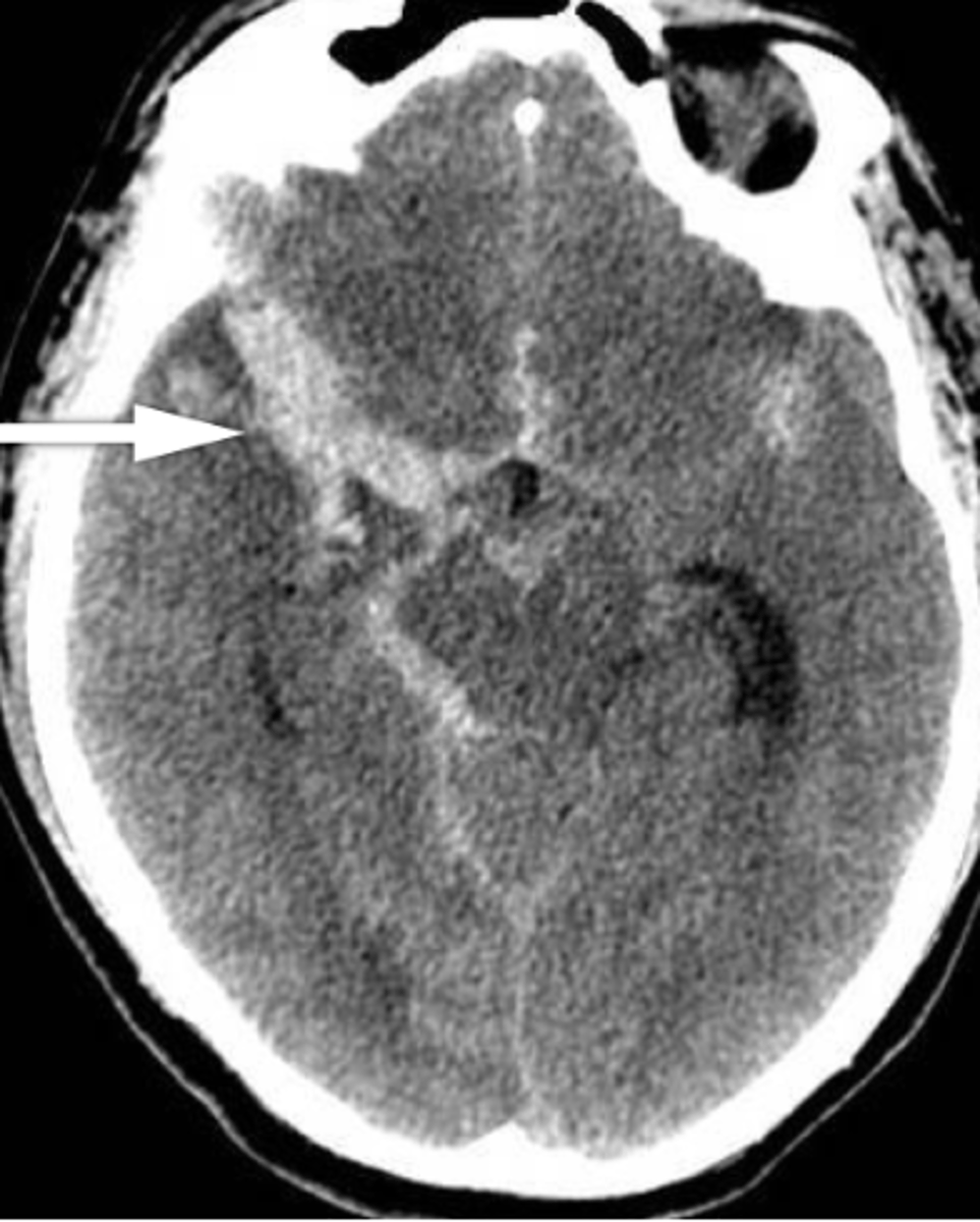
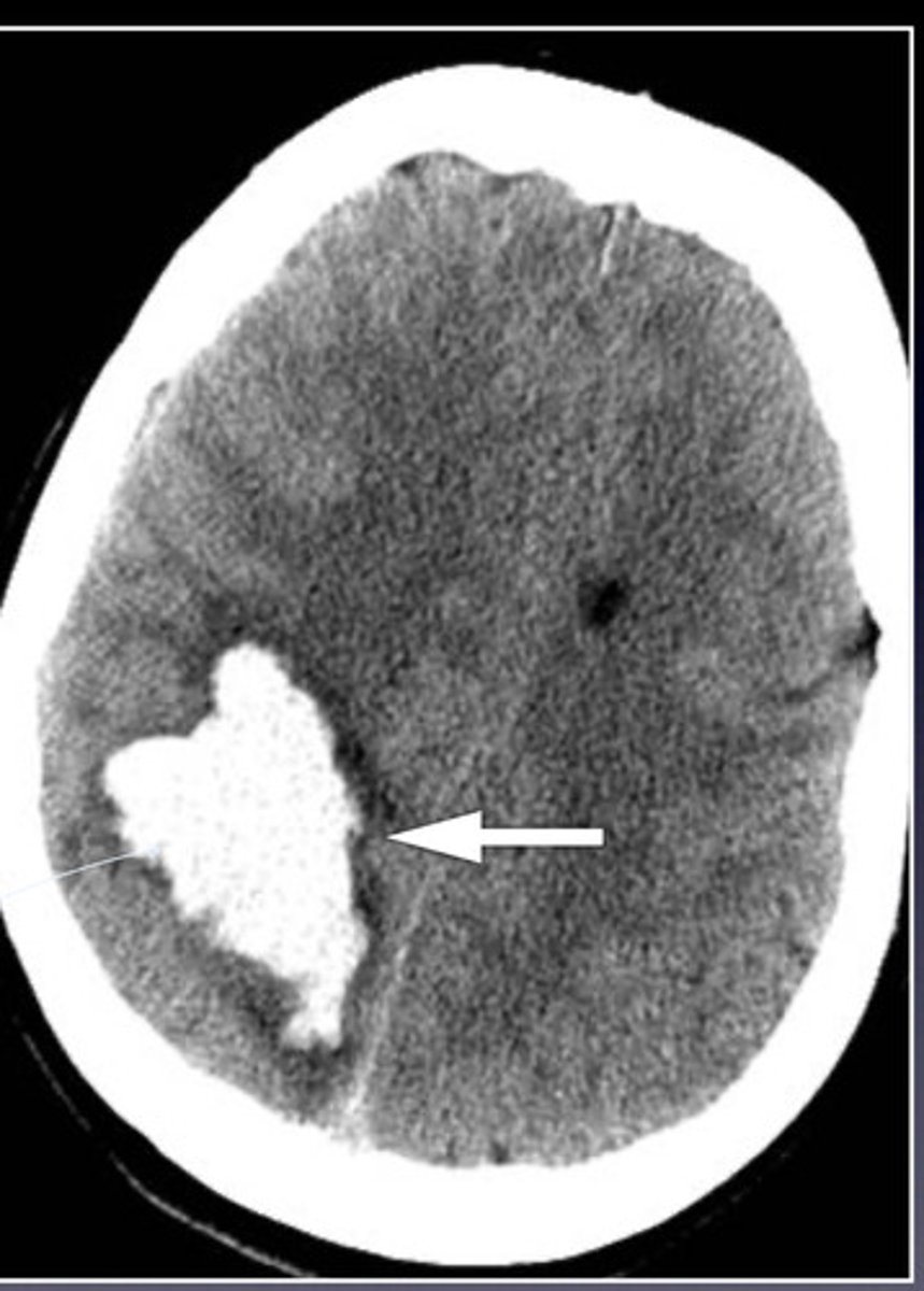
subdural hemorrhage
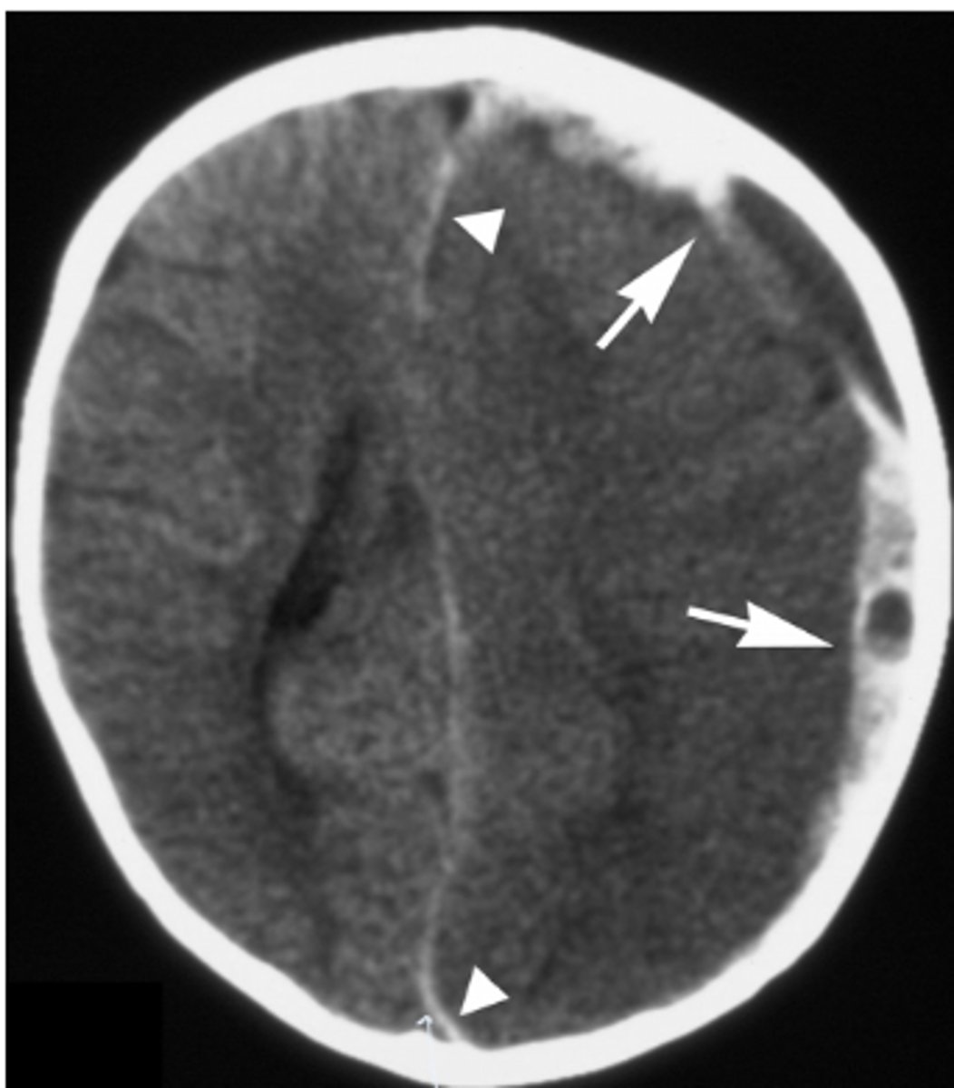
dura; brain; sutures; midline
subdural hemorrhage is bleeding between _______ and ______; it crosses _______, but not _______
epidural hemorrhage
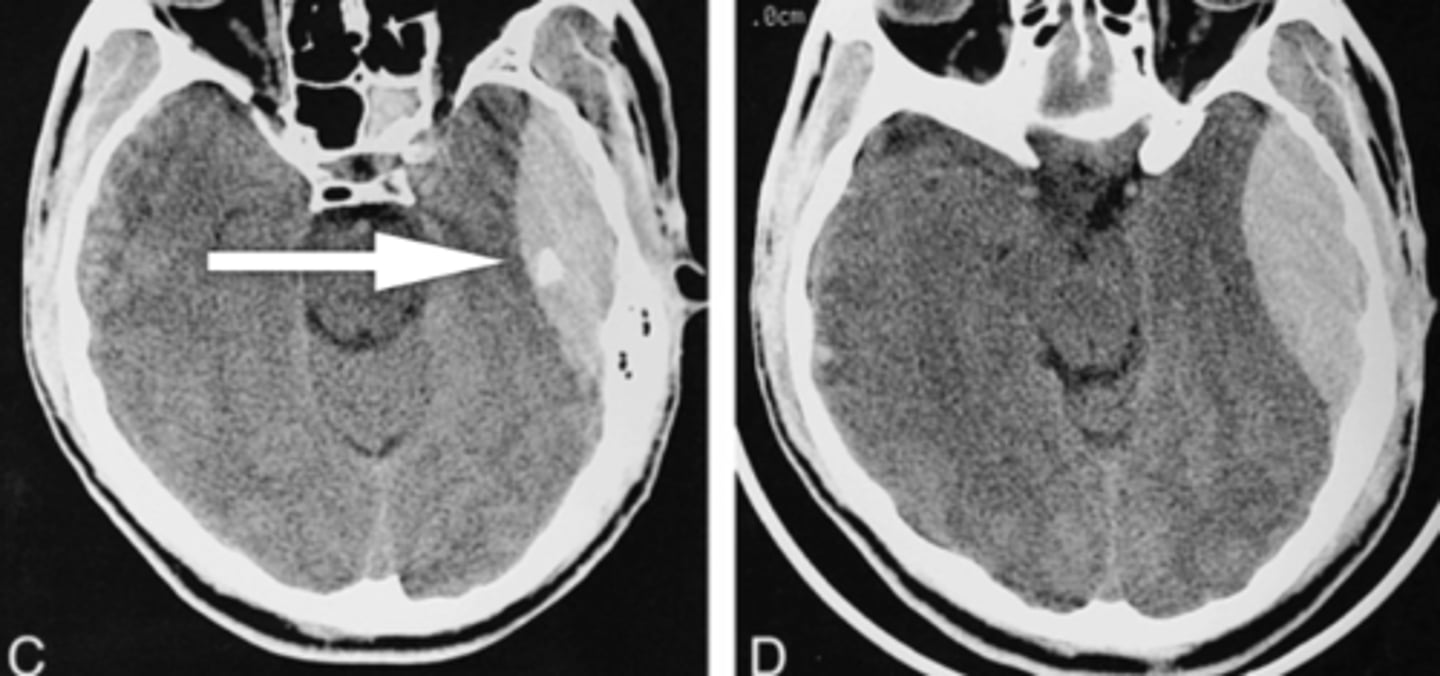
dura; skull; midline; sutures
epidural hemorrhage is bleeding between ________ and ________; it crosses __________ but not _________
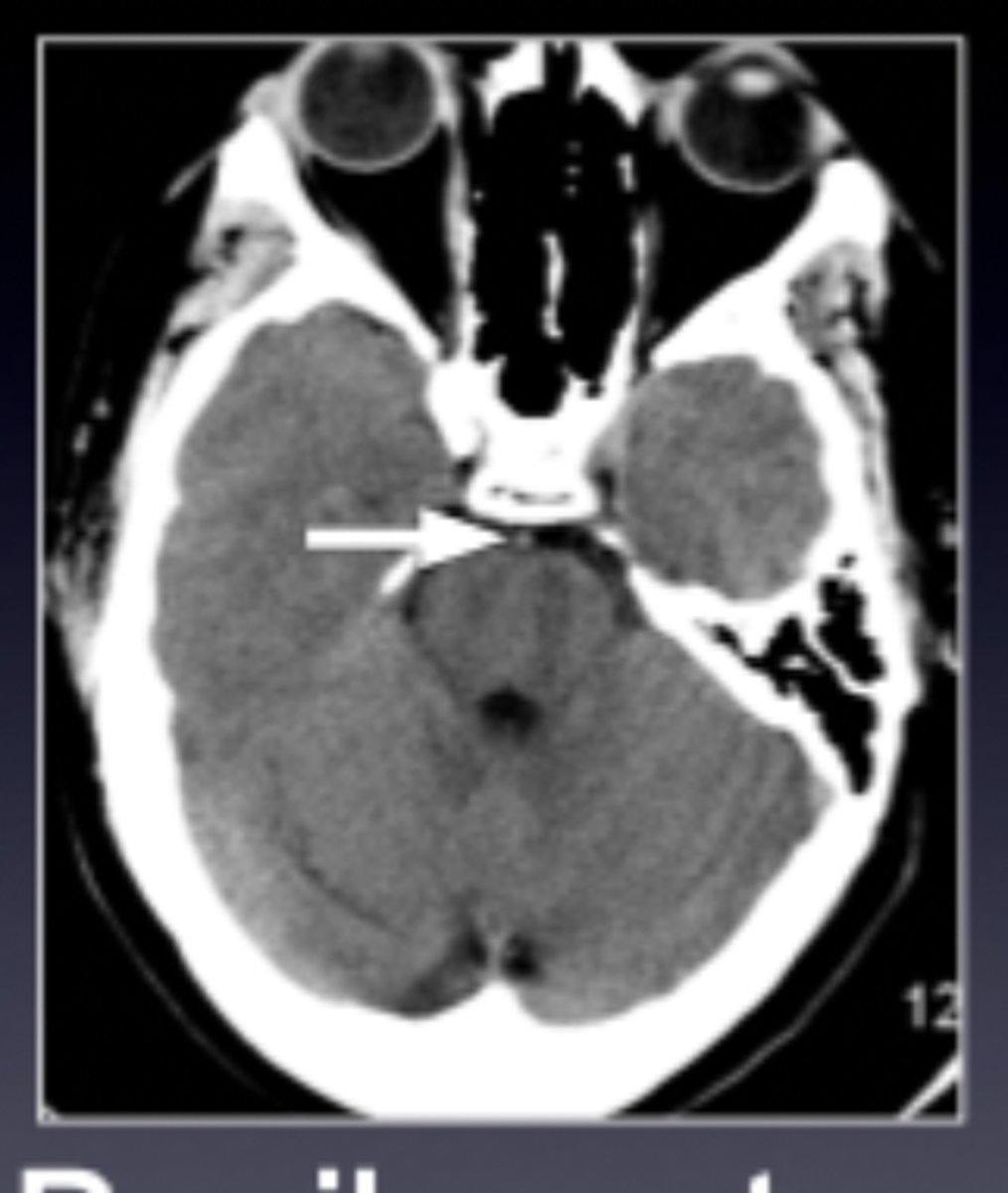
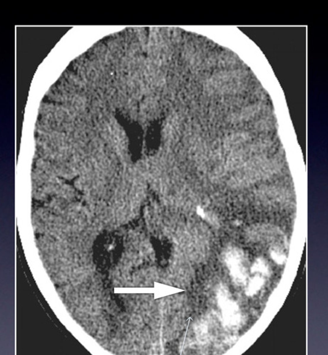
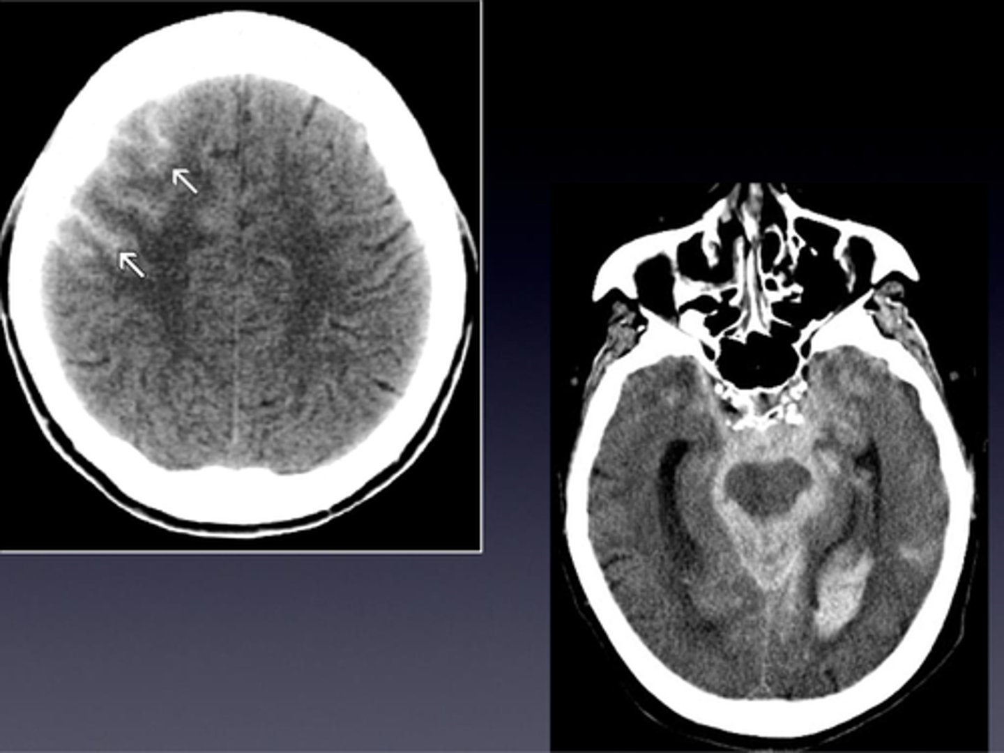
subarachnoid hemorrhage
subarachnoid hemorrhage
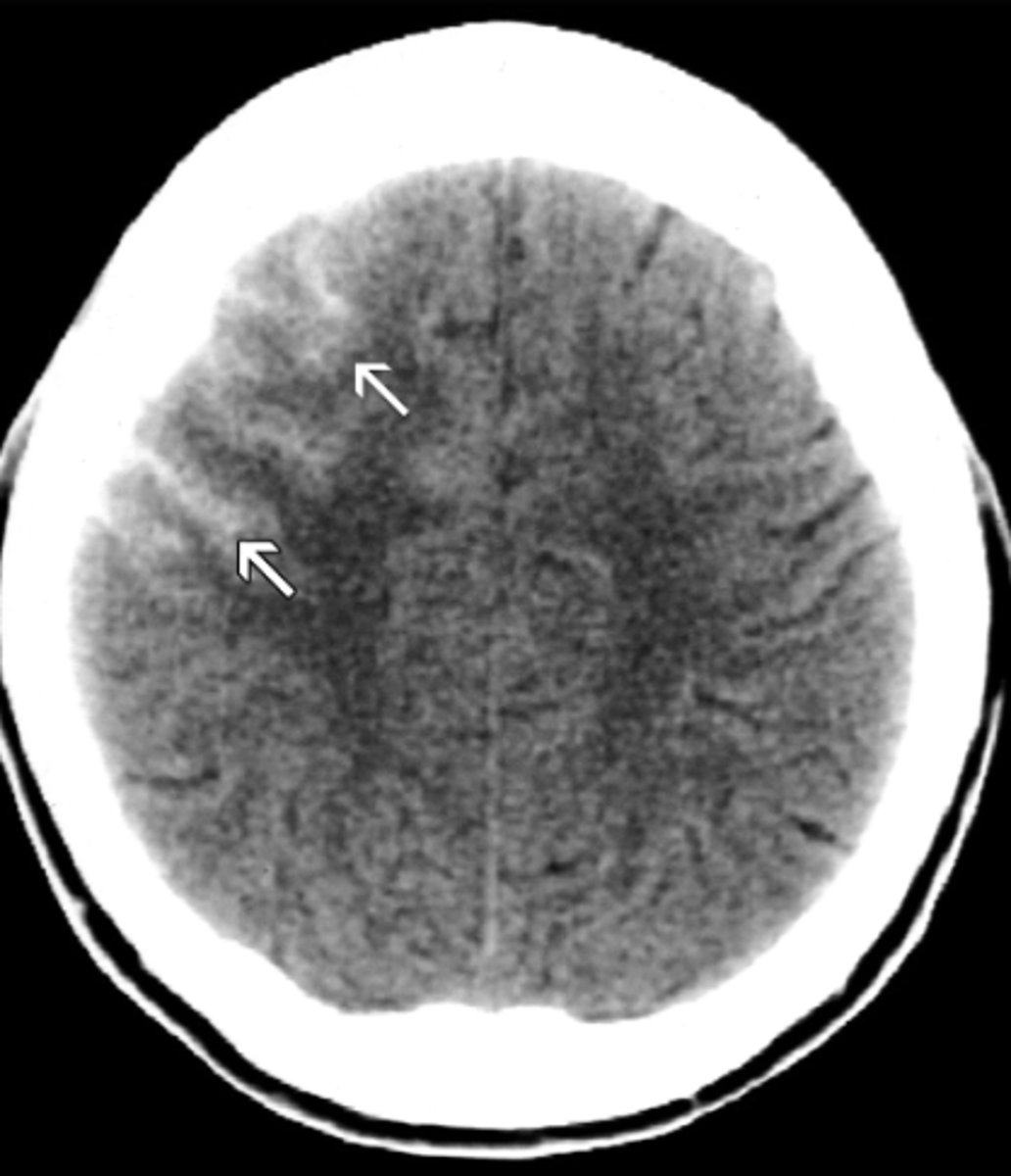
subarachnoid hemorrhage
lobes
subarachnoid hemorrhage is bleeding between _______
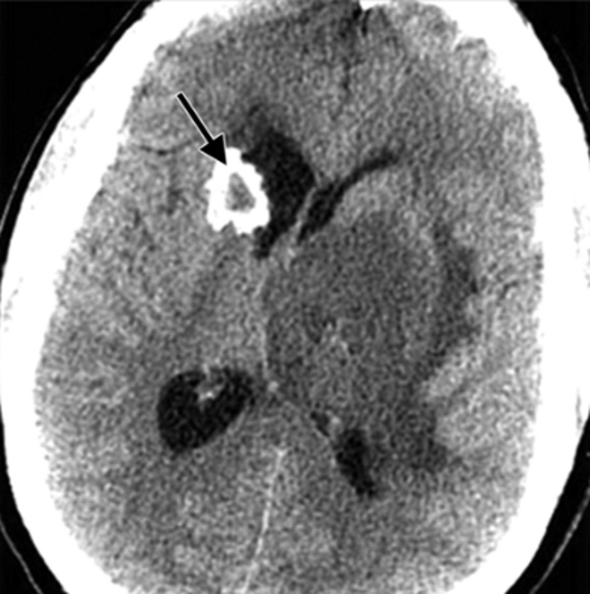
mass effect and midline shift
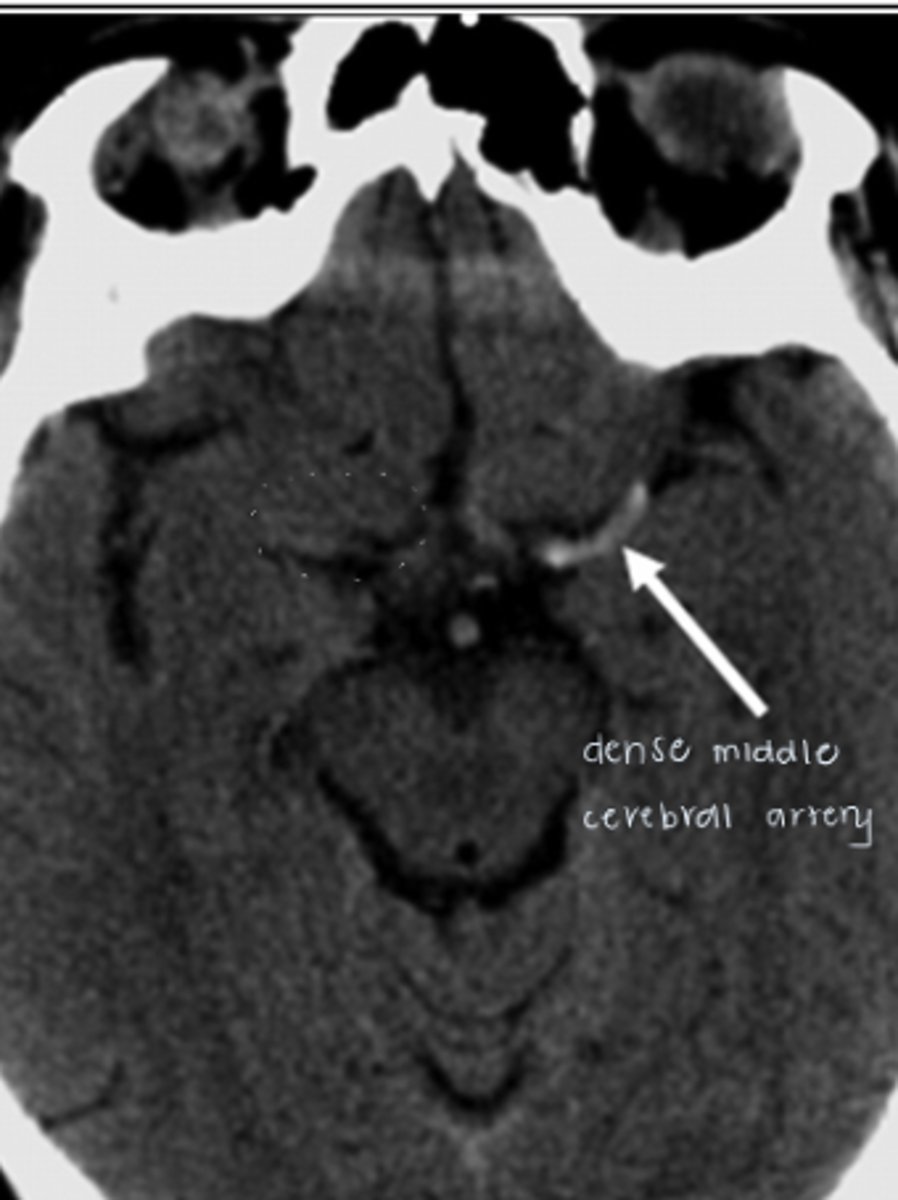
dense middle cerebral artery; early sign of stroke
what is this and what does it suggest?
hemorrhagic stroke
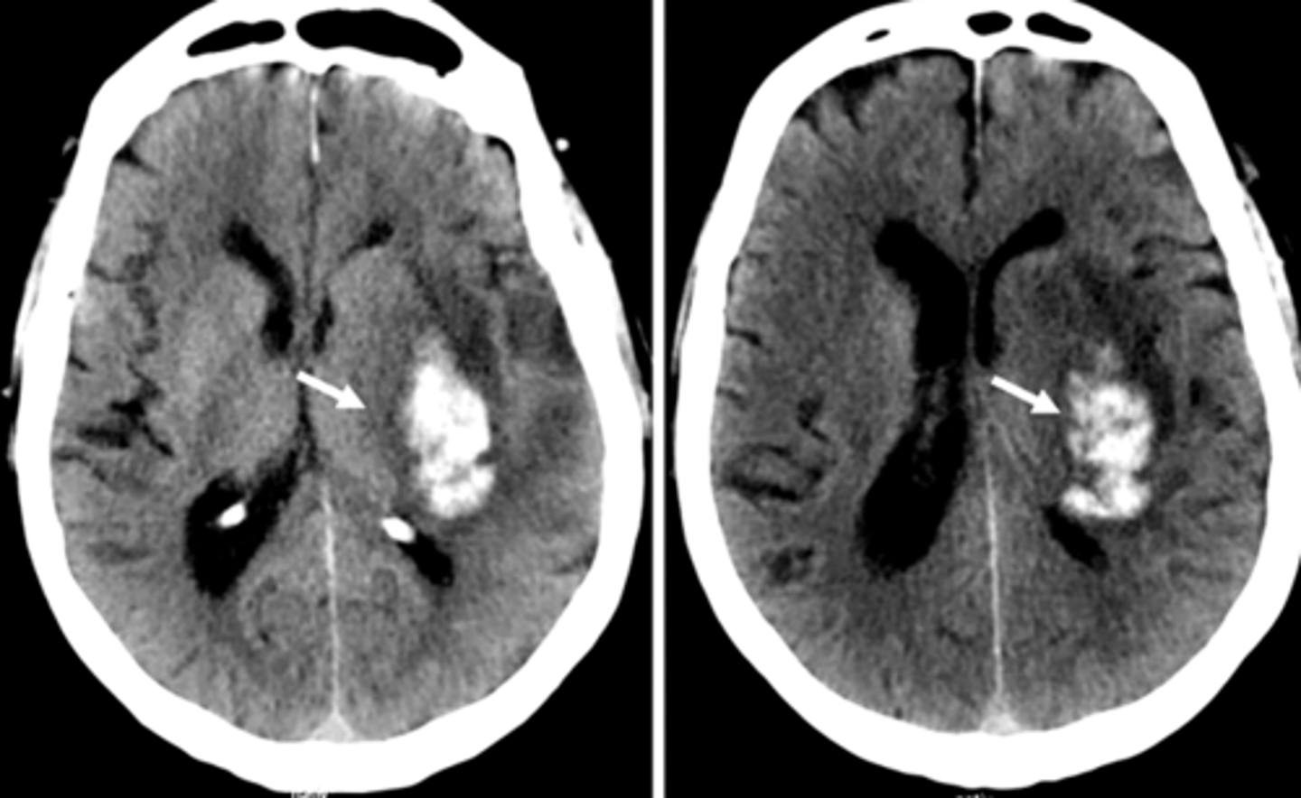
middle cerebral artery
most common artery involved in a stroke
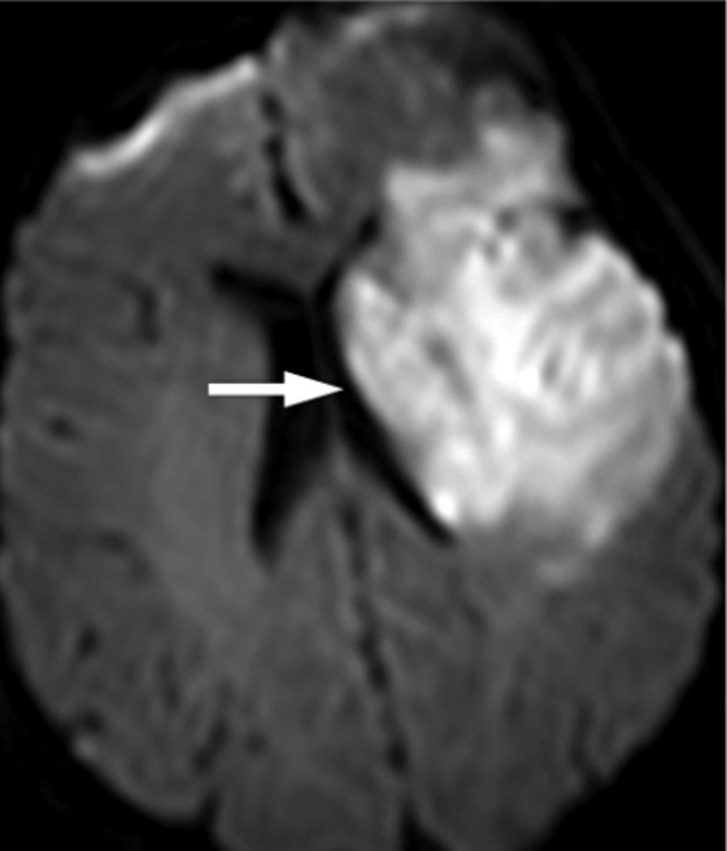
dwi; new cva's look bright
_______ mri is the best for stroke identification; what is a key finding?
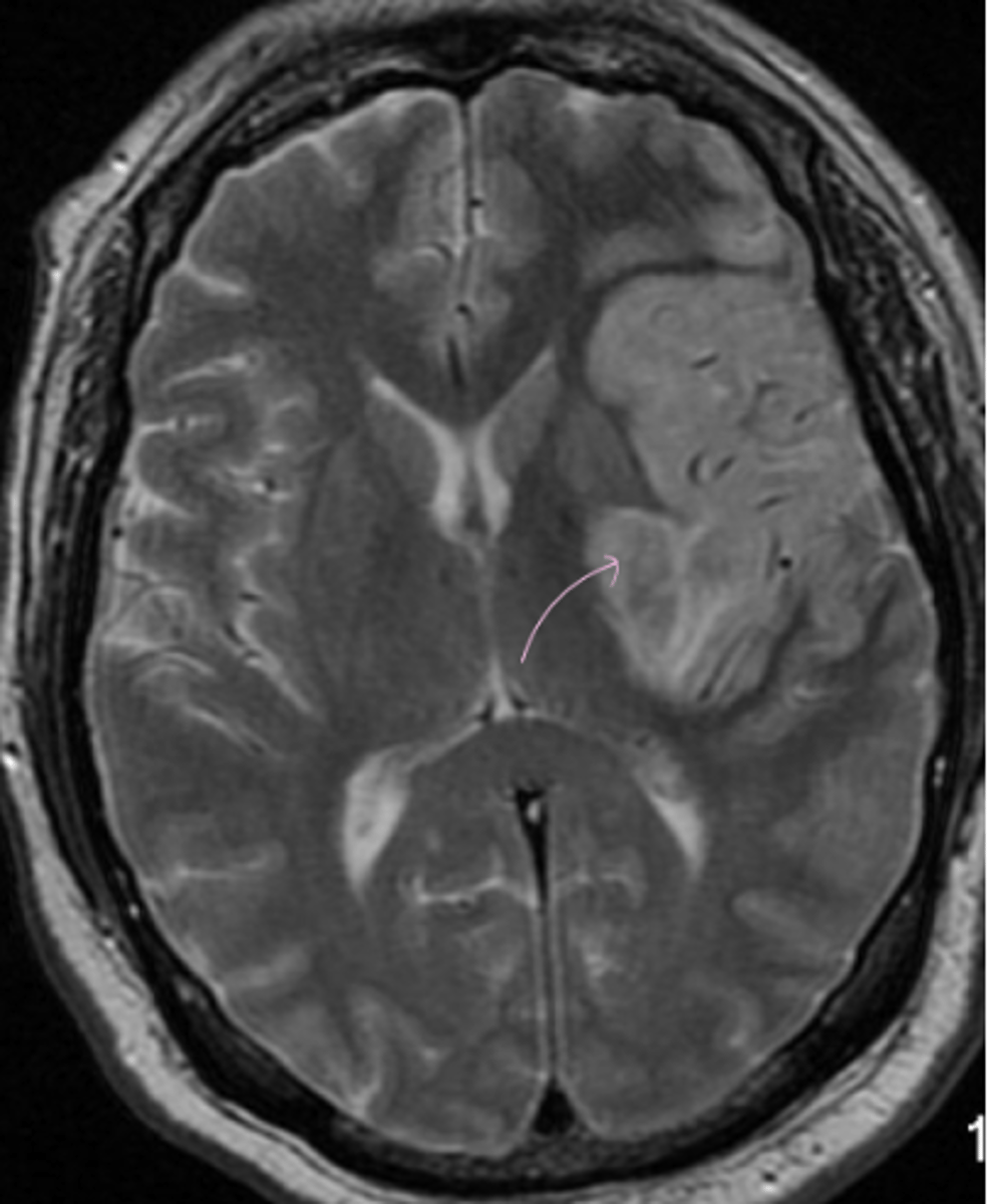
DWI (diffusion weighted image) of stroke that occurred 10-14 days ago
core: where the brain died; penumbra: area of the brain where there is not enough blood flow but if clot is dissolved quickly, brain can be salvaged and deficits resolved
what is penumbra and core in CVA
DWI (diffusion weighted image) of stroke that occurred 10-14 days ago
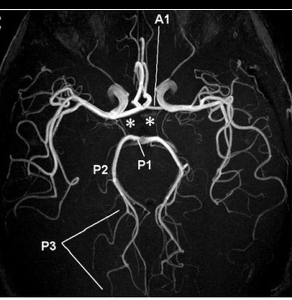
cerebral aneurysm via angiogram
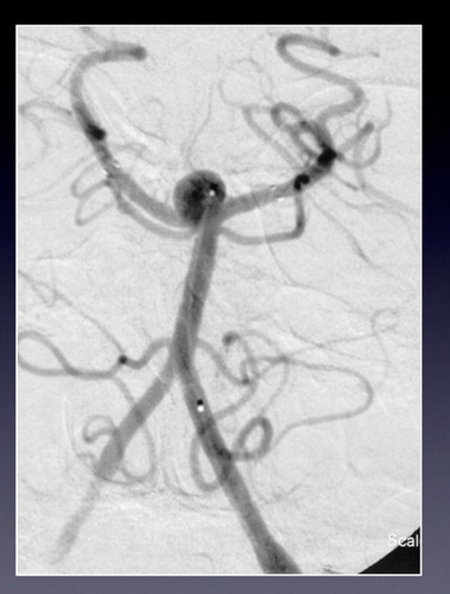
cerebral clot on angiogram
intracranial hemorrhage
amyloid angiopathy, hypertension, trauma
a common cause of this hemorrhage in the brain is:
CT; MRI
____ is used for acute intracranial bleed/hemorrhage, while ____ is used for everything else
true
T/F: contrast is not necessary most of the time, but is used for a suspected tumor or infection
intracranial hemorrhage; without
CT is the best test for acute ______________ _____________ and is ordered with/without contrast 99% of the time
mri
an ______ is best to view strokes, MS, malignancy, infection, or a non-emergent seizure workup
darker; brighter
white matter is ________ on CT while gray matter is __________ on CT
parenchymal, subarachnoid, subdural, epidural, intraventricular
there are 5 types of intracranial hemorrhage:
acute stroke
an ______ _____ is associated with mass effect
ruptured cerebral (intracranial) aneurysm
is the most common cause of non-traumatic subarachnoid hemorrhage
left:
- trauma (finger-like projections between gyri)
right:
- aneurysm
left vs. right
CT
best initial test for evaluating bleeding in the brain
CT
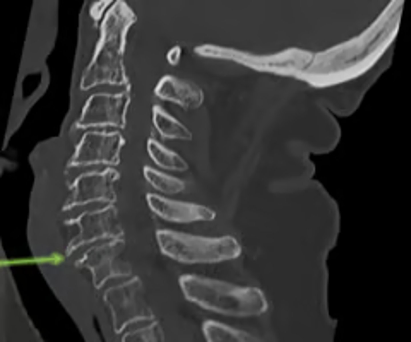
____ is first line for spinal trauma
spinal trauma imaging criteria
anterior longitudinal ligament, anterior 2/3 vertebral body and disc
anterior column of spine (contents)
posterior longitudinal ligament, posterior 1/3 vertebral body and disc
middle column of spine contents
articular facets, lamina, spinous process, ligamentum flavum, interspinous ligament
posterior column of spine contents
spondylosis
- osteophytes
this is _________, which is what?
spondylolithesis
- translation
this is _________, which is what?
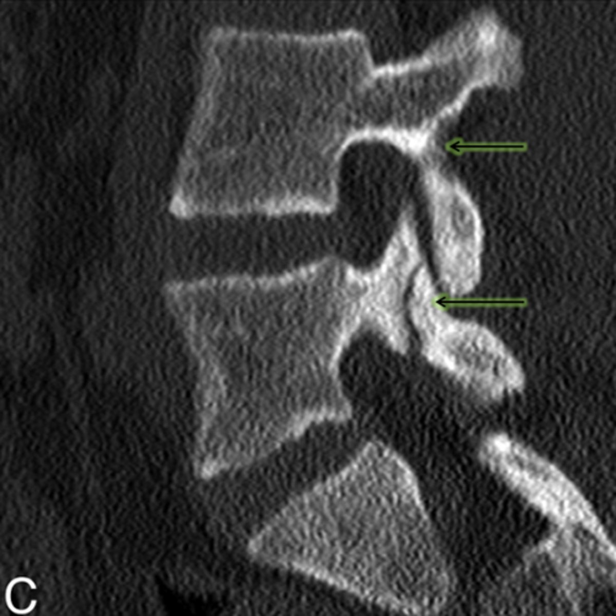
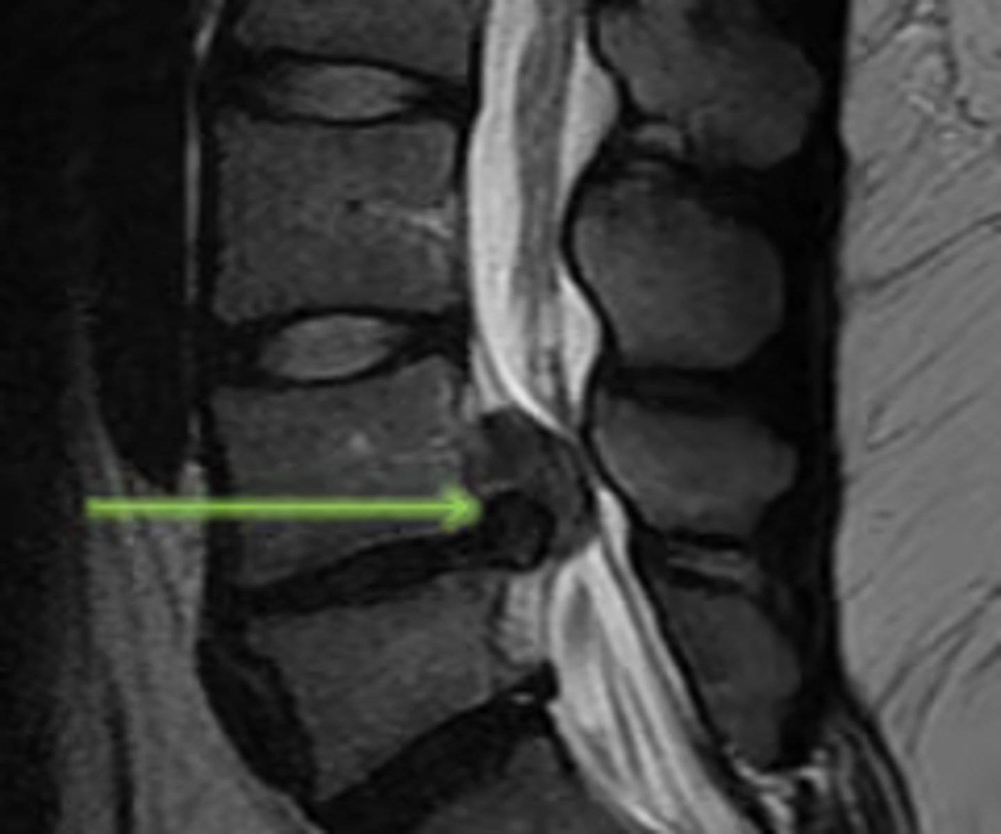
spondylolysis
- defect thru pars interarticularis
image: normal pars articularis, broken pars articularis
this is ______, which is what?
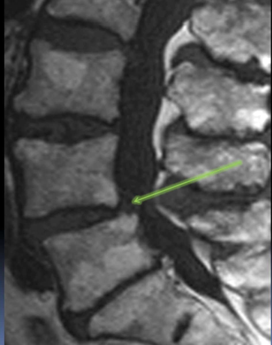
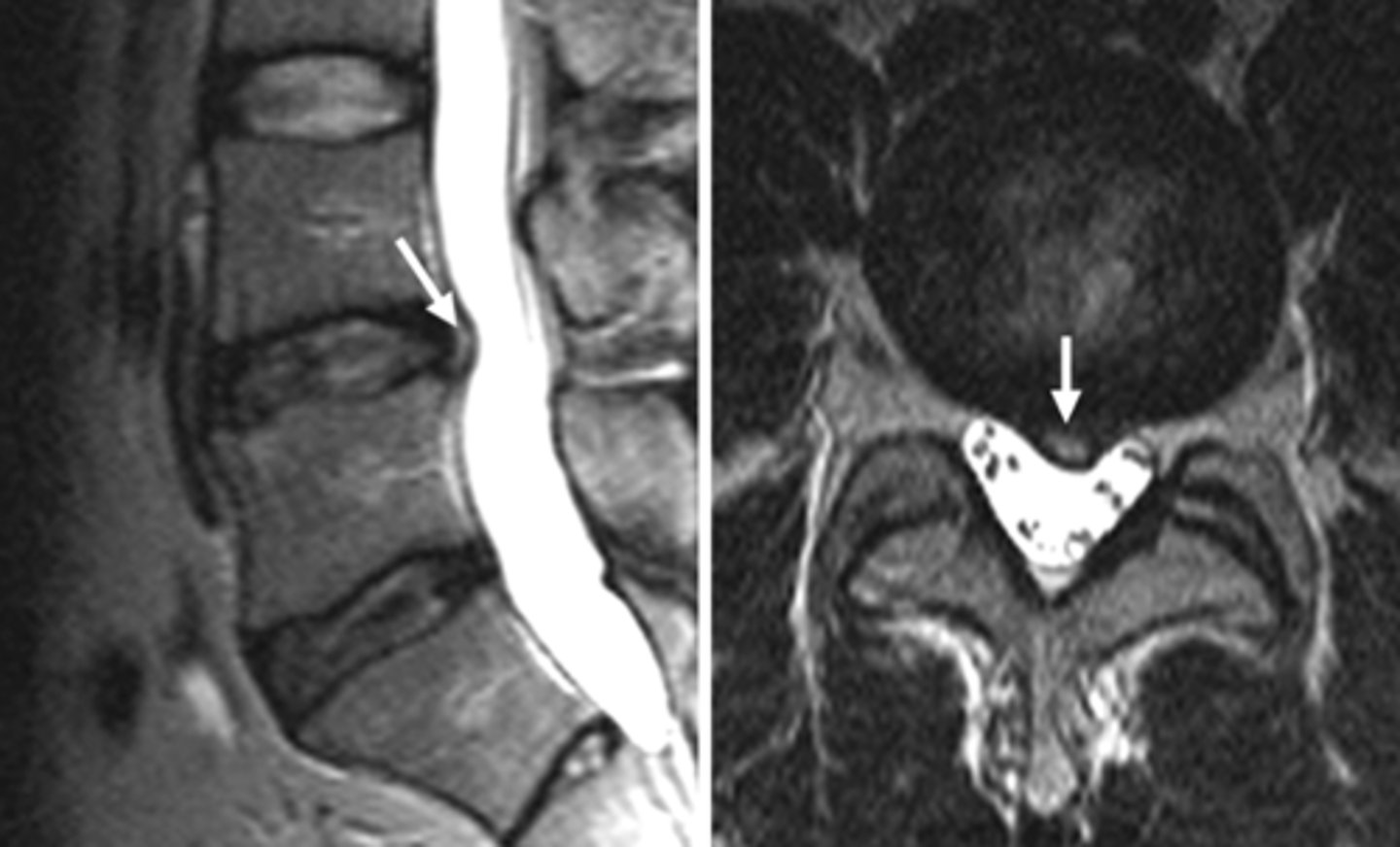
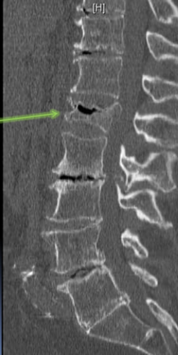
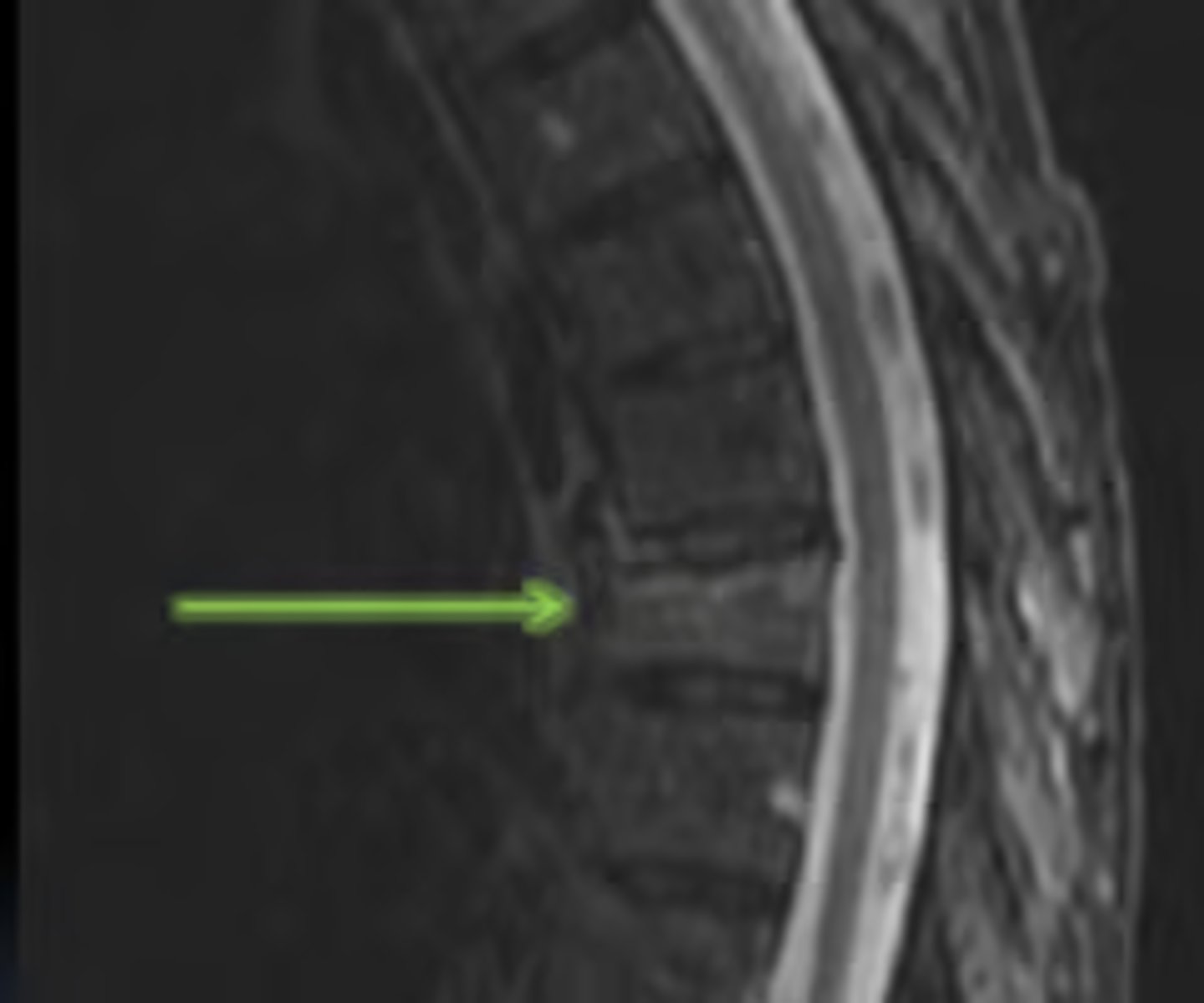
degenerative disc (characteristic finding: dark discs on mri)
what is this?

disc bulge
herniation (protrusion)
disc herniation
disc herniation (extrusion)
- protrusion: a little bit is coming out
- extrusion: comes out and it starts to go up or down
extrusion vs. protrusion disc herniation?
a- protrusion
b- extrusion
c- extrusion
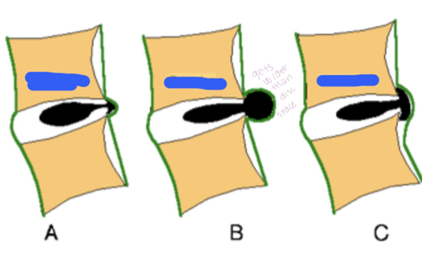
bulging gets wider, but herniated has a "mickey mouse ear" coming out of the intervertebral disc space
herniated vs. bulging discs
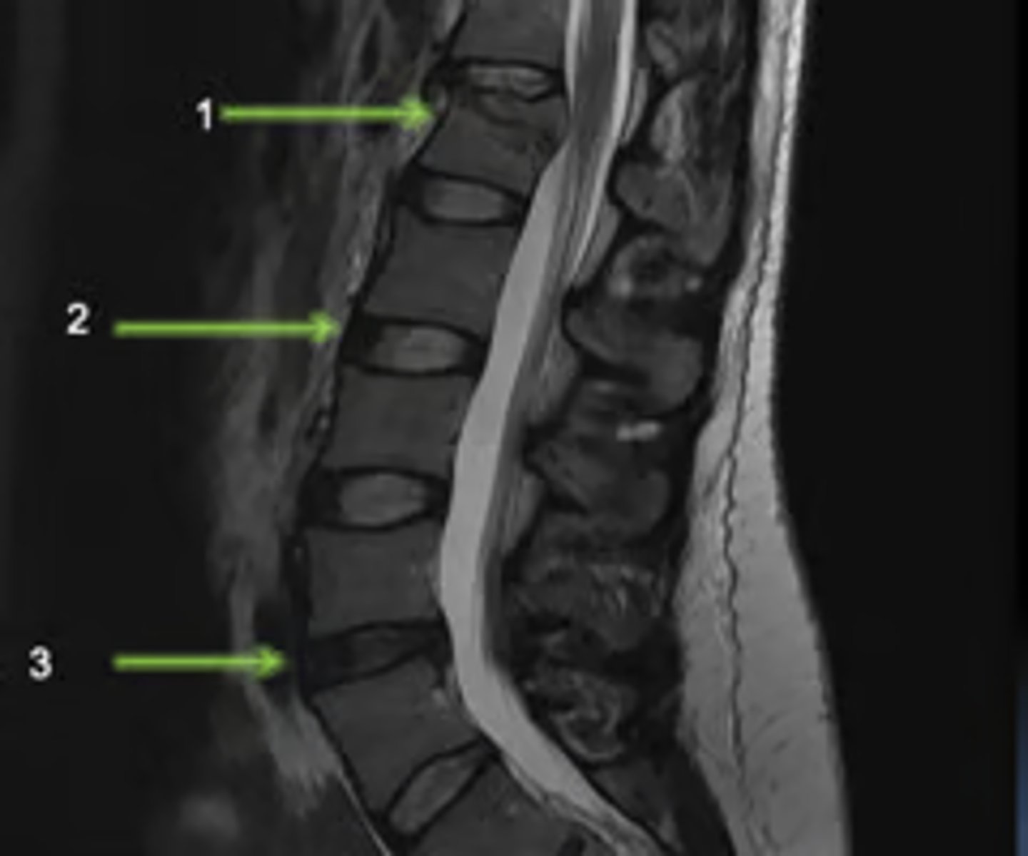
1- compression fracture
2- normal disc
3- abnormal disc
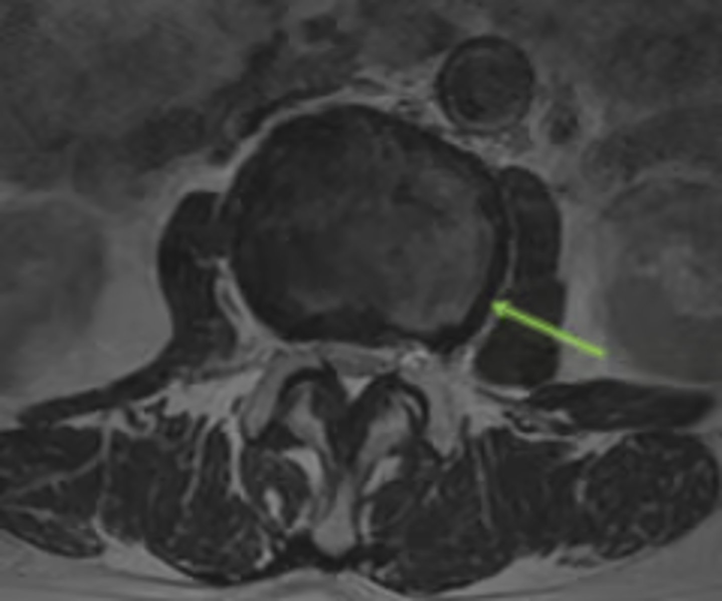
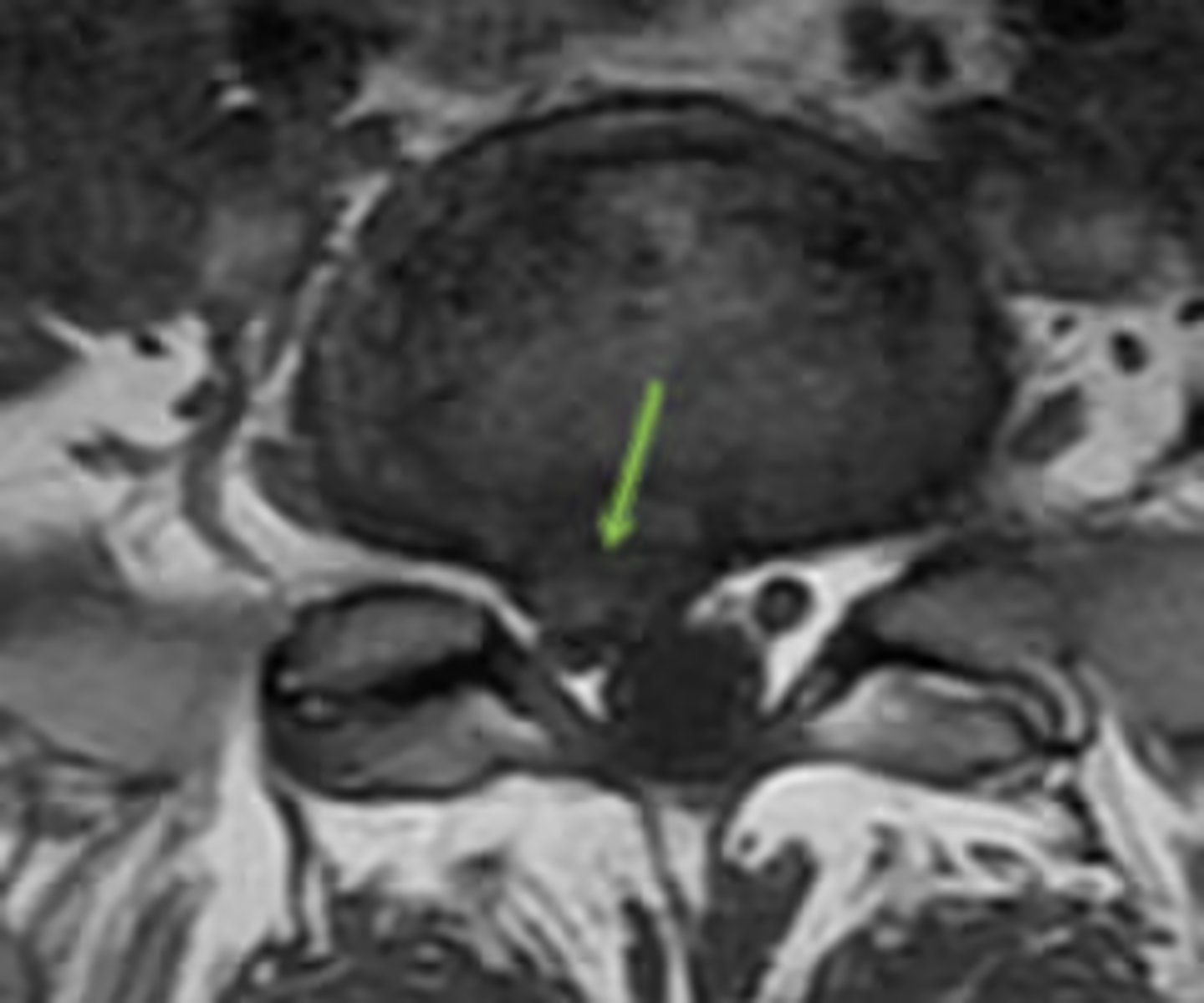
the area between the superior and inferior articulations of the vertebrae; common site of fractures due to spondylolysis
pars interarticularis
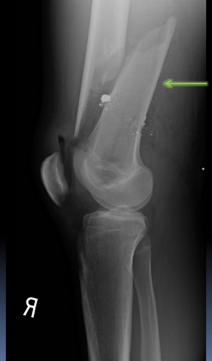
compression fracture
compression fracture
compression fracture with edema
typically a loss of height in the vertebral body, and a wedge-shaped or collapsed appearance
how to identify a compression fracture
x-ray
image modality of choice in msk trauma
epiphysis
metaphysis
diaphysis
scaphoid and navicular fracture
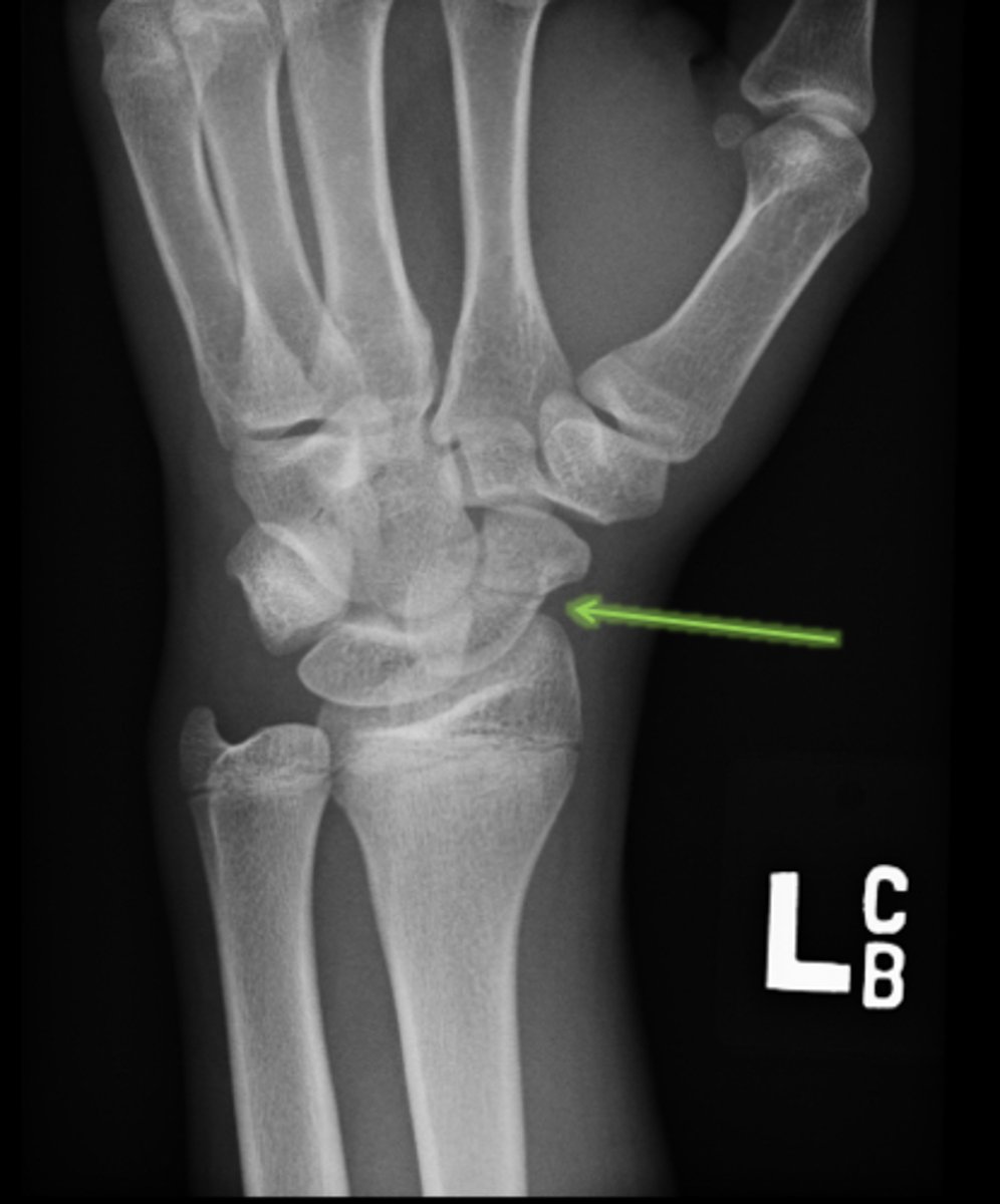
osteonecrosis
complication that can arise from a scaphoid/navicular fracture
7-10 days
(for persistent pain & point tenderness ---> CT & MRI for further evaluation; MRI for ligament or tendon injuries)
when to do follow up with x-ray in patients
open fracture
when a fracture extends through the skin
comminuted
more than two fragments
interarticular fracture
cartilage involvement of fracture
displacement fracture
distal fragment will move relative to proximal fragment in _______ _______
lateral displacement of distal fracture fragment

posterior displacement of distal fracture fragment
anterior displacement of distal fracture fragment
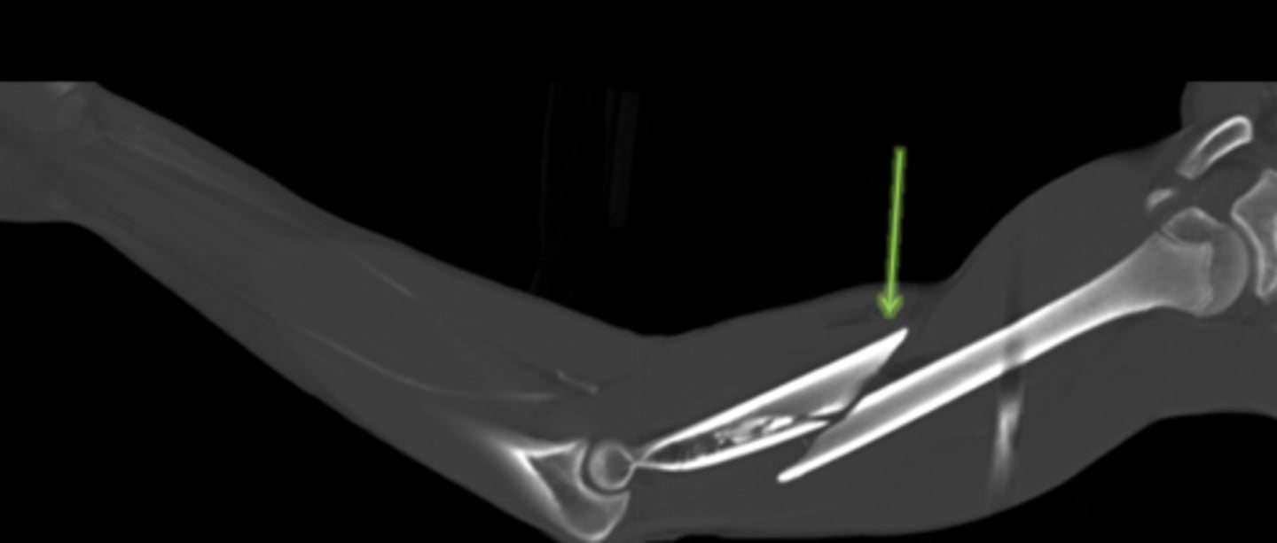
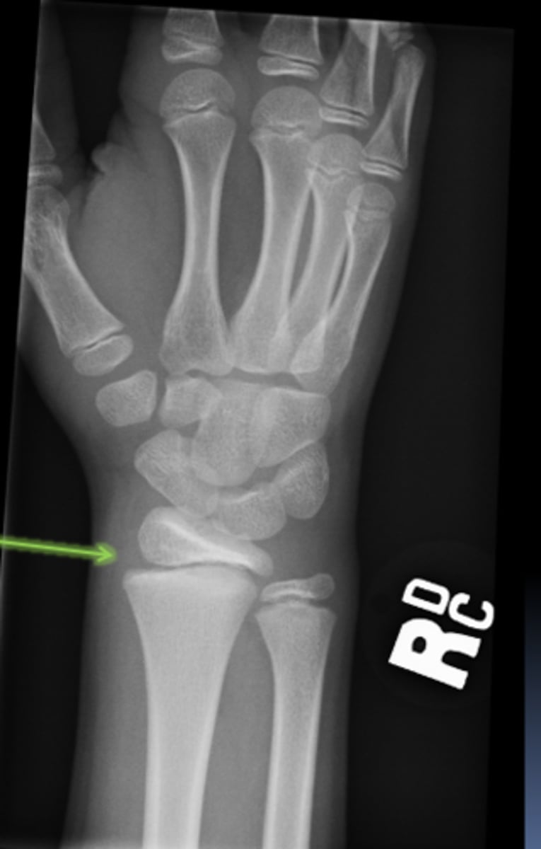
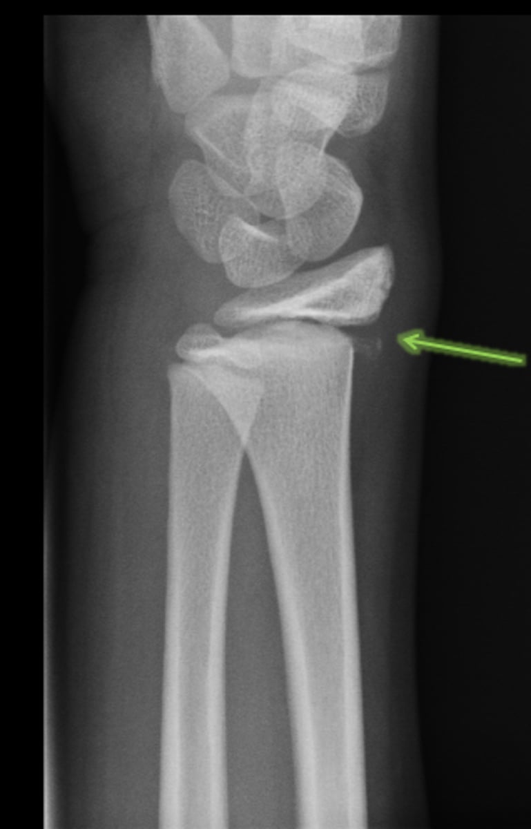
1. ulnar styloid fracture
2. torus/buckle fracture at radius
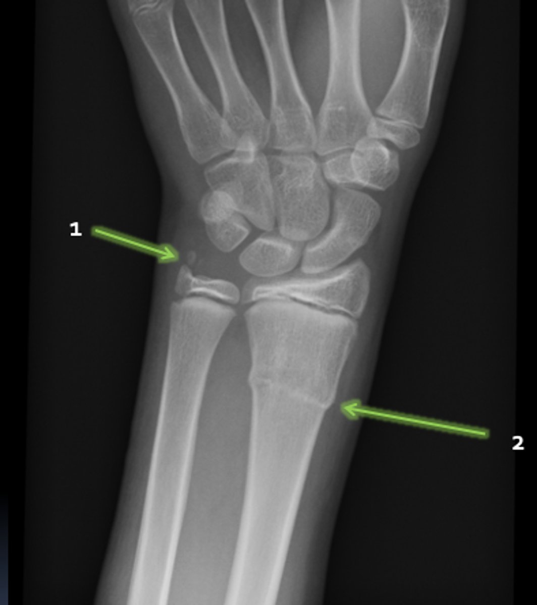
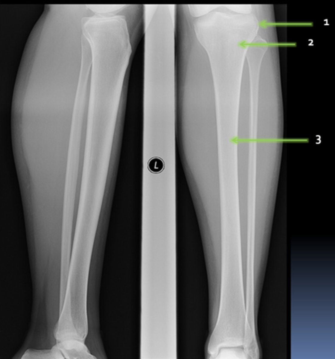
torus/buckle fracture
common pediatric long bone fracture:
salter harris
use what to describe pediatric fractures:
fracture through physeal (growth) plate only

with metaphyseal fracture

with epiphyseal fracture

with metaphyseal and epiphyseal fracture

crush injury

type 1 salter harris
type 2 salter harris
a shoulder dislocation occurs between the glenohumeral joint;
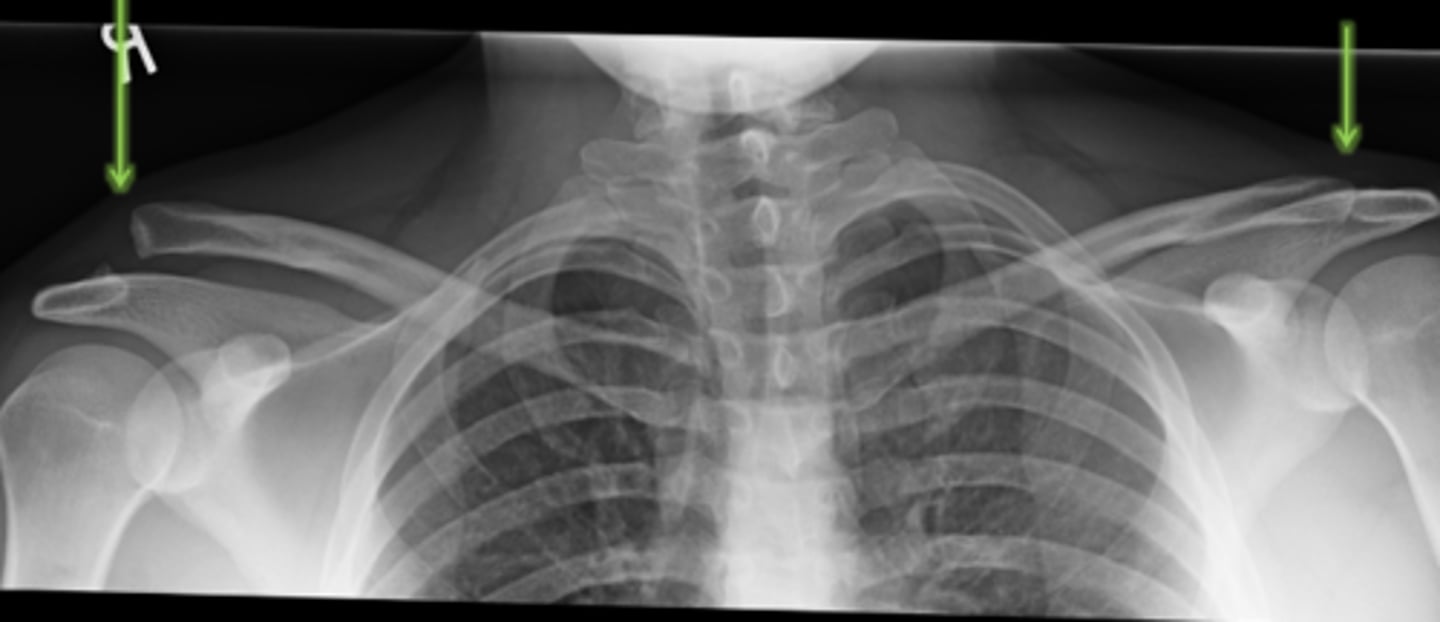
a shoulder separation occurs between the acromioclavicular joint
shoulder separation vs. dislocation
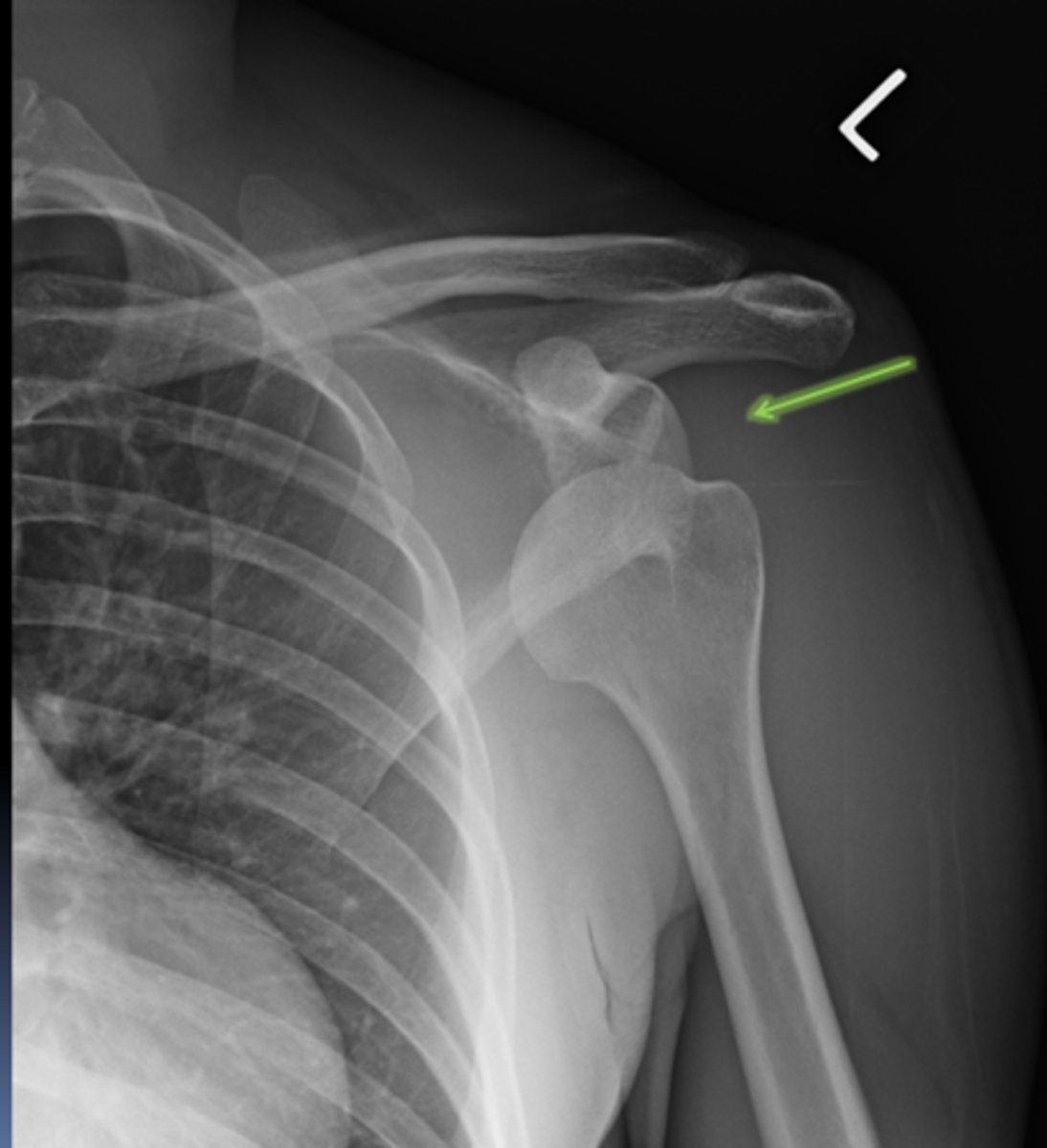
shoulder dislocation
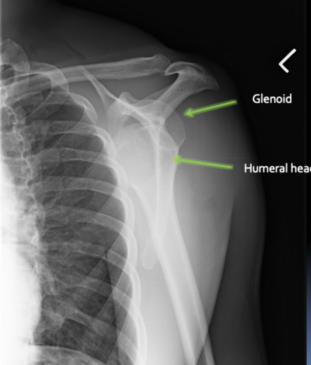
shoulder dislocation
separated AC joint, normal AC joint
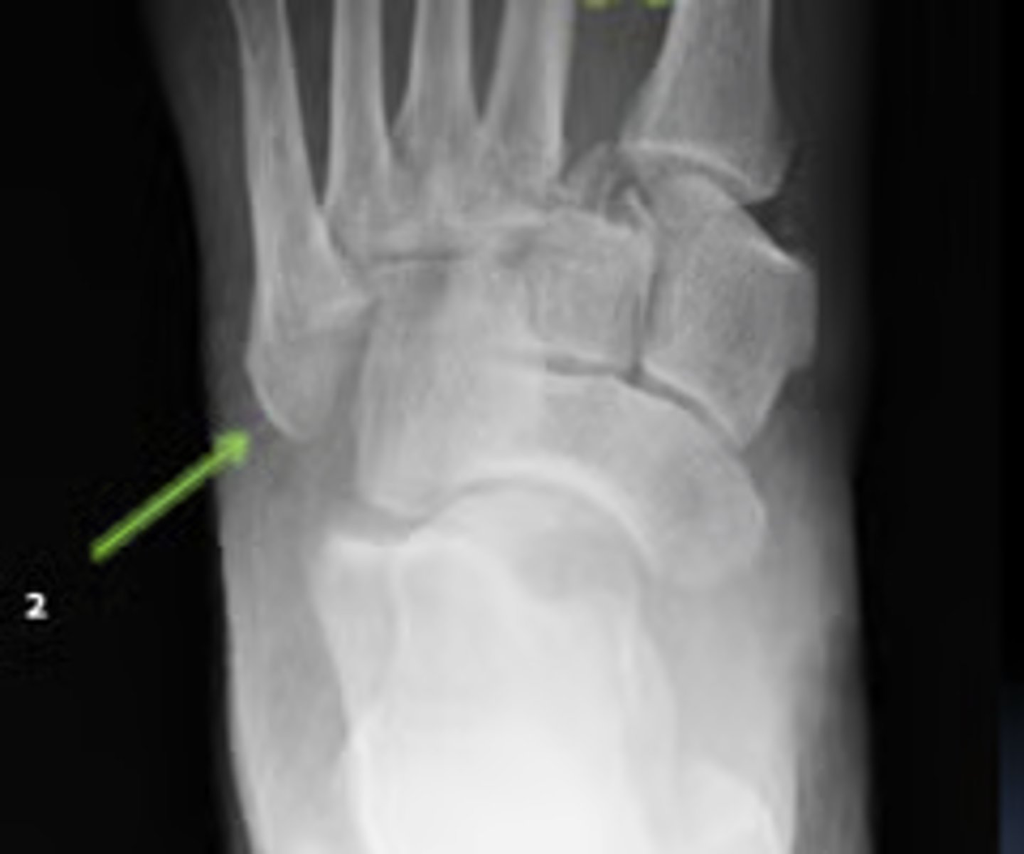
lisfranc fracture
fracture-dislocation of the tarsometatarsal joint/ midfoot
can slide laterally or "split in half" if left untreated
why lisfranc fractures are so important to catch
lisfranc fracture